Abstract
Deletion of the 17p13 chromosomal region [del(17p)] is associated with a poor outcome in multiple myeloma. Most of the studies have targeted the TP53 gene for deletion analyses, although no study showed that this gene is the deletion target. In order to address this issue, we sequenced the TP53 gene in 92 patients with multiple myeloma at diagnosis, 54 with a del(17p) and 38 lacking del(17p). At least one mutation was found in 20 patients, all of them presenting a del(17p). The analysis of the mutation location showed that virtually all of them occurred in highly conserved domains involved in the DNA-protein interactions. In conclusion, we showed that 37% of the myeloma patients with del(17p) present a TP53 mutation versus 0% in patients lacking the del(17p). The prognostic significance of these mutations remains to be evaluated.
Keywords: mutations, TP53, del(17p), multiple myeloma
Introduction
The prevalence of TP53 mutations differs considerably between tumor types and stages of cancer, and approximately 50% of all tumors present mutations. In multiple myeloma (MM), mutations of the TP53 gene is rarely detected at diagnosis, although it becomes more frequent in advanced disease1 and human myeloma cell lines.2 In other hematologic malignancies, like diffuse large B-cell lymphomas (DLBCL),3,4 follicular lymphoma5 or chronic lymphocytic leukemia (CLL)6 mutations in TP53 correlate with unfavorable prognosis and chemotherapy resistance, especially when located in DNA binding domain. Furthermore, a strong correlation between 17p deletions and TP53 mutations has been shown in CLL.
In multiple myeloma, we previously showed that deletion of the TP53 gene (located at 17p13) was present in 7% of the patients enrolled in the IFM99 trials and tested by FISH. After a median follow up of 56 months, univariate statistical analyses showed that del(17p) negatively impacted both the event free survival and the overall survival.7 However, it is unknown whether p53 signaling is still functional in those myeloma cells or if p53 is totally inactivated through mutations on the other allele. We therefore set out to clarify the prevalence of TP53 mutations in del17p MM patients and compared it to prevalence in a series of non-del(17p) MM patients.
Design and Methods
Patients
Primary myeloma cells were obtained from bone marrow aspirates after Ficoll density gradient centrifugation followed by separation of myeloma cells with CD138 microbeads (StemCell Technologies, Vancouver, Canada). Cytospins of purified samples stained according to the MGG method routinely confirmed plasma cell morphology for more than 90% of cells. All primary cells were obtained from routine diagnostic samples after informed consent was provided by the patients. Fluorescence in situ hybridization (FISH) analysis using a TP53-specific probe (Abbott, Rungis, France) was routinely performed for patients enrolled in the IFM99 trials. RNA from 92 untreated patients was obtained from purified plasma cells. This study was conducted in accordance with the Declaration of Helsinki.
TP53 mutation analysis
We screened cDNA samples (exons 2-11) for TP53 mutations by direct sequencing as described previously.8 Two overlapping fragments spanning the TP53 coding region were amplified by PCR, purified, and were bidirectionally sequenced, using the same primers and the Big Dye Terminator kit on the Applied Biosystems 3130xl Genetic Analyser (Applied Biosystems, Foster City, CA, USA). Data were analyzed by visual inspection of electropherograms on Seqscanner software and compared to reference sequence NM_000546.2 (NCBI Nucleotide) using Seqscape software (Applied Biosystems, Foster City, CA, USA). TP53 mutations found in patients were compared to the UMD p53 Web site (http://p53.free.fr/)9 and analyzed using MUT-MAT 2.0, a verification spreadsheet to certify p53 mutations.10
Results and Discussion
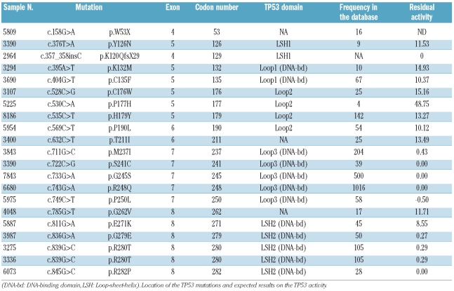
Deletion of the short arm of chromosome 17 was detected in 11% of newly diagnosed patients.7 However, we did show that a short survival was predicted only in patients with a deletion present in at least 60% of the plasma cells (7% of the patients at the time of diagnosis). We sequenced cDNA coding for the TP53 gene in 54 of those patients. Twenty-one hemizygous TP53 mutations were detected in 20 of these 54 cases (37%) of MM with del(17p) (Table 1) including 19 single nucleotide missense mutations, one single nucleotide nonsense mutation and one single nucleotide nonsense insertion (Table 2), unlike Chng et al. who found a majority of deletions and insertions.11 We compared these cases with 38 patients lacking del(17p). No TP53 mutation was found in those cases of newly diagnosed MM without del(17p) (P<0.0001).
Table 1.
Results of TP53 mutation analysis.
Table 2.
Description of the 21 TP53 mutations in 20 patients according to Ref Seq for p53: GenBank: NM_000546.2.
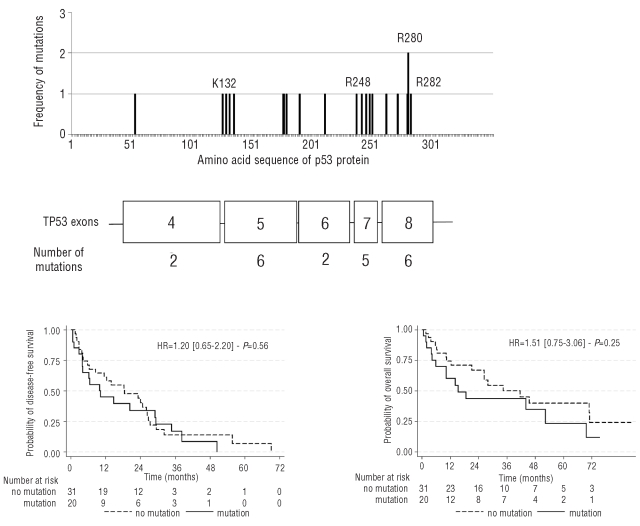
The distribution of the 21 mutations was one insertion and one point mutation in exon 4, 6 point mutations in exon 5, 2 in exon 6, 5 in exon 7, and 6 in exon 8 (Figure 1). No mutations were identified in exons 2, 3, 9, 10, or 11. All missense mutations were previously reported in the UMD TP53 mutation database, most of them were frequent and 5 were infrequent. When the TP53 mutation distribution pattern was analyzed, these included 2 mutations in LSH1 (loop-sheet-helix1), 2 mutations in Loop-L1, 4 mutations in Loop-L2, 5 mutations in Loop-L3, 5 mutations in the LSH 2 regions (Table 2); more than half of the TP53 mutations found in our series were located to DNA-binding domain, as described in diffuse large B-cell lymphomas (DLBCL).3,4
Figure 1.
TP53 mutations are distributed in exons 4 to 8, with no recurrent mutation.
Indeed, as described by Levine, loops 1 and 3 and the Loop-Sheet-Helix (LSH) region from codons 272 to 287 make direct contacts with the DNA adjacent to a gene that is regulated by the p53 transcription factor, while loop 2 is required for folding and stabilization of this DNA-binding domain and is not in direct contact with DNA.12
As recently reported by Young et al. in DLBCL,4 position of the mutation seems to determine the outcome. Such correlation has not yet been described in MM. In 2 cases, the mutations were localized to codons involved in the zinc-binding site (Cys176 and Cys 179). Seventeen mutations were localized to highly conserved areas: 4 mutations in area II (codons 117-142), 3 mutations in area III (codons 171-181), 5 mutations in area IV (codons 234-258), and 5 mutations in area V (codons 270-286). According to the UMD TP53 mutation database, virtually all p53 mutants had low remaining activity (Table 2). Survival analyses did not reveal any difference between patients presenting a TP53 mutation and those with a germline TP53, in patients with del(17p) (Figure 2). However, the numbers are too small to draw any definitive conclusion.
Figure 2.
Patients with TP53 mutations do not present different outcome.
We did not find TP53 mutations in the vast majority (63%) of del(17p) hemizygous patients, suggesting that normal p53 protein is still present and still functional in these non-mutated patients and potentially overcome the poor effect of del(17p). However, epigenetic modifications may occur that alter the p53 pathway. Absence of response to treatment in patients with del(17p), especially in chronic lymphocytic leukemia (CLL) treated with fludarabine, is strongly in favor of the implication of the TP53 pathway. Also in CLL, TP53 mutations have as poor a prognosis as del(17p), suggesting a similar mechanism, probably because of the high incidence of TP53 mutations in patients with del(17p)6.
We may have underestimated the frequency of TP53 mutations: as we sequenced cDNA, we could not estimate frequency of mutations in introns. However, very few mutations have been described in introns so far3 and we doubt many intron mutations have been missed. However, direct sequencing (similarly to PCR-SSCP, DHPLC, or HRM assay frequently used to detect TP53 gene mutations) has low sensitivity and may miss some minor mutant subclones which could subsequently be responsible for relapse and explain poor prognosis of del(17p). Indeed, some studies have demonstrated a mutation in up to 40% of patients with end-stage disease and plasma cell leukemia.1,13,14
As a result, more sensitive technologies such as Next-Generation Sequencing technology could help in finding some minor mutant subclones, and functional impairment of p53 in del(17p) patients has to be explored through other effectors of the p53 DNA repair pathway (like MDM215 or ATM16). Recently, Xiong et al. reported on genes regulated by p53, and confirmed that low expression of p53 was associated with a poor outcome.17 In our small series (92 patients, 54 [del(17p)]), it was not possible to explore the prognostic impact of TP53 mutations. Analysis of mutations in purified plasma cells in other larger series is required to address this issue.
Acknowledgments
we thank Fabienne Perrault-Hu, Laetitia Ergand, Marie-Christine Boursier and Amandine Sebie for technical assistance.
Footnotes
Authorship and Disclosures
The information provided by the authors about contributions from persons listed as authors and in acknowledgments is available with the full text of this paper at www.haematologica.org.
Financial and other disclosures provided by the authors using the ICMJE (www.icmje.org) Uniform Format for Disclosure of Competing Interests are also available at www.haematologica.org.
References
- 1.Neri A, Baldini L, Trecca D, Cro L, Polli E, Maiolo AT. p53 gene mutations in multiple myeloma are associated with advanced forms of malignancy. Blood. 1993;81(1):128–35. [PubMed] [Google Scholar]
- 2.Mazars GR, Portier M, Zhang XG, Jourdan M, Bataille R, Theillet C, et al. Mutations of the p53 gene in human myeloma cell lines. Oncogene. 1992;7(5):1015–8. [PubMed] [Google Scholar]
- 3.Young KH, Weisenburger DD, Dave BJ, Smith L, Sanger W, Iqbal J, et al. Mutations in the DNA-binding codons of TP53, which are associated with decreased expression of TRAILreceptor-2, predict for poor survival in diffuse large B-cell lymphoma. Blood. 2007;110(13):4396–405. doi: 10.1182/blood-2007-02-072082. [DOI] [PMC free article] [PubMed] [Google Scholar]
- 4.Young KH, Leroy K, Moller MB, Colleoni GW, Sanchez-Beato M, Kerbauy FR, et al. Structural profiles of TP53 gene mutations predict clinical outcome in diffuse large B-cell lymphoma: an international collaborative study. Blood. 2008;112(8):3088–98. doi: 10.1182/blood-2008-01-129783. [DOI] [PMC free article] [PubMed] [Google Scholar]
- 5.O'Shea D, O'Riain C, Taylor C, Waters R, Carlotti E, Macdougall F, et al. The presence of TP53 mutation at diagnosis of follicular lymphoma identifies a high-risk group of patients with shortened time to disease progression and poorer overall survival. Blood. 2008;112(8):3126–9. doi: 10.1182/blood-2008-05-154013. [DOI] [PMC free article] [PubMed] [Google Scholar]
- 6.Zenz T, Krober A, Scherer K, Habe S, Buhler A, Benner A, et al. Monoallelic TP53 inactivation is associated with poor prognosis in chronic lymphocytic leukemia: results from a detailed genetic characterization with long-term follow-up. Blood. 2008;112(8):3322–9. doi: 10.1182/blood-2008-04-154070. [DOI] [PubMed] [Google Scholar]
- 7.Avet-Loiseau H, Attal M, Moreau P, Charbonnel C, Garban F, Hulin C, et al. Genetic abnormalities and survival in multiple myeloma: the experience of the Intergroupe Francophone du Myelome. Blood. 2007;109(8):3489–95. doi: 10.1182/blood-2006-08-040410. [DOI] [PubMed] [Google Scholar]
- 8.Stuhmer T, Chatterjee M, Hildebrandt M, Herrmann P, Gollasch H, Gerecke C, et al. Nongenotoxic activation of the p53 pathway as a therapeutic strategy for multiple myeloma. Blood. 2005;106(10):3609–17. doi: 10.1182/blood-2005-04-1489. [DOI] [PubMed] [Google Scholar]
- 9.Soussi T, Asselain B, Hamroun D, Kato S, Ishioka C, Claustres M, et al. Meta-analysis of the p53 mutation database for mutant p53 biological activity reveals a methodologic bias in mutation detection. Clin Cancer Res. 2006;12(1):62–9. doi: 10.1158/1078-0432.CCR-05-0413. [DOI] [PubMed] [Google Scholar]
- 10.Soussi T, Hamroun D, Hjortsberg L, Rubio-Nevado JM, Fournier JL, Béroud C. MUT-TP53 2.0: a novel versatile matrix for statistical analysis of TP53 mutations in human cancer. Hum Mutat. 2010;31(9):1020–5. doi: 10.1002/humu.21313. [DOI] [PubMed] [Google Scholar]
- 11.Chng WJ, Price-Troska T, Gonzalez-Paz N, Van Wier S, Jacobus S, Blood E, et al. Clinical significance of TP53 mutation in myeloma. Leukemia. 2007;21(3):582–4. doi: 10.1038/sj.leu.2404524. [DOI] [PubMed] [Google Scholar]
- 12.Levine AJ, Vosburgh E. P53 mutations in lymphomas: position matters. Blood. 2008;112(8):2997–8. doi: 10.1182/blood-2008-07-167718. [DOI] [PubMed] [Google Scholar]
- 13.Portier M, Moles JP, Mazas GR, Jeanteur P, Bataille R, et al. p53 and RAS gene mutations in multiple myeloma. Oncogene. 1992;7(12):2539–43. [PubMed] [Google Scholar]
- 14.Corradini P, Inghirami G, Astolfi M, Ladetto M, Voena C, et al. Inactivation of tumor suppressor genes, p53 and Rb1, in plasma cell dyscrasias. Leukemia. 1994;8 (5):758–67. [PubMed] [Google Scholar]
- 15.Gryshchenko I, Hofbauer S, Stoecher M, Daniel PT, Steurer M, Gaiger A, et al. MDM2 SNP309 is associated with poor outcome in B-cell chronic lymphocytic leukemia. J Clin Oncol. 2008;26(14):2252–7. doi: 10.1200/JCO.2007.11.5212. [DOI] [PubMed] [Google Scholar]
- 16.Pettitt AR, Sherrington PD, Stewart G, Cawley JC, Taylor AM, Stankovic T. p53 dysfunction in B-cell chronic lymphocytic leukemia: inactivation of ATM as an alternative to TP53 mutation. Blood. 2001;98 (3):814–22. doi: 10.1182/blood.v98.3.814. [DOI] [PubMed] [Google Scholar]
- 17.Xiong W, Wu X, Starnes S, Johnson SK, Haessler J, Wang S, et al. An analysis of the clinical and biologic significance of TP53 loss and the identification of potential novel transcriptional targets of TP53 in multiple myeloma. Blood. 2008;112(10):4235–46. doi: 10.1182/blood-2007-10-119123. [DOI] [PMC free article] [PubMed] [Google Scholar]


