Abstract
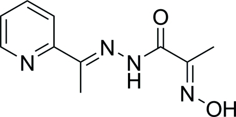
The title compound, C10H12N4O2, features an intramolecular N—H⋯N hydrogen bond formed between the imine NH and oxime N atoms. The oxime group and the amide C=O bond are anti to each other. In the crystal, molecules are connected by O—H⋯O hydrogen bonds into supramolecular zigzag chains along the c axis.
Related literature
For oxime and pyridine derivatives, see: Sliva et al. (1997b
▶); Mokhir et al. (2002 ▶); Krämer et al. (2002 ▶); Kovbasyuk et al. (2004 ▶). For 2-hydroxyiminopropanamide and amide derivatives of 2-hydroxyiminopropanoic acid, see: Onindo et al. (1995 ▶); Duda et al. (1997 ▶); Sliva et al. (1997a
▶). For the preparation and characterization of 3d-metal complexes with 2-hydroxyimino-N′-[1-(2-pyridyl)ethylidene]propanohydrazone, see: Moroz et al. (2008a
▶,b
▶). For typical bond lengths, see: Bürgi & Dunitz (1994 ▶).
Experimental
Crystal data
C10H12N4O2
M r = 220.24
Monoclinic,

a = 4.4498 (11) Å
b = 22.833 (7) Å
c = 10.955 (3) Å
β = 97.47 (2)°
V = 1103.7 (5) Å3
Z = 4
Mo Kα radiation
μ = 0.10 mm−1
T = 293 K
0.15 × 0.10 × 0.05 mm
Data collection
Oxford Diffraction KM-4/Xcalibur diffractometer with a Sapphire3 detector
Absorption correction: multi-scan (CrysAlis CCD; Oxford Diffraction, 2006 ▶) T min = 0.986, T max = 0.995
3899 measured reflections
958 independent reflections
793 reflections with I > 2σ(I)
R int = 0.059
Refinement
R[F 2 > 2σ(F 2)] = 0.046
wR(F 2) = 0.097
S = 1.10
958 reflections
155 parameters
2 restraints
H atoms treated by a mixture of independent and constrained refinement
Δρmax = 0.13 e Å−3
Δρmin = −0.16 e Å−3
Data collection: CrysAlis CCD (Oxford Diffraction, 2006 ▶); cell refinement: CrysAlis RED (Oxford Diffraction, 2006 ▶); data reduction: CrysAlis RED; program(s) used to solve structure: SHELXS97 (Sheldrick, 2008 ▶); program(s) used to refine structure: SHELXL97 (Sheldrick, 2008 ▶); molecular graphics: ORTEP-3 for Windows (Farrugia, 1997 ▶); software used to prepare material for publication: WinGX (Farrugia, 1999 ▶).
Supplementary Material
Crystal structure: contains datablocks I, global. DOI: 10.1107/S1600536809033352/tk2523sup1.cif
Structure factors: contains datablocks I. DOI: 10.1107/S1600536809033352/tk2523Isup2.hkl
Additional supplementary materials: crystallographic information; 3D view; checkCIF report
Table 1. Hydrogen-bond geometry (Å, °).
| D—H⋯A | D—H | H⋯A | D⋯A | D—H⋯A |
|---|---|---|---|---|
| O2—H2OA⋯O1i | 0.84 (5) | 1.88 (5) | 2.709 (4) | 170 (5) |
| N3—H3NA⋯N4 | 0.87 (4) | 2.30 (4) | 2.640 (4) | 104 (3) |
Symmetry code: (i)  .
.
Acknowledgments
The authors thank the Ministry of Education and Science of Ukraine for financial support (grant No. F28/241–2009).
supplementary crystallographic information
Comment
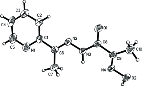
As a part of our on-going work, we would like to report the structure of the title compound (I), Fig. 1, which comprises several groups capable of forming hydrogen bonding interactions: oxime, hydrazone, azomethine, and pyridine. Molecule (I) has been shown previously to form mono- and tetra-nuclear grid-like complexes with 3d-metals (Moroz et al., 2008a,b).
The C—N and N—O bond lengths in the oxime group, i.e. 1.285 (5) and 1.388 (4) Å, respectively, adopt typical values (Sliva et al., 1997b; Mokhir et al., 2002). The oxime group is in an anti- position with respect to the amide group, an observation consistent with the structures of 2-hydroxyiminopropanamide and other amide derivatives of 2-hydroxyiminopropanoic acid (Onindo et al., 1995; Duda et al., 1997; Sliva et al., 1997a). This conformation is stabilised by an N3—H···N4 intramolecular interaction, Table 1. The CH3C(=NOH)C(O)NH fragment deviates from planarity as seen in a twist between the oxime and amide groups about the C8—C9 bond; the O1-C8-C9-N4 torsion angle is -164.0 (4)°. The flattened geometry of molecule results in the appearance of short intramolecular contacts H10···O2 is 2.34 Å and H7C···H3N 2.28 Å. The C—N bond distance in the azomethine group is 1.277 (4) Å, and the N2—C6—C1 angle is 115.7 (3)°. The pyridine-N atom is situated in an anti- position with respect to the azomethine group. Finally, the C—N and C—C bond lengths within the pyridine ring are normal for 2-substituted pyridine derivatives (Krämer et al., 2002; Kovbasyuk et al., 2004).
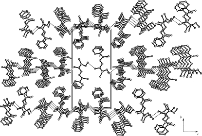
In the crystal packing molecules are united by O2—H···O1 hydrogen bonds, Table 1, where oximic-oxygen atom acts as donor and the hydrazone-oxygen atom acts as an acceptor (Fig. 2). This interaction probably results in the elongation of the C8—O1 bond to 1.233 (4) Å as compared with its mean value 1.210 Å (Bürgi & Dunitz, 1994). Due to the presence of the O2—H···O1 hydrogen bonds, zig-zag supramolecular chains are formed along the c axis.
Experimental
Compound (I) was prepared according to the reported procedure (Moroz et al., 2008b).
Refinement
All H atoms were observed in a difference Fourier map, but C—H hydrogen atoms were placed at calculated positions and treated as riding on their parent atoms [C—H = 0.93-0.96 Å and Uiso(H) = 1.2- 1.5Ueq(C)]. The N—H and O—H hydrogen atoms were fully refined; O-H = 0.84 (5) Å and N-H = 0.87 (4) Å. In the absence of significant anomalous scattering effects, 766 Friedel pairs were averaged in the final refinement.
Figures
Fig. 1.
A view of (I), with displacement ellipsoids shown at the 40% probability level and atom labelling.
Fig. 2.
A packing diagram for (I) viewed in projection down the a axis. Hydrogen bonds are indicated by dashed lines; H atoms are omitted for clarity.
Crystal data
| C10H12N4O2 | F(000) = 464 |
| Mr = 220.24 | Dx = 1.325 Mg m−3 |
| Monoclinic, Cc | Mo Kα radiation, λ = 0.71073 Å |
| Hall symbol: C -2yc | Cell parameters from 5860 reflections |
| a = 4.4498 (11) Å | θ = 3.6–32.0° |
| b = 22.833 (7) Å | µ = 0.10 mm−1 |
| c = 10.955 (3) Å | T = 293 K |
| β = 97.47 (2)° | Needles, white |
| V = 1103.7 (5) Å3 | 0.15 × 0.10 × 0.05 mm |
| Z = 4 |
Data collection
| Oxford Diffraction KM-4/Xcalibur diffractometer with a Sapphire3 detector | 958 independent reflections |
| Radiation source: Enhance (Mo) X-ray Source | 793 reflections with I > 2σ(I) |
| graphite | Rint = 0.059 |
| Detector resolution: 16.1827 pixels mm-1 | θmax = 25.0°, θmin = 3.6° |
| φ scans and ω scans with κ offset | h = −5→5 |
| Absorption correction: multi-scan (CrysAlis CCD; Oxford Diffraction, 2006) | k = −25→26 |
| Tmin = 0.986, Tmax = 0.995 | l = −12→12 |
| 3899 measured reflections |
Refinement
| Refinement on F2 | Secondary atom site location: difference Fourier map |
| Least-squares matrix: full | Hydrogen site location: inferred from neighbouring sites |
| R[F2 > 2σ(F2)] = 0.046 | H atoms treated by a mixture of independent and constrained refinement |
| wR(F2) = 0.097 | w = 1/[σ2(Fo2) + (0.0453P)2 + 0.2337P] where P = (Fo2 + 2Fc2)/3 |
| S = 1.10 | (Δ/σ)max < 0.001 |
| 958 reflections | Δρmax = 0.13 e Å−3 |
| 155 parameters | Δρmin = −0.16 e Å−3 |
| 2 restraints | Absolute structure: nd |
| Primary atom site location: structure-invariant direct methods |
Special details
| Geometry. All e.s.d.'s (except the e.s.d. in the dihedral angle between two l.s. planes) are estimated using the full covariance matrix. The cell e.s.d.'s are taken into account individually in the estimation of e.s.d.'s in distances, angles and torsion angles; correlations between e.s.d.'s in cell parameters are only used when they are defined by crystal symmetry. An approximate (isotropic) treatment of cell e.s.d.'s is used for estimating e.s.d.'s involving l.s. planes. |
| Refinement. Refinement of F2 against ALL reflections. The weighted R-factor wR and goodness of fit S are based on F2, conventional R-factors R are based on F, with F set to zero for negative F2. The threshold expression of F2 > σ(F2) is used only for calculating R-factors(gt) etc. and is not relevant to the choice of reflections for refinement. R-factors based on F2 are statistically about twice as large as those based on F, and R- factors based on ALL data will be even larger. |
Fractional atomic coordinates and isotropic or equivalent isotropic displacement parameters (Å2)
| x | y | z | Uiso*/Ueq | ||
| N1 | −0.4385 (8) | 0.29326 (15) | 0.1660 (4) | 0.0601 (10) | |
| N2 | −0.0269 (7) | 0.41668 (13) | 0.0900 (3) | 0.0411 (8) | |
| N3 | 0.1840 (7) | 0.45542 (13) | 0.1431 (3) | 0.0418 (8) | |
| N4 | 0.6418 (7) | 0.52027 (13) | 0.2434 (3) | 0.0402 (8) | |
| O1 | 0.1686 (7) | 0.50958 (13) | −0.0301 (3) | 0.0624 (9) | |
| O2 | 0.8544 (6) | 0.55993 (14) | 0.2979 (3) | 0.0559 (8) | |
| C1 | −0.3464 (9) | 0.33542 (16) | 0.0958 (4) | 0.0439 (10) | |
| C2 | −0.4547 (11) | 0.3399 (2) | −0.0272 (4) | 0.0624 (13) | |
| H2 | −0.3891 | 0.3701 | −0.0743 | 0.075* | |
| C3 | −0.6589 (11) | 0.2998 (2) | −0.0793 (5) | 0.0763 (16) | |
| H3 | −0.7310 | 0.3019 | −0.1628 | 0.092* | |
| C4 | −0.7553 (11) | 0.2573 (2) | −0.0090 (5) | 0.0706 (15) | |
| H4 | −0.8962 | 0.2296 | −0.0423 | 0.085* | |
| C5 | −0.6403 (13) | 0.2558 (2) | 0.1128 (6) | 0.0750 (15) | |
| H5 | −0.7085 | 0.2265 | 0.1614 | 0.090* | |
| C6 | −0.1176 (8) | 0.37676 (17) | 0.1582 (3) | 0.0425 (10) | |
| C7 | −0.0053 (11) | 0.3692 (2) | 0.2922 (4) | 0.0607 (13) | |
| H7A | −0.0416 | 0.4044 | 0.3357 | 0.091* | |
| H7B | 0.2082 | 0.3611 | 0.3022 | 0.091* | |
| H7C | −0.1103 | 0.3372 | 0.3246 | 0.091* | |
| C8 | 0.2702 (8) | 0.50043 (16) | 0.0783 (3) | 0.0405 (9) | |
| C9 | 0.4981 (9) | 0.54051 (15) | 0.1430 (3) | 0.0404 (10) | |
| C10 | 0.5493 (13) | 0.59805 (19) | 0.0889 (5) | 0.0755 (16) | |
| H10A | 0.3634 | 0.6119 | 0.0440 | 0.113* | |
| H10B | 0.7001 | 0.5945 | 0.0342 | 0.113* | |
| H10C | 0.6177 | 0.6253 | 0.1534 | 0.113* | |
| H3NA | 0.239 (8) | 0.4565 (15) | 0.222 (4) | 0.035 (10)* | |
| H2OA | 0.932 (11) | 0.5383 (19) | 0.355 (5) | 0.061 (14)* |
Atomic displacement parameters (Å2)
| U11 | U22 | U33 | U12 | U13 | U23 | |
| N1 | 0.063 (2) | 0.045 (2) | 0.069 (3) | −0.013 (2) | −0.0028 (19) | 0.0015 (19) |
| N2 | 0.0370 (18) | 0.0396 (16) | 0.0439 (18) | −0.0042 (15) | −0.0056 (14) | −0.0027 (15) |
| N3 | 0.0436 (19) | 0.0436 (19) | 0.0353 (19) | −0.0083 (16) | −0.0062 (15) | −0.0009 (14) |
| N4 | 0.0361 (18) | 0.0403 (17) | 0.0408 (18) | −0.0022 (15) | −0.0078 (14) | −0.0055 (14) |
| O1 | 0.071 (2) | 0.0603 (18) | 0.0478 (17) | −0.0208 (16) | −0.0238 (15) | 0.0105 (14) |
| O2 | 0.0555 (18) | 0.0570 (18) | 0.0478 (17) | −0.0045 (15) | −0.0220 (13) | −0.0013 (14) |
| C1 | 0.036 (2) | 0.041 (2) | 0.054 (2) | 0.0004 (18) | 0.0032 (19) | −0.0047 (18) |
| C2 | 0.068 (3) | 0.071 (3) | 0.046 (2) | −0.025 (3) | 0.002 (2) | −0.007 (2) |
| C3 | 0.072 (4) | 0.089 (4) | 0.063 (3) | −0.026 (3) | −0.007 (3) | −0.020 (3) |
| C4 | 0.051 (3) | 0.058 (3) | 0.100 (4) | −0.020 (2) | 0.000 (3) | −0.028 (3) |
| C5 | 0.078 (4) | 0.052 (3) | 0.093 (4) | −0.028 (3) | 0.004 (3) | −0.003 (3) |
| C6 | 0.045 (3) | 0.038 (2) | 0.043 (2) | 0.0000 (18) | 0.0022 (19) | −0.0043 (17) |
| C7 | 0.075 (3) | 0.061 (3) | 0.044 (2) | −0.017 (2) | −0.005 (2) | −0.0039 (19) |
| C8 | 0.040 (2) | 0.038 (2) | 0.039 (2) | 0.0029 (18) | −0.0085 (17) | 0.0004 (17) |
| C9 | 0.047 (3) | 0.0368 (19) | 0.0342 (19) | 0.0021 (18) | −0.0066 (17) | −0.0001 (16) |
| C10 | 0.087 (4) | 0.059 (3) | 0.069 (3) | −0.024 (3) | −0.034 (3) | 0.015 (3) |
Geometric parameters (Å, °)
| N1—C5 | 1.320 (6) | C3—C4 | 1.345 (7) |
| N1—C1 | 1.330 (5) | C3—H3 | 0.9300 |
| N2—C6 | 1.277 (4) | C4—C5 | 1.366 (8) |
| N2—N3 | 1.364 (4) | C4—H4 | 0.9300 |
| N3—C8 | 1.334 (5) | C5—H5 | 0.9300 |
| N3—H3NA | 0.87 (4) | C6—C7 | 1.498 (5) |
| N4—C9 | 1.285 (5) | C7—H7A | 0.9600 |
| N4—O2 | 1.388 (4) | C7—H7B | 0.9600 |
| O1—C8 | 1.233 (4) | C7—H7C | 0.9600 |
| O2—H2OA | 0.84 (5) | C8—C9 | 1.477 (5) |
| C1—C2 | 1.374 (6) | C9—C10 | 1.471 (5) |
| C1—C6 | 1.488 (5) | C10—H10A | 0.9600 |
| C2—C3 | 1.362 (6) | C10—H10B | 0.9600 |
| C2—H2 | 0.9300 | C10—H10C | 0.9600 |
| C5—N1—C1 | 117.2 (4) | N2—C6—C1 | 115.7 (3) |
| C6—N2—N3 | 117.7 (3) | N2—C6—C7 | 124.4 (4) |
| C8—N3—N2 | 120.1 (3) | C1—C6—C7 | 119.9 (4) |
| C8—N3—H3NA | 116 (2) | C6—C7—H7A | 109.5 |
| N2—N3—H3NA | 122 (2) | C6—C7—H7B | 109.5 |
| C9—N4—O2 | 111.7 (3) | H7A—C7—H7B | 109.5 |
| N4—O2—H2OA | 97 (3) | C6—C7—H7C | 109.5 |
| N1—C1—C2 | 121.7 (4) | H7A—C7—H7C | 109.5 |
| N1—C1—C6 | 115.9 (3) | H7B—C7—H7C | 109.5 |
| C2—C1—C6 | 122.4 (4) | O1—C8—N3 | 123.3 (4) |
| C3—C2—C1 | 119.4 (5) | O1—C8—C9 | 120.0 (3) |
| C3—C2—H2 | 120.3 | N3—C8—C9 | 116.7 (3) |
| C1—C2—H2 | 120.3 | N4—C9—C10 | 125.5 (4) |
| C4—C3—C2 | 119.3 (5) | N4—C9—C8 | 115.0 (3) |
| C4—C3—H3 | 120.3 | C10—C9—C8 | 119.5 (3) |
| C2—C3—H3 | 120.3 | C9—C10—H10A | 109.5 |
| C3—C4—C5 | 118.1 (4) | C9—C10—H10B | 109.5 |
| C3—C4—H4 | 121.0 | H10A—C10—H10B | 109.5 |
| C5—C4—H4 | 121.0 | C9—C10—H10C | 109.5 |
| N1—C5—C4 | 124.2 (5) | H10A—C10—H10C | 109.5 |
| N1—C5—H5 | 117.9 | H10B—C10—H10C | 109.5 |
| C4—C5—H5 | 117.9 | ||
| C6—N2—N3—C8 | −175.2 (3) | C2—C1—C6—N2 | −0.3 (6) |
| C5—N1—C1—C2 | −0.3 (6) | N1—C1—C6—C7 | 0.2 (5) |
| C5—N1—C1—C6 | −179.7 (4) | C2—C1—C6—C7 | −179.2 (4) |
| N1—C1—C2—C3 | −0.9 (7) | N2—N3—C8—O1 | −0.1 (6) |
| C6—C1—C2—C3 | 178.5 (4) | N2—N3—C8—C9 | 179.6 (3) |
| C1—C2—C3—C4 | 1.3 (8) | O2—N4—C9—C10 | 0.7 (6) |
| C2—C3—C4—C5 | −0.7 (8) | O2—N4—C9—C8 | 179.1 (3) |
| C1—N1—C5—C4 | 0.9 (8) | O1—C8—C9—N4 | −164.0 (4) |
| C3—C4—C5—N1 | −0.5 (9) | N3—C8—C9—N4 | 16.3 (5) |
| N3—N2—C6—C1 | −179.7 (3) | O1—C8—C9—C10 | 14.5 (6) |
| N3—N2—C6—C7 | −0.8 (5) | N3—C8—C9—C10 | −165.2 (4) |
| N1—C1—C6—N2 | 179.1 (3) |
Hydrogen-bond geometry (Å, °)
| D—H···A | D—H | H···A | D···A | D—H···A |
| O2—H2OA···O1i | 0.84 (5) | 1.88 (5) | 2.709 (4) | 170 (5) |
| N3—H3NA···N4 | 0.87 (4) | 2.30 (4) | 2.640 (4) | 104 (3) |
Symmetry codes: (i) x+1, −y+1, z+1/2.
Footnotes
Supplementary data and figures for this paper are available from the IUCr electronic archives (Reference: TK2523).
References
- Bürgi, H.-D. & Dunitz, J. D. (1994). Structure Correlation, Vol. 2., Weinhiem: VCH.
- Duda, A. M., Karaczyn, A., Kozłowski, H., Fritsky, I. O., Głowiak, T., Prisyazhnaya, E. V., Sliva, T. Yu. & Świątek-Kozłowska, J. (1997). J. Chem. Soc. Dalton Trans. pp. 3853–3859.
- Farrugia, L. J. (1997). J. Appl. Cryst.30, 565.
- Farrugia, L. J. (1999). J. Appl. Cryst.32, 837–838.
- Kovbasyuk, L., Pritzkow, H., Krämer, R. & Fritsky, I. O. (2004). Chem. Commun. pp. 880–881. [DOI] [PubMed]
- Krämer, R., Fritsky, I. O., Pritzkow, H. & Kovbasyuk, L. A. (2002). J. Chem. Soc. Dalton Trans. pp. 1307–1314.
- Mokhir, A. A., Gumienna-Kontecka, E., Świątek-Kozłowska, J., Petkova, E. G., Fritsky, I. O., Jerzykiewicz, L., Kapshuk, A. A. & Sliva, T. Yu. (2002). Inorg. Chim. Acta, 329, 113–121.
- Moroz, Yu. S., Kulon, K., Haukka, M., Gumienna-Kontecka, E., Kozłowski, H., Meyer, F. & Fritsky, I. O. (2008a). Inorg. Chem.47, 5656–5665. [DOI] [PubMed]
- Moroz, Y. S., Sliva, T. Yu., Kulon, K., Kozłowski, H. & Fritsky, I. O. (2008b). Acta Cryst. E64, m353–m354. [DOI] [PMC free article] [PubMed]
- Onindo, C. O., Sliva, T. Yu., Kowalik-Jankowska, T., Fritsky, I. O., Buglyo, P., Pettit, L. D., Kozłowski, H. & Kiss, T. (1995). J. Chem. Soc. Dalton Trans. pp. 3911–3915.
- Oxford Diffraction (2006). CrysAlis CCD and CrysAlis RED Oxford Diffraction Ltd, Abingdon, England.
- Sheldrick, G. M. (2008). Acta Cryst. A64, 112–122. [DOI] [PubMed]
- Sliva, T. Yu., Duda, A. M., Glowiak, T., Fritsky, I. O., Amirkhanov, V. M., Mokhir, A. A. & Kozłowski, H. (1997a). J. Chem. Soc. Dalton Trans. pp. 273–276.
- Sliva, T. Yu., Kowalik-Jankowska, T., Amirkhanov, V. M., Głowiak, T., Onindo, C. O., Fritsky, I. O. & Kozłowski, H. (1997b). J. Inorg. Biochem.65, 287–294.
Associated Data
This section collects any data citations, data availability statements, or supplementary materials included in this article.
Supplementary Materials
Crystal structure: contains datablocks I, global. DOI: 10.1107/S1600536809033352/tk2523sup1.cif
Structure factors: contains datablocks I. DOI: 10.1107/S1600536809033352/tk2523Isup2.hkl
Additional supplementary materials: crystallographic information; 3D view; checkCIF report


