Abstract
Pancreatic cancers generally respond poorly to chemotherapy, prompting a need to identify agents that could sensitize tumors to treatment. In this study, we investigated the response of human pancreatic cells to gamma-tocotrienol (γ-T3), a novel, unsaturated form of vitamin E found in palm oil and rice bran oil, to determine whether it could potentiate the effects of gemcitabine, a standard of care in clinical treatment of pancreatic cancer. γ-T3 inhibited the in vitro proliferation of pancreatic cancer cell lines with variable p53 status and potentiated gemcitabine-induced apoptosis. These effects correlated with an inhibition of NF-κB activation by γ-T3 and a suppression of key cellular regulators including cyclin D1, c-Myc, COX-2, Bcl-2, cIAP, survivin, VEGF, ICAM-1, and CXCR4. In an orthotopic nude mouse model of human pancreatic cancer, oral administration of γ-T3 inhibited tumor growth and enhanced the antitumor properties of gemcitabine. Immunohistochemical analysis indicated a correlation between tumor growth inhibition and reduced expression of Ki-67, COX-2, MMP-9, NF-κB p65 and VEGF in the tissue. Combination treatment also downregulated NF-κB activity along with the NF-κB-regulated gene products cyclin D1, c-Myc, VEGF, MMP-9, CXCR4. Consistent with an enhancement of tumor apoptosis caspase activation was observed in tumor tissues. Overall, Our findings suggest that γ-T3 can inhibit the growth of human pancreatic tumors and sensitize them to gemcitabine by suppressing of NF-κB-mediated inflammatory pathways linked to tumorigenesis.
Keywords: tocotrienol, pancreatic cancer, NF-κB, inflammation
Introduction
The National Cancer Institute estimated that 42,470 men and 35,240 women in the United States would die of pancreatic cancer in 2009 (1). The only agents approved by the U.S. Food and Drug Administration to treat pancreatic cancer are gemcitabine and erlotinib, both of which produce responses in <10% of patients and are associated with multiple adverse events and drug resistance. Therefore, the need for novel strategies to treat this lethal disease is imperative. The transcription factor nuclear factor-kappaB (NF-κB) has been linked with cell proliferation, invasion, angiogenesis, metastasis, antiapoptosis, and chemoresistance in multiple tumors (2, 3). In addition, numerous lines of evidence suggest that NF-κB plays a major role in the growth and chemoresistance of pancreatic cancer. NF-κB is constitutively active in pancreatic cancer cells (4) but not in immortalized, nontumorigenic pancreatic ductal epithelial cells (5).Thus, NF-κB activation has been reported in animal models of pancreatic cancer (6) and in human pancreatic cancer tissue (4). NF-κB promotes pancreatic cancer growth via antiapoptosis (4, 7) and mediates the induction of mitogenic gene products, such as c-Myc and cyclin D1 (8), the latter of which is overexpressed in human pancreatic cancer tissue and inversely correlated with patient survival (9). Additionally, NF-κB enhances the angiogenic potential of pancreatic cancer cells via increased expression of proangiogenic factors, including vascular endothelial growth factor (VEGF) (10), while other NF-κB-regulated gene products promote the migration and invasion of pancreatic cancer cells (11). Finally, NF-κB plays a pivotal role in promoting gemcitabine resistance in pancreatic cancer (3). Together, this evidence implicates NF-κB in pancreatic cancer and suggests that agents that block NF-κB activation could reduce the cells’ chemoresistance to gemcitabine and perhaps be used in combination with gemcitabine as a novel therapeutic regimen for pancreatic cancer.
Vitamin E, as represented by tocopherols, was first discovered in 1922 (12). Tocotrienols, unsaturated derivatives of vitamin E found in rice, barley, oats, and palm, were first identified in 1965 and first found to prevent cancer in 1989 (13). Although tocopherols have been studied extensively (over 30,000 citations), very little is known about tocotrienols. Limited studies have revealed that tocotrienols are more potent antioxidant agents than tocopherols (14) and exhibit activities superior to those of tocopherols. For instance, studies from our laboratory and others have shown that gamma-tocotrienol (γ-T3), but not tocopherol, can inhibit both constitutive and inducible NF-κB activation in various cancer cell lines (15, 16). This activity correlated with downregulation of the NF-κB-regulated gene products associated with survival, proliferation, invasion, and angiogenesis (16). Tocotrienols have been shown to suppress the in vitro proliferation of a wide variety of tumor cells, including human breast, colon, prostate, and pancreatic (17). γ-T3 has been reported to potentiate the effect of gefitinib, celecoxib and statins against various human cancers (12, 18–20). This dietary agent can also inhibit ErbB3-dependent PI3K/Akt mitogenic signaling in cancer cells (21). Moreover, tocotrienols have been shown to inhibit angiogenesis (22, 23) by suppressing hypoxia-inducible factor-1 alpha (24) and to inhibit proinflammatory markers and cyclooxygenase-2 (COX-2) expression in vitro (25). Other studies have demonstrated that γ-T3 inhibits human cancer cell progression by downregulating cyclin D1 and E (26). Recent studies have shown that tocotrienols can suppress the proliferation of various human pancreatic cancer cells (17) and, when given orally to mice, significant levels accumulate in the pancreatic tissues. Another recent study indicate that γ-T3 can sensitize androgen-independent prostate cancer to docetaxel in vivo through downregulation of proliferating cell nuclear antigen (PCNA), Ki-67 and Id1; and upregulation of cleaved caspase-3 and PARP (27).
The possibility that dietary agents can potentiate the effects of chemotherapeutic agents such as gemcitabine is an attractive strategy. In the present study, we investigated whether γ-T3 could sensitize human pancreatic tumors to gemcitabine in vitro and in an orthotopic mouse model. We demonstrated that γ-T3 inhibited the in vitro proliferation of various pancreatic cancer cells, enhanced gemcitabine-induced apoptosis, and potentiated the antitumor activity of gemcitabine against orthotopically implanted human pancreatic tumors through the downregulation of NF-κB and NF-κB-regulated gene products.
Materials and Methods
Materials
γ-T3 (Fig. 1A) was kindly supplied by Davos Life Science (Singapore). The following polyclonal antibodies against p65 (recognizing the epitope within the NH2-terminal domain of human NF-κB p65) were obtained from Santa Cruz Biotechnology (Santa Cruz, CA): intercellular adhesion molecule-1 (ICAM-1), cyclin D1, matrix metalloproteinase 9 (MMP-9), survivin, cellular inhibitor of apoptosis protein 1 (cIAP-1), procaspase-3, and procaspase-9. Also obtained from Santa Cruz Biotechnology were monoclonal antibodies against COX-2, c-Myc, Bcl-2, and Bcl-xL. Antiobodies against VEGF and Ki-67 were purchased from Thermo Fisher Scientific (Fremont, CA). The liquid DAB+ Substrate Chromogen System-HRP used for immunocytochemistry was obtained from Dako (Carpinteria, CA). Penicillin, streptomycin, RPMI 1640, and fetal bovine serum (FBS) were obtained from Invitrogen (Grand Island, NY). Tris, glycine, NaCl, sodium dodecyl sulfate (SDS), and bovine serum albumin (BSA) were obtained from Sigma Chemical (St. Louis, MO). Gemcitabine (Gemzar; kindly supplied by Eli Lilly, Indianapolis, IN) was stored at 4°C and dissolved in sterile phosphate buffered saline (PBS) on the day of use.
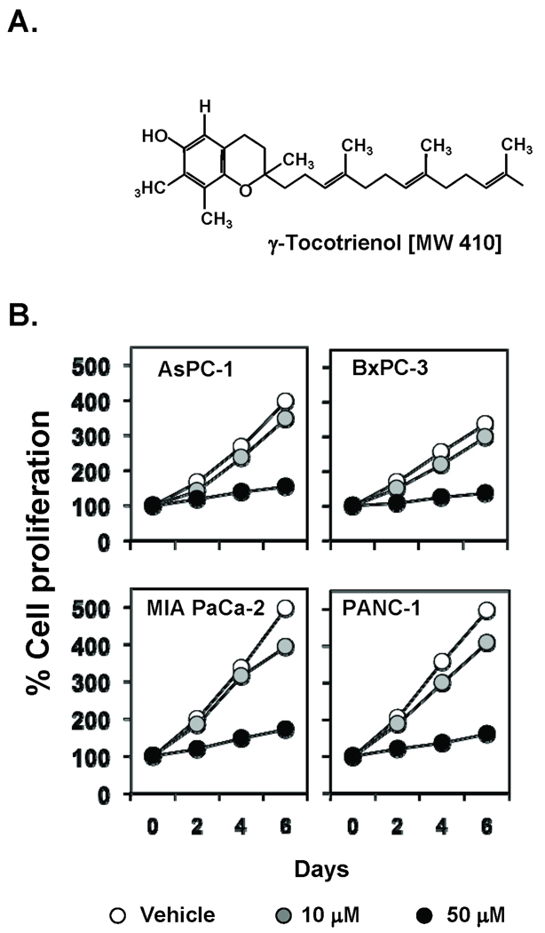
Figure 1. γ-T3 inhibits proliferation in pancreatic cancer cells in vitro.
(A), Structure of γ-T3. (B), MTT assay results showed dose-dependent suppression of cell proliferation in all four pancreatic cancer cell lines tested. Points, mean of triplicate; bars, SE. Data are representative of three independent experiments.
Cell lines
The pancreatic cancer cell lines BxPC-3, MIA PaCa-2, and Panc-1 were obtained from the American Type Culture Collection (Manassas, VA). MPanc-96 was a kind gift from Dr. C. Logsdon (The University of Texas MD Anderson Cancer Center, Houston, TX). All cell lines were cultured in RPMI 1640 supplemented with 10% FBS, 100 units/mL penicillin, and 100 µg/mL streptomycin. The above-mentioned cell lines were procured more than 6 months ago and have not been tested recently for authentication in our laboratory.
Proliferation assay
The effect of γ-T3 on cell proliferation was determined by the 3-(4,5-dimethylthiazol-2-yl)-2,5-diphenyltetrazolium bromide (MTT) uptake method as described previously (28). The cells (2,000/well) were incubated with γ-T3 in triplicate in a 96-well plate and then incubated for 2, 4, or 6 days at 37°C. A MTT solution was added to each well and incubated for 2 h at 37°C. An extraction buffer (20% SDS and 50% dimethylformamide) was added, and the cells were incubated overnight at 37°C. The absorbance of the cell suspension was measured at 570 nm with an MRX Revelation 96-well multiscanner (Dynex Technologies, Chantilly, VA).
Apoptosis assay
To investigate whether γ-T3 could potentiate the apoptotic effects of gemcitabine in pancreatic cancer cells, we used a LIVE/DEAD cell viability assay kit (Invitrogen), which is used to determine intracellular esterase activity and plasma membrane integrity. This assay uses calcein, a polyanionic, green fluorescent dye that is retained within live cells, and a red fluorescent ethidium homodimer dye that can enter cells through damaged membranes and bind to nucleic acids but is excluded by the intact plasma membranes of live cells (29). Briefly, cells (5,000/well) were incubated in chamber slides, pretreated with γ-T3 for 4 h, and treated with gemcitabine for 24 h. Cells were then stained with the assay reagents for 30 min at room temperature. Cell viability was determined under a fluorescence microscope by counting live (green) and dead (red) cells.
Animals
Male athymic nu/nu mice (4 weeks old) were obtained from the breeding colony of the Department of Experimental Radiation Oncology at MD Anderson. The animals were housed in standard plexiglass cages (five per cage) in a room maintained at constant temperature and humidity and in a 12 h:12 h light-dark cycle. Their diet consisted of regular autoclave chow and water ad libitum. None of the mice exhibited any lesions, and all were tested pathogen-free. Before initiating the experiment, we acclimatized all of the mice to a pulverized diet for 3 days. Our experimental protocol was reviewed and approved by the Institutional Animal Care and Use Committee at MD Anderson.
Orthotopic implantation of MIA PaCa-2 cells
MIA PaCa-2 cells were stably transfected with luciferase as previously described for Panc-1 cells (30). The MIA PaCa-2 cells were orthotopically implanted as described previously (29). Briefly, luciferase-transfected MIA PaCa-2 cells were harvested from subconfluent cultures after a brief exposure to 0.25% trypsin and 0.2% ethylenediaminetetraacetic acid (EDTA). Trypsinization was stopped with medium containing 10% FBS. The cells were washed once in serum-free medium and resuspended in PBS. Only suspensions consisting of single cells, with >90% viability, were used for the injections. After mice were anesthetized with ketamine-xylazine solution, a small incision was made in the left abdominal flank, and MIA PaCa-2 cells (1 × 10⁶) in 100 µL PBS were injected into the subcapsular region of the pancreas with a 27-gauge needle and a calibrated push button-controlled dispensing device (Hamilton Syringe Co., Reno, NV). A cotton swab was held cautiously for 1 min over the site of injection to prevent leakage. The abdominal wound was closed in one layer with wound clips (Braintree Scientific, Inc., Braintree, MA).
Experimental protocol
One week after implantation, the mice were randomized into the following treatment groups (n = 6/group) based on the bioluminescence first measured with an in vivo imaging system (IVIS 200, Xenogen Corp., Alameda, CA): (a) untreated control (olive oil, 100 µL daily); (b) tocotrienol (400 mg/kg once daily orally [p.o.]); (c) gemcitabine alone (25 mg/kg twice weekly by intraperitoneal [i.p.] injection); and (d) combination (tocotrienol, 400 mg/kg once daily p.o., and gemcitabine, 25 mg/kg twice weekly by i.p. injection). Tumor volumes were monitored weekly with the bioluminescence IVIS, which includes a cryogenic cooling unit, and a data acquisition computer running Living Image software (Xenogen Corp.). Before imaging, the animals were anesthetized in an acrylic chamber with 2.5% isoflurane/air mixture and injected i.p. with 40 mg/mL D-luciferin potassium salt in PBS at a dose of 150 mg/kg body weight. After 10 min of incubation with luciferin, each mouse was placed in a right lateral decubitus position and a digital grayscale animal image was acquired, followed by the acquisition and overlay of a pseudocolor image representing the spatial distribution of detected photons emerging from active luciferase within the animal. Signal intensity was quantified as the sum of all detected photons within the region of interest per second. The mice were subjected to imaging on days 0, 7, 17, 24, and 29 of treatment. Therapy was continued for 4 weeks, and the animals were euthanized 1 week later. Primary tumors in the pancreas were excised and the final tumor volume was measured as V = 2/3πr3, where r is the mean of the three dimensions (length, width, and depth). Half of the tumor tissue was fixed in formalin and embedded in paraffin for immunohistochemistry and routine hematoxylin and eosin (H&E) staining. The other half was snap frozen in liquid nitrogen and stored at −80°C. H&E staining confirmed the presence of tumor(s) in each pancreas.
Preparation of nuclear extract from tumor samples
Pancreatic tumor tissues (75–100 mg/mouse) from mice in the control and treatment groups were minced and incubated on ice for 30 min in 0.5 mL of ice-cold buffer A [10 mmol/L 4-(2-hydroxyethyl)-1-piperazineethanesulfonic acid (HEPES; pH 7.9), 1.5 mmol/L KCl, 10 mmol/L MgCl2, 0.5 mmol/L dithiothreitol (DTT), 0.5 mmol/L phenylmethylsulfonyl fluoride (PMSF)]. The minced tissue was homogenized using a Dounce homogenizer and centrifuged at 16,000 × g at 4°C for 10 min. The resulting nuclear pellet was suspended in 0.2 mL of buffer B [20 mmol/L HEPES (pH 7.9), 25% glycerol, 1.5 mmol/L MgCl2, 420 mmol/L NaCl, 0.5 mmol/L DTT, 0.2 mmol/L EDTA, 0.5 mmol/L PMSF, 2 µg/mL leupeptin] and incubated on ice for 2 h with intermittent mixing. The suspension was then centrifuged at 16,000 × g at 4°C for 30 min. The supernatant (nuclear extract) was collected and stored at −70°C until used as previously described (28). Protein concentration was measured by the Bradford assay with BSA as the standard.
NF-κB activation in pancreatic cancer cells and tumor samples
To assess NF-κB activation, we isolated nuclei from pancreatic cancer cell lines and tumor samples and carried out electrophoretic mobility shift assays (EMSA) essentially as described previously (29). Briefly, nuclear extracts prepared from pancreatic cancer cells (1 × 10⁶/mL) and tumor samples were incubated with 32P-end-labeled 45-mer double-stranded NF-κB oligonucleotide (4 µg of protein with 16 fmol of DNA) from the HIV long terminal repeat (5'-TTGTTACAAGGGACTTTCCGCTGGGGACTTTCCAGGGGGAGGCGTGG-3'; underline indicates NF-κB–binding sites) for 15 min at 37°C. The resulting DNA-protein complex was separated from free oligonucleotide on 6.6% native polyacrylamide gels. The dried gels were visualized, and radioactive bands were quantitated with a PhosphorImager and ImageQuant software (GE Healthcare, Piscataway, NJ).
Immunolocalization of NF-κB p65, VEGF, and COX-2 in tumor samples
The nuclear localization of p65 and the expression of COX-2 and VEGF were examined via an immunohistochemical method described previously (28). Briefly, pancreatic cancer tumor samples were embedded in paraffin and fixed with paraformaldehyde. After being washed in PBS, the slides were blocked with protein block solution (Dako) for 20 min and then incubated overnight with rabbit polyclonal anti-human p65, mouse monoclonal anti-human VEGF, and anti-COX-2 antibodies (1:100, 1:50, and 1:75, respectively). After incubation with the antibodies, the slides were washed and then incubated with biotinylated link universal antiserum followed by horseradish peroxidase-streptavidin detection with the LSAB+ kit (Dako). The slides were rinsed, and color was developed with 3,3'-diaminobenzidine hydrochloride used as a chromogen. Finally, sections were rinsed in distilled water, counterstained with Mayer’s hematoxylin, and mounted with DPX mounting medium (Sigma) for evaluation. Pictures were captured with a Photometrics CoolSNAP CF color camera (Nikon, Lewisville, TX) and MetaMorph software version 4.6.5 (Universal Imaging, Downingtown, PA).
Ki-67 immunohistochemistry
Formalin-fixed, paraffin-embedded sections (5 µm) were stained with anti-Ki-67 (rabbit monoclonal clone SP6) antibody as described previously (29). Results were expressed as the percentage of Ki-67+ cells ± standard error per × 40 magnification. A total of ten 40× fields were examined and counted from three tumors of each of the treatment groups.
Western blot analysis
Pancreatic tumor tissues (75–100 mg/mouse) from mice in the control and treatment groups were minced and incubated on ice for 30 min in 0.5 mL of ice-cold whole-cell lysate buffer (10% NP40, 5 mol/L NaCl, 1 mol/L HEPES, 0.1 mol/L EGTA, 0.5 mol/L EDTA, 0.1 mol/L PMSF, 0.2 mol/L sodium orthovanadate, 1 mol/L NaF, 2 µg/mL aprotinin, 2 µg/mL leupeptin). The minced tissue was homogenized with a Dounce homogenizer and centrifuged at 16,000 × g at 4°C for 10 min. The proteins were then fractionated by SDS-PAGE, electrotransferred to nitrocellulose membranes, blotted with each antibody, and detected by enhanced chemiluminescence reagents (GE Healthcare).
High-performance liquid chromatography analysis
The extraction of γ-T3 was performed according to the method described earlier (31). Frozen tissue (100 mg) was thawed, 400 µL of ethanol was added and homogenized for 30 sec. Next 500 µL of water was added, homogenized for 15 sec. And then, 500 µL of hexane was added and homogenized for 15 sec. The samples were centrifuged at 5,000 rpm for 5 min at 4°C. The organic layer was evaporated to dryness under purified air at 40°C. The pellets were reconstituted with mobile phase and estimated by HPLC (Waters separation module Alliance 2695, Waters, Milford, MA). Silica-based C18 column and mobile phase that was a mixture of acetonitrile: methanol: acetic acid (10%) in a ratio of 45:45:10, respectively, were used for separation. Analyses were run at a flow-rate of 0.4 ml/min with the detector operating at wavelength of 291 nm. Tissues from vehicle fed mice are set as controls that reflect the base line.
Statistics
In vitro experiments were repeated twice, and statistical analysis was performed. The values were initially subjected to one-way analysis of variance (ANOVA), which revealed significant differences between groups, and then later compared between groups with an unpaired Student’s t-test, which revealed significant differences between two sample means. In vivo experiments were done as at least three independent assays. The values were initially subjected to one-way ANOVA and then later compared among groups with an unpaired Student’s t-test.
Results
The objective of this study was to (a) determine whether γ-T3 can inhibit the growth of human pancreatic cancer cells; (b) determine whether γ-T3 can enhance the antitumor effects of gemcitabine in vitro; (c) determine whether γ-T3 can potentiate the effects of a chemotherapeutic agent in vivo in models of human pancreatic cancer; and (d) investigate the mechanism by which γ-T3 enhances the effects of the chemotherapeutic agent. We used four different human pancreatic cancer cell lines-AsPC-1, BxPC3, MIA PaCa-2, and Panc-1- of different origin for this investigation. These cell lines differ with respect to K-Ras and p53 mutations. To monitor tumor growth in vivo, we used noninvasive imaging of the luciferase-transfected MIA PaCa-2 cell line.
γ-T3 inhibits the proliferation of pancreatic cancer cells in vitro
We examined whether γ-T3 could inhibit the proliferation of human pancreatic cancer cell lines AsPC-1, Panc-1, BxPC3, and MIA PaCa-2, all of which exhibit K-Ras and p53 mutations, except BxPC-3, which harbors wild-type K-Ras. We treated these cell lines with different doses of γ-T3 for various time periods and determined the inhibition of proliferation by examining mitochondrial activity via the MTT uptake method. As Fig. 1B shows, γ-T3 suppressed the proliferation of all four pancreatic cancer cell lines in a dose- and time-dependent manner, irrespective of the genetic background (Fig. 1B). Proliferation of pancreatic cancer cells was almost completely suppressed with a 50 µM dose.
γ-T3 sensitizes human pancreatic cancer cells to gemcitabine
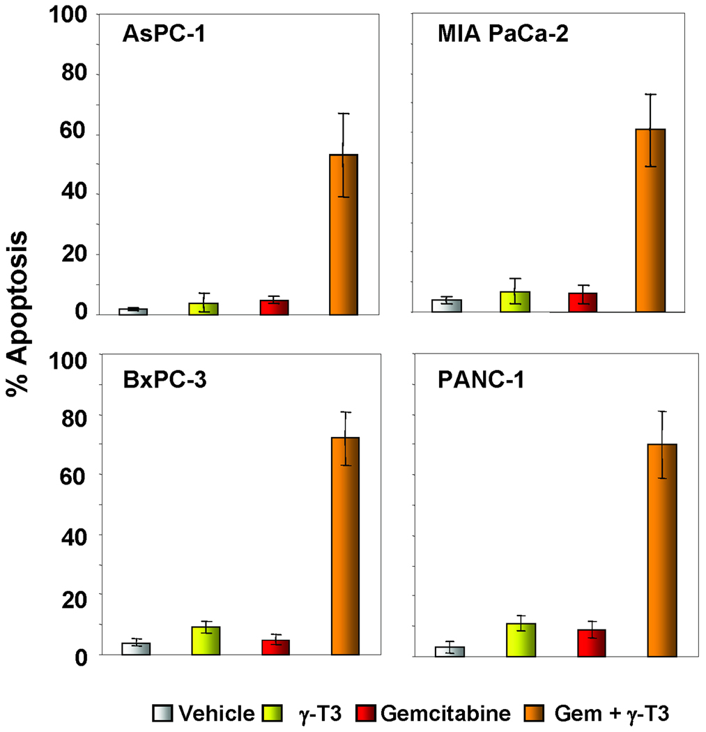
Gemcitabine is a standard treatment for patients with pancreatic cancer. To determine whether γ-T3 could enhance the apoptotic effects of gemcitabine in these cell lines, we performed Live/Dead assay that measured the esterase activity. At a dose at which γ-T3 (10 µM) and gemcitabine (200 nM) alone had minimally toxic effects, the two together were highly effective for inducing apoptosis in pancreatic cancer cell lines in vitro (Fig. 2). No significant difference was observed between the cell lines with respect to their sensitivity to the combination.
Figure 2. γ-T3 potentiates the apoptotic effects of gemcitabine in pancreatic cancer cells in vitro.
Live/Dead assay results indicated that γ-T3 potentiates gemcitabine (Gem)-induced cytotoxicity. Percentages, proportions of apoptotic pancreatic cancer cells. Data are representative of two independent experiments.
γ-T3 inhibits constitutive NF-κB activation and proteins associated with inflammation, proliferation, invasion, and angiogenesis in pancreatic cancer cells
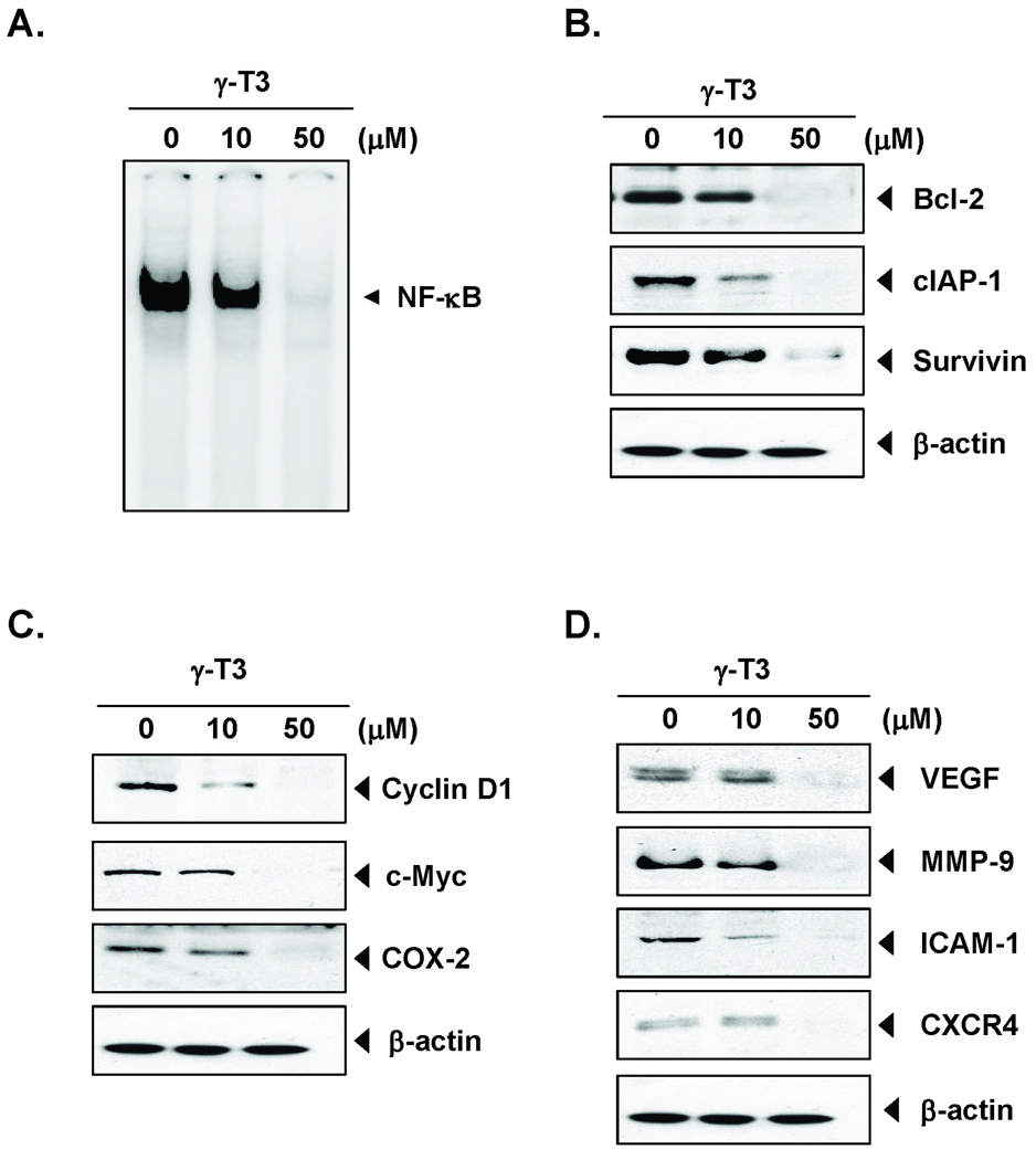
Because NF-κB has been linked with both proliferation and chemoresistance, we next examined whether γ-T3 could inhibit constitutive NF-κB activation in MIA PaCa-2 cell lines. Our results showed that γ-T3 inhibited constitutive NF-κB activation (Fig. 3A).
Figure 3. γ-T3 inhibits constitutive activation of NF-κB and NF-κB-regulated gene products in vitro.
(A), MIA PaCa-2 (1 × 10⁶) cells were treated with γ-T3 (10 and 50 µmol/L) for 4 h; nuclear extracts were prepared and then assayed for NF-κB activation by EMSA. (B–D), Western blot analysis showed that γ-T3 inhibited constitutive expression of NF-κB-regulated gene products that regulate antiapoptosis (B), proliferation (C), and metastasis and angiogenesis (D) in pancreatic cancer cells. The MIA PaCa-2 (1 × 10⁶) cells were treated with with γ-T3 (10 and 50 µmol/L) for 24 h. Whole-cell lysates were prepared and assayed for NF-κB-regulated gene products by Western blotting. Data represent two independent experiments.
We next investigated the effects of γ-T3 on the constitutive expression of antiapoptotic proteins Bcl-2, cIAP-1, and survivin. The expression of all these proteins was downregulated by γ-T3 in a dose-dependent manner (Fig. 3B). When we examined whether γ-T3 could modulate the constitutive expression of proliferative proteins cyclin D1, c-Myc, and COX-2, we found that γ-T3 suppressed the expression of all these proteins in a dose-dependent manner (Fig. 3C). Likewise, γ-T3 downregulated the expression of proteins linked to invasion, adhesion, and angiogenesis--MMP-9, ICAM-1, VEGF, and CXCR4--in a dose-dependent manner (Fig. 3D).
γ-T3 potentiates the antitumor activity of gemcitabine in an orthotopic pancreatic tumor model in nude mice
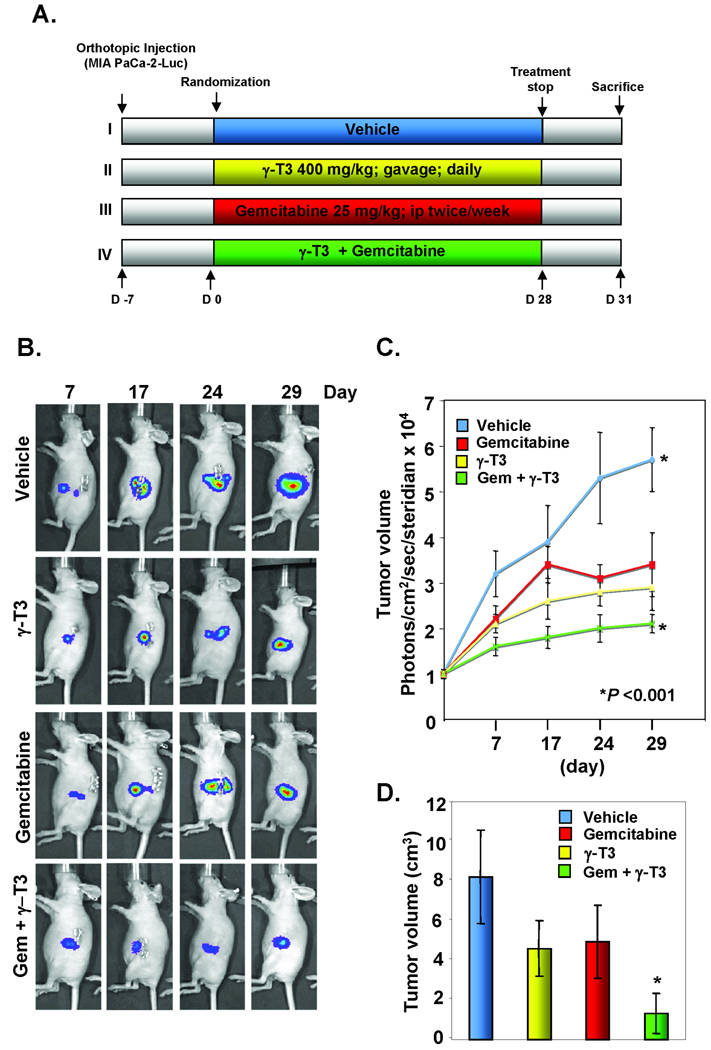
Based on our in vitro results, we designed studies to determine the effects of γ-T3 on gemcitabine in orthotopically implanted human pancreatic tumors in nude mice (Fig. 4A). The dose of γ-T3 employed has been used previously for radiosensitization of tumors in mice (32).
Figure 4. γ-T3 potentiates the effect of gemcitabine in blocking the growth of pancreatic cancer in nude mice.
(A), Schematic of experimental protocol described in Materials and Methods. Group I was given olive oil (100 µL, p.o., daily) and PBS (100 µL, i.p., twice weekly), group II was given γ-T3 (400 mg/kg, p.o., daily), group III was given gemcitabine (25 mg/kg, i.p., twice weekly), and group IV was given γ-T3 (400 mg/kg, p.o., daily) and gemcitabine (25 mg/kg, i.p., twice weekly; n = 6). (B), Bioluminescence IVIS images of orthotopically implanted pancreatic tumors in live, anesthetized mice; (C), Measurements of photons per second depicting tumor volume at various time points using live IVIS imaging at the indicated times (n = 6). Points, mean; bars, SE. D, Tumor volumes in mice measured on the last day of the experiment at autopsy with vernier calipers and calculated using the formula V = 2 / 3πr3 (n = 6). Columns, mean; bars, SE.
Luciferase-transfected MIA PaCa-2 cells were implanted in the tails of the pancreas in nude mice. Based on the IVIS imaging, 1 week later, the mice were randomized into four groups. The treatment was started 1 week after the tumor cell implantation and continued per experimental protocol for 4 weeks. The animals were euthanized 6 weeks after tumor cell injection and 5 weeks from the date of treatment (Fig. 4A). To determine tumor development and the effects of γ-T3 and gemcitabine, we assessed the tumors with the bioluminescence IVIS on days 7, 17, 24, and 29 after the start of treatment. The bioluminescence imaging results (Fig. 4B and C) indicated a gradual increase in tumor volume in the control group compared with the three treatment groups. The imaging results showed that the tumor volume in the combination group was significantly lower than in the group treated with tocotrienol or gemcitabine alone as well as in the vehicle-treated control group (P < 0.001 versus gemcitabine; P < 0.001 versus control). On the 42nd day, we euthanized the mice and measured the tumor volume with vernier calipers. The results were in concordance with those from bioluminescence imaging and showed that the combination reduced the tumor volume more than in the other three groups (P < 0.001 versus control) (Fig. 4D).
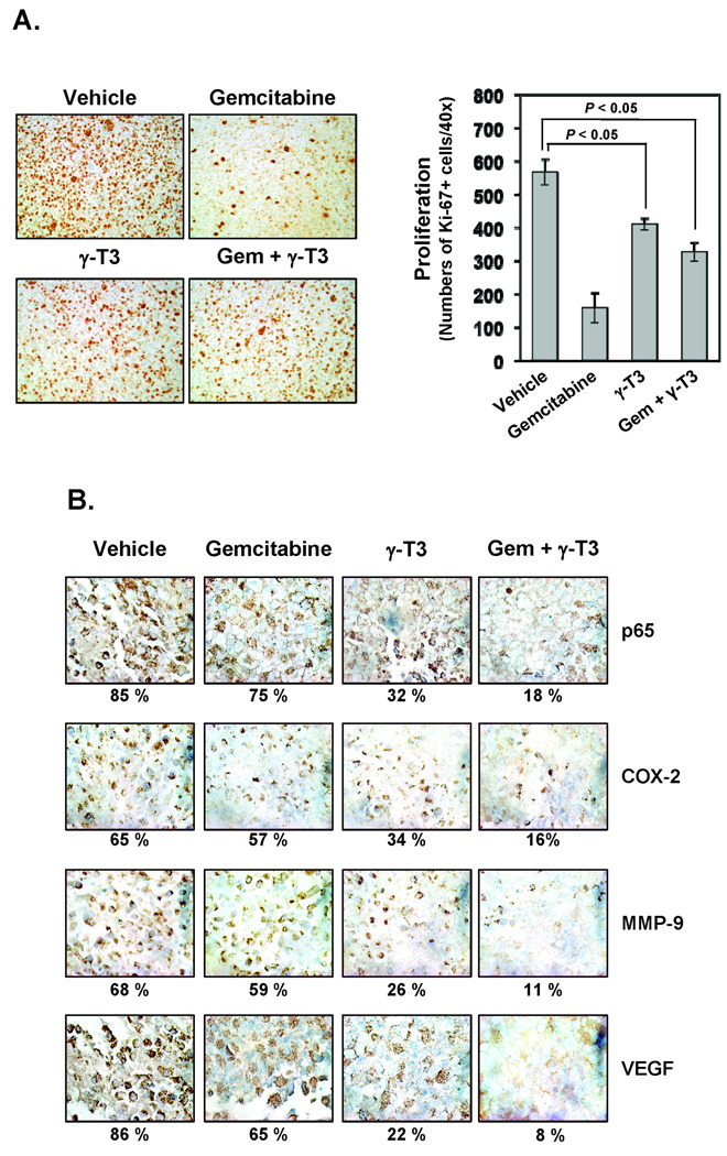
We next examined the expression of the cell proliferation marker Ki-67 in tumor tissues from the four groups. The results in Fig. 5A show that γ-T3 in gemcitabine significantly downregulated the expression of Ki-67 in tumor tissues compared with the control group (P < 0.05 versus control). The results also showed that γ-T3 alone significantly suppressed the expression of Ki-67 (P < 0.05 versus control; Fig. 5B).
Figure 5. γ-T3 enhances the effect of gemcitabine against tumor cell proliferation and the expression of NF-κB and NF-κB-regulated genes in pancreatic cancer.
(A) Left, Immunohistochemical analysis of proliferation maker Ki-67. Right, Quantification of Ki-67+ cells, as described in Materials and Methods section. Values are represented as the mean ± SE of triplicate. (B), Immunohistochemical analysis of NF-κB and NF-κB-regulated genes COX-2, MMP-9, and VEGF in pancreatic tumor tissues from mice. Percentages indicate positive staining for the given biomarker. Samples from three animals in each group were analyzed, and representative data are shown.
γ-T3 inhibits NF-κB activation and NF-κB-regulated gene products in orthotopic pancreatic tumors
We next investigated whether the effects of γ-T3 on tumor growth in mice were associated with the inhibition of NF-κB activation. The results of immunohistochemical analysis showed that γ-T3 alone completely suppressed NF-κB activation in orthotopically grown human pancreatic tumor samples (Fig. 5B). γ-T3 also further suppressed NF-κB activation in gemcitabine-treated tissues. Immunohistochemical analysis further demonstrated that γ-T3 suppressed the expression of COX-2 (Fig. 5B, top panel), MMP-9 (Fig. 5B, second panel), and VEGF (Fig. 5B, bottom panel) in human pancreatic tumor tissues and this effect was enhanced in tissues treated with gemcitabine.
γ-T3 potentiates the effects of gemcitabine in downregulating the expression of NF-κB–regulated gene products
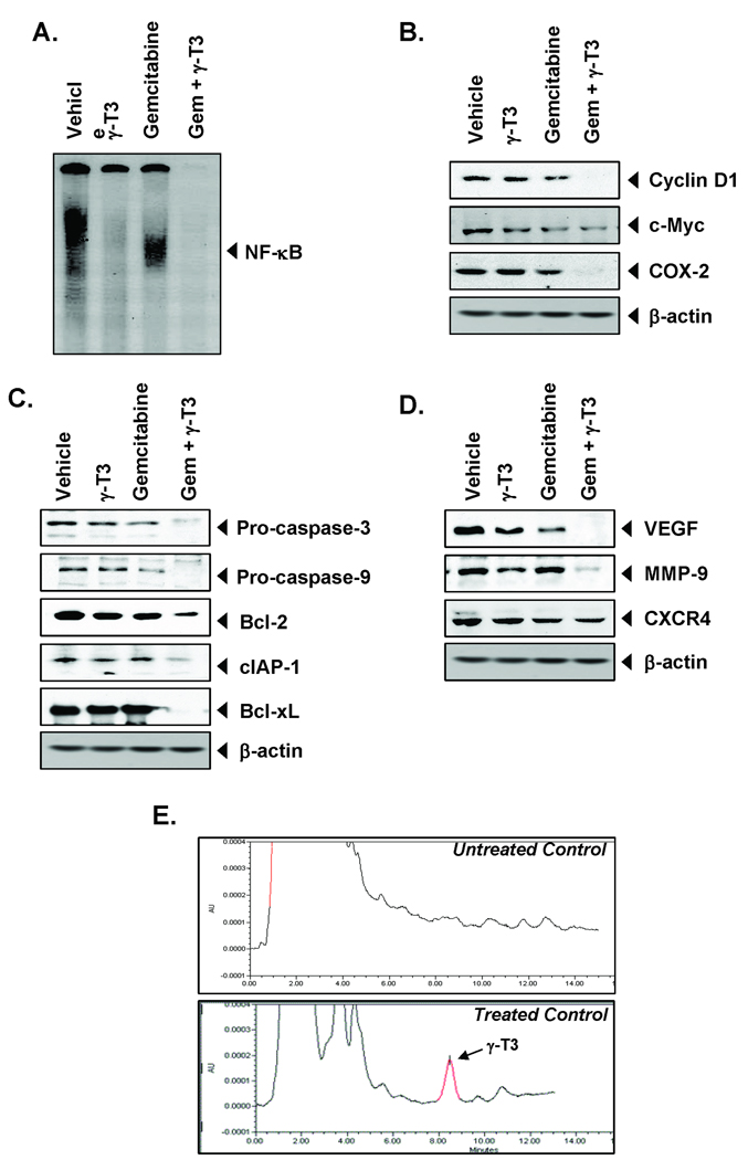
We also examined NF-κB activation in tumor tissues by EMSA. The results showed that γ-T3 alone abolished the constitutive activation of NF-κB in the pancreatic tumor tissue and this downmodulation was maintained in tissue obtained from animals treated with gemcitabine (Fig. 6A). Thus, EMSA results confirmed those obtained from immunohistochemical analysis. NF-κB is known to regulate the expression of proteins involved in proliferation: cyclin D1, c-Myc, and COX-2. The expression of all these proteins was found to be maximally downregulated in pancreatic tissue obtained from animals treated with both agents together (Fig. 6B). We also examined the expression of survival proteins Bcl-xL, cIAP-1, and Bcl-2 by Western blot. Maximum downregulation was noted when both agents were used together (Fig. 6C). When activation of caspase-9 and caspase-3 were examined, maximum activation was also noted when both agents were used together (Fig. 6C). Our results also showed that γ-T3 and gemcitabine together decreased the expression of proteins linked to metastasis (MMP-9), invasion (ICAM-1), and angiogenesis (VEGF) (Fig. 6D). Thus, γ-T3 in combination with gemcitabine inhibited the expression of various gene products in pancreatic tumors from mice.
Figure 6. γ-T3 enhances the effect of gemcitabine against the expression of NF-κB and NF-κB-regulated gene products in pancreatic cancer samples.
(A), Detection of NF-κB by DNA binding in orthotopic tumor tissue samples showed the inhibition of NF-κB by γ-T3. (B), Western blot analysis showed that γ-T3 and gemcitabine together inhibited the expression of NF-κB-dependent proliferative gene products cyclin D1, c-Myc, and COX-2. (C), Western blot analysis showed that γ-T3 and gemcitabine together inhibited the expression of caspase-3, caspase-9, Bcl-2, cIAP-1, and Bcl-xL in pancreatic tumor tissues. (D), Western blot analysis showed that γ-T3 and gemcitabine together inhibited the expression of metastatic and angiogenic genes VEGF, MMP-9, and CXCR4 in pancreatic tumor tissues. Samples from three animals in each group were analyzed, and representative data are shown. (E), Uptake of γ-T3 by pancreatic tissue in mice HPLC chromatogram of γ-T3 mobile phase.
γ-T3 can be detected in pancreatic tumor tissue
In order to determine whether γ-T3 is directly detectable in pancreatic tissue when administered orally, we extracted tissues and analyzed them by high-performance liquid chromatography for γ-T3. Purified γ-T3 was used as a control. We also used tissues from untreated animals as a control. The results in Fig. 6E show a significant presence of γ-T3 in human pancreatic tumor tissue (94.12 ng/g tissue) as compared to untreated control. γ-T3 estimation by HPLC was linear in this range (see Supplement Fig. 1).
Discussion
Pancreatic cancer is the most lethal type of cancer, with an estimated 5-year survival rate of around 4% despite the best treatment available today. Agents that are nontoxic and yet potentiate the effects of chemotherapy are highly desirable. In the current study, we investigated whether γ-T3, an analogue of vitamin E, could enhance the antitumor activity of gemcitabine against human pancreatic cancer. We found that γ-T3 inhibited the proliferation of various pancreatic cancer cell lines in vitro, potentiated gemcitabine-induced apoptosis, and inhibited constitutively active NF-κB and NF-κB-regulated gene products. When administered alone, γ-T3 significantly inhibited the growth of PaCa in established orthotopically grown human pancreatic tumors in nude mice. In addition, γ-T3 further potentiated the effects of gemcitabine against human pancreatic tumors in nude mice.
We found that γ-T3 can suppress the proliferation of various pancreatic cancer cell lines. Our results agree with those from Hussein et al. (17) that showed that δ-T3 could suppress the proliferation of Panc-1, MIA PaCa-2, and BxPC-3 cells. This suppression of proliferation was due to cell cycle arrest at G1. However, they did not investigate the mechanism in detail(17). We found that downregulation of cell proliferative gene products such as cyclin D1, c-Myc, and COX-2 could account for the suppression of cell proliferation by γ-T3.
Our study is the first report to suggest that γ-T3 can enhance the apoptotic effect of gemcitabine in various pancreatic cancer cell lines in vitro. Previously, it has been reported that γ-T3 can enhance the effect of gefitinib, docetaxel, celecoxib and statins against various human cancers (18–20, 27). Although different cancer has its own property and therapeutic strategy, the synergistic action of γ-T3 is partially confirmed.
We found that this effect may be mediated through the downregulation of cell survival proteins such as Bcl-2, cIAP-1, and survivin by γ-T3. Moreover, constitutive NF-κB expressed in human pancreatic cancer cell lines has been linked with chemoresistance (3). We found that this NF-κB activation in cell lines was also suppressed by γ-T3. These results are consistent with those previously reported for curcumin (29) and resveratrol (33). Hussein and Mo showed that mevalonate reversed the effect of δ-T3, thus implicating the role of HMG-CoA in action of δ-T3; furthermore, lovastatin, an inhibitor of HMG-CoA reductase, potentiated the antiproliferative effect of δ-T3 (17, 34). These results suggest that γ-T3 can be used in combination with gemcitabine and lovastatin, as γ-T3 is known to inhibit HMG-CoA reductase also (35–37). Similarly, we have shown previously that both statins and γ-T3 are potent inhibitors of NF-κB activation (16, 38).
We found for the first time that oral administration of γ-T3 alone inhibited the growth of human pancreatic tumors when examined in vivo in an orthotopic nude mice model. Tumor growth was inhibited by almost 50% by 400 mg/kg γ-T3 alone, and this level of inhibition was comparable to that reduced by gemcitabine alone. γ-T3 was very well tolerated by the animals. When the two agents were used in combination, they were found to be much more effective. How γ-T3 exhibited its effects in vivo were also investigated in several ways. First, analysis of Ki-67 revealed that γ-T3 inhibited its proliferation in tumor tissues. Likewise, analysis of NF-κB in pancreatic tumors showed that γ-T3 alone inhibited constitutive activation of NF-κB. γ-T3 significantly downregulated the expression of proinflammatory marker COX-2, suppressed the expression of invasion biomarker MMP-9, and inhibited the angiogenic biomarker VEGF in the tissues. All of these effects were further enhanced by gemcitabine. The synergistic interaction between the two agents for down-modulation of various biomarkers was more apparent by Western blot analysis than by immunohistochemical analysis. All these data indicate for the first time the mechanisms by which γ-T3 could exhibit its effects against human pancreatic cancer. We also found a significant presence of intact γ-T3 in the pancreatic tumor tissue (94.12 ng/g). These results agree with those reported by Husain et al. (39) in a previous study of δ-T3. They showed that δ-T3 when given orally at 100 mg/kg to mice, a peak plasma concentration of 57 µM at 2 h, plasma T1/2 of 3.5 h. Six week feeding of δ-T3 gave a tissue levels of 41 nM/g tissue; levels were 10 times higher in pancreas than in tumor or liver tissue (39).
Vitamin E consists of four different isoforms of tocopherols and of tocotrienols (Aggarwal, 2010). Among the tocotrienols, it seems that anticancer activities are limited to only gamma and delta forms. Why alfa and beta forms of tocotrienols, exhibit minimal anticancer activities, is not understood at present. Differential cellular uptake, membrane insertion, bioavailabilty, anti-antioxidant or anti-inflammatory potential may account for these differences.
Overall, our results show for the first time that γ-T3 potentiates the antitumor activity of gemcitabine by inhibiting NF-κB and NF-κB-regulated gene products, leading to the inhibition of proliferation, angiogenesis, and invasion. However, further studies are necessary to confirm our findings in patients with pancreatic cancer. Considering that tocotrienols, although structurally homologous to tocopherols, exhibit distinct molecular targets, they merit further exploration.
Supplementary Material
Acknowledgments
We thank Markeda Wade for carefully proof-reading the manuscript and providing valuable comments. Dr. Aggarwal is the Ransom Horne, Jr., Professor of Cancer Research. This work was supported by grant from Malaysian Palm Oil Board, a core grant from the National Institutes of Health (CA-16 672), a program project grant from National Institutes of Health (NIH CA-124787-01A2), and grant from Center for Targeted Therapy of M.D. Anderson Cancer Center.
References
- 1. http://www.cancer.gov/cancertopics/types/pancreatic.
- 2.Aggarwal BB. Nuclear factor-kappaB: the enemy within. Cancer Cell. 2004;6:203–208. doi: 10.1016/j.ccr.2004.09.003. [DOI] [PubMed] [Google Scholar]
- 3.Arlt A, Gehrz A, Muerkoster S, et al. Role of NF-kappaB and Akt/PI3K in the resistance of pancreatic carcinoma cell lines against gemcitabine-induced cell death. Oncogene. 2003;22:3243–3251. doi: 10.1038/sj.onc.1206390. [DOI] [PubMed] [Google Scholar]
- 4.Liptay S, Weber CK, Ludwig L, et al. Mitogenic and antiapoptotic role of constitutive NF-kappaB/Rel activity in pancreatic cancer. Int J Cancer. 2003;105:735–746. doi: 10.1002/ijc.11081. [DOI] [PubMed] [Google Scholar]
- 5.Wang W, Abbruzzese JL, Evans DB, et al. The nuclear factor-kappa B RelA transcription factor is constitutively activated in human pancreatic adenocarcinoma cells. Clin Cancer Res. 1999;5:119–127. [PubMed] [Google Scholar]
- 6.Fujioka S, Sclabas GM, Schmidt C, et al. Function of nuclear factor kappaB in pancreatic cancer metastasis. Clin Cancer Res. 2003;9:346–354. [PubMed] [Google Scholar]
- 7.Greten FR, Weber CK, Greten TF, et al. Stat3 and NF-kappaB activation prevents apoptosis in pancreatic carcinogenesis. Gastroenterology. 2002;123:2052–2063. doi: 10.1053/gast.2002.37075. [DOI] [PubMed] [Google Scholar]
- 8.Yamamoto Y, Gaynor RB. Therapeutic potential of inhibition of the NF-kappaB pathway in the treatment of inflammation and cancer. J Clin Invest. 2001;107:135–142. doi: 10.1172/JCI11914. [DOI] [PMC free article] [PubMed] [Google Scholar]
- 9.Kornmann M, Ishiwata T, Itakura J, et al. Increased cyclin D1 in human pancreatic cancer is associated with decreased postoperative survival. Oncology. 1998;55:363–369. doi: 10.1159/000011879. [DOI] [PubMed] [Google Scholar]
- 10.Xiong HQ, Abbruzzese JL, Lin E, et al. NF-kappaB activity blockade impairs the angiogenic potential of human pancreatic cancer cells. Int J Cancer. 2004;108:181–188. doi: 10.1002/ijc.11562. [DOI] [PubMed] [Google Scholar]
- 11.Yebra M, Filardo EJ, Bayna EM, et al. Induction of carcinoma cell migration on vitronectin by NF-kappa B-dependent gene expression. Mol Biol Cell. 1995;6:841–850. doi: 10.1091/mbc.6.7.841. [DOI] [PMC free article] [PubMed] [Google Scholar]
- 12.Aggarwal BB, Sundaram C, Prasad S, Kannappan R. Tocotrienols, the Vitamin E of the 21(st) Century: It's Potential Against Cancer and Other Chronic Diseases. Biochem Pharmacol. 2010 doi: 10.1016/j.bcp.2010.07.043. [DOI] [PMC free article] [PubMed] [Google Scholar]
- 13.Sundram K, Khor HT, Ong AS, Pathmanathan R. Effect of dietary palm oils on mammary carcinogenesis in female rats induced by 7,12-dimethylbenz(a)anthracene. Cancer Res. 1989;49:1447–1451. [PubMed] [Google Scholar]
- 14.Kamal-Eldin A, Appelqvist LA. The chemistry and antioxidant properties of tocopherols and tocotrienols. Lipids. 1996;31:671–701. doi: 10.1007/BF02522884. [DOI] [PubMed] [Google Scholar]
- 15.Shah SJ, Sylvester PW. Gamma-tocotrienol inhibits neoplastic mammary epithelial cell proliferation by decreasing Akt and nuclear factor kappaB activity. Exp Biol Med (Maywood) 2005;230:235–241. doi: 10.1177/153537020523000402. [DOI] [PubMed] [Google Scholar]
- 16.Ahn KS, Sethi G, Krishnan K, Aggarwal BB. Gamma-tocotrienol inhibits nuclear factor-kappaB signaling pathway through inhibition of receptor-interacting protein and TAK1 leading to suppression of antiapoptotic gene products and potentiation of apoptosis. J Biol Chem. 2007;282:809–820. doi: 10.1074/jbc.M610028200. [DOI] [PubMed] [Google Scholar]
- 17.Hussein D, Mo H. d-Dlta-tocotrienol-mediated suppression of the proliferation of human PANC-1, MIA PaCa-2, and BxPC-3 pancreatic carcinoma cells. Pancreas. 2009;38:e124–e136. doi: 10.1097/MPA.0b013e3181a20f9c. [DOI] [PubMed] [Google Scholar]
- 18.Bachawal SV, Wali VB, Sylvester PW. Enhanced antiproliferative and apoptotic response to combined treatment of gamma-tocotrienol with erlotinib or gefitinib in mammary tumor cells. BMC Cancer. 2010;10:84. doi: 10.1186/1471-2407-10-84. [DOI] [PMC free article] [PubMed] [Google Scholar]
- 19.Shirode AB, Sylvester PW. Synergistic anticancer effects of combined gamma-tocotrienol and celecoxib treatment are associated with suppression in Akt and NFkappaB signaling. Biomed Pharmacother. 2010;64:327–332. doi: 10.1016/j.biopha.2009.09.018. [DOI] [PMC free article] [PubMed] [Google Scholar]
- 20.Wali VB, Bachawal SV, Sylvester PW. Combined treatment of gamma-tocotrienol with statins induce mammary tumor cell cycle arrest in G1. Exp Biol Med (Maywood) 2009;234:639–650. doi: 10.3181/0810-RM-300. [DOI] [PubMed] [Google Scholar]
- 21.Samant GV, Sylvester PW. gamma-Tocotrienol inhibits ErbB3-dependent PI3K/Akt mitogenic signalling in neoplastic mammary epithelial cells. Cell Prolif. 2006;39:563–574. doi: 10.1111/j.1365-2184.2006.00412.x. [DOI] [PMC free article] [PubMed] [Google Scholar]
- 22.Mizushina Y, Nakagawa K, Shibata A, et al. Inhibitory effect of tocotrienol on eukaryotic DNA polymerase lambda and angiogenesis. Biochem Biophys Res Commun. 2006;339:949–955. doi: 10.1016/j.bbrc.2005.11.085. [DOI] [PubMed] [Google Scholar]
- 23.Wells SR, Jennings MH, Rome C, et al. alpha-, gamma- and delta-tocopherols reduce inflammatory angiogenesis in human microvascular endothelial cells. J Nutr Biochem. 2009 doi: 10.1016/j.jnutbio.2009.03.006. [DOI] [PubMed] [Google Scholar]
- 24.Shibata A, Nakagawa K, Sookwong P, et al. Tocotrienol inhibits secretion of angiogenic factors from human colorectal adenocarcinoma cells by suppressing hypoxia-inducible factor-1alpha. J Nutr. 2008;138:2136–2142. doi: 10.3945/jn.108.093237. [DOI] [PubMed] [Google Scholar]
- 25.Yam ML, Abdul Hafid SR, Cheng HM, Nesaretnam K. Tocotrienols suppress proinflammatory markers and cyclooxygenase-2 expression in RAW264.7 macrophages. Lipids. 2009;44:787–797. doi: 10.1007/s11745-009-3326-2. [DOI] [PubMed] [Google Scholar]
- 26.Gysin R, Azzi A, Visarius T. Gamma-tocopherol inhibits human cancer cell cycle progression and cell proliferation by down-regulation of cyclins. FASEB J. 2002;16:1952–1954. doi: 10.1096/fj.02-0362fje. [DOI] [PubMed] [Google Scholar]
- 27.Yap WN, Zaiden N, Luk SY, et al. In vivo evidence of gamma-tocotrienol as a chemosensitizer in the treatment of hormone-refractory prostate cancer. Pharmacology. 2010;85:248–258. doi: 10.1159/000278205. [DOI] [PubMed] [Google Scholar]
- 28.Kunnumakkara AB, Diagaradjane P, Anand P, et al. Curcumin sensitizes human colorectal cancer to capecitabine by modulation of cyclin D1, COX-2, MMP-9, VEGF and CXCR4 expression in an orthotopic mouse model. Int J Cancer. 2009;125:2187–2197. doi: 10.1002/ijc.24593. [DOI] [PubMed] [Google Scholar]
- 29.Kunnumakkara AB, Guha S, Krishnan S, et al. Curcumin potentiates antitumor activity of gemcitabine in an orthotopic model of pancreatic cancer through suppression of proliferation, angiogenesis, and inhibition of nuclear factor-kappaB-regulated gene products. Cancer Res. 2007;67:3853–3861. doi: 10.1158/0008-5472.CAN-06-4257. [DOI] [PubMed] [Google Scholar]
- 30.Khanbolooki S, Nawrocki ST, Arumugam T, et al. Nuclear factor-kappaB maintains TRAIL resistance in human pancreatic cancer cells. Mol Cancer Ther. 2006;5:2251–2260. doi: 10.1158/1535-7163.MCT-06-0075. [DOI] [PubMed] [Google Scholar]
- 31.Yap SP, Julianto T, Wong JW, Yuen KH. Simple high-performance liquid chromatographic method for the determination of tocotrienols in human plasma. J Chromatogr B Biomed Sci Appl. 1999;735:279–283. doi: 10.1016/s0378-4347(99)00385-0. [DOI] [PubMed] [Google Scholar]
- 32.Kumar KS, Raghavan M, Hieber K, et al. Preferential radiation sensitization of prostate cancer in nude mice by nutraceutical antioxidant gamma-tocotrienol. Life Sci. 2006;78:2099–2104. doi: 10.1016/j.lfs.2005.12.005. [DOI] [PubMed] [Google Scholar]
- 33.Harikumar KB, Kunnumakkara AB, Sethi G, et al. Resveratrol, a multitargeted agent, can enhance antitumor activity of gemcitabine in vitro and in orthotopic mouse model of human pancreatic cancer. Int J Cancer. 127:257–268. doi: 10.1002/ijc.25041. [DOI] [PMC free article] [PubMed] [Google Scholar]
- 34.McAnally JA, Gupta J, Sodhani S, Bravo L, Mo H. Tocotrienols potentiate lovastatin-mediated growth suppression in vitro and in vivo. Exp Biol Med (Maywood) 2007;232:523–531. [PubMed] [Google Scholar]
- 35.Song BL, DeBose-Boyd RA. Insig-dependent ubiquitination and degradation of 3-hydroxy-3-methylglutaryl coenzyme a reductase stimulated by delta- and gamma-tocotrienols. J Biol Chem. 2006;281:25054–25061. doi: 10.1074/jbc.M605575200. [DOI] [PubMed] [Google Scholar]
- 36.Mo H, Elson CE. Studies of the isoprenoid-mediated inhibition of mevalonate synthesis applied to cancer chemotherapy and chemoprevention. Exp Biol Med (Maywood) 2004;229:567–585. doi: 10.1177/153537020422900701. [DOI] [PubMed] [Google Scholar]
- 37.Parker RA, Pearce BC, Clark RW, Gordon DA, Wright JJ. Tocotrienols regulate cholesterol production in mammalian cells by post-transcriptional suppression of 3-hydroxy-3-methylglutaryl-coenzyme A reductase. J Biol Chem. 1993;268:11230–11238. [PubMed] [Google Scholar]
- 38.Ahn KS, Sethi G, Aggarwal BB. Simvastatin potentiates TNF-alpha-induced apoptosis through the down-regulation of NF-kappaB-dependent antiapoptotic gene products: role of IkappaBalpha kinase and TGF-beta-activated kinase-1. J Immunol. 2007;178:2507–2516. doi: 10.4049/jimmunol.178.4.2507. [DOI] [PubMed] [Google Scholar]
- 39.Husain K, Francois RA, Hutchinson SZ, et al. Vitamin E delta-tocotrienol levels in tumor and pancreatic tissue of mice after oral administration. Pharmacology. 2009;83:157–163. doi: 10.1159/000190792. [DOI] [PMC free article] [PubMed] [Google Scholar]
Associated Data
This section collects any data citations, data availability statements, or supplementary materials included in this article.


