Abstract
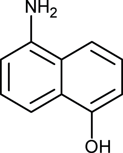
In the title compound, C10H9NO, the amino and the hydroxy groups act both as a single donor and a single acceptor in hydrogen bonding. In the crystal, molecules are connected via chains of intermolecular ⋯N—H⋯O—H⋯ interactions, forming a two-dimensional polymeric structure resembling the hydrogen-bonded molecular assembly found in the crystal structure of naphthalene-1,5-diol. Within this layer, molecules related by a translation along the a axis are arranged into slipped stacks via π–π stacking interactions [interplanar distance = 3.450 (4) Å]. The amino N atom shows sp 3 hybridization and the two attached H atoms are located on the same side of the aromatic ring.
Related literature
For the crystal structure of 1,5-dihydroxynaphthalene, see: Belskii et al. (1990 ▶). For amino-hydroxy group recognition and packing motifs of aminols, see: Ermer & Eling (1994 ▶); Hanessian et al. (1994 ▶); Allen et al. (1997 ▶); Dey et al. (2005 ▶).
Experimental
Crystal data
C10H9NO
M r = 159.18
Orthorhombic,

a = 4.8607 (2) Å
b = 12.3175 (6) Å
c = 13.0565 (5) Å
V = 781.71 (6) Å3
Z = 4
Mo Kα radiation
μ = 0.09 mm−1
T = 130 K
0.30 × 0.15 × 0.02 mm
Data collection
Kuma KM-4-CCD κ-geometry diffractometer
Absorption correction: none
8484 measured reflections
963 independent reflections
726 reflections with I > 2σ(I)
R int = 0.037
Refinement
R[F 2 > 2σ(F 2)] = 0.036
wR(F 2) = 0.091
S = 1.06
963 reflections
121 parameters
H atoms treated by a mixture of independent and constrained refinement
Δρmax = 0.17 e Å−3
Δρmin = −0.21 e Å−3
Data collection: CrysAlis CCD (Oxford Diffraction, 2007 ▶); cell refinement: CrysAlis RED (Oxford Diffraction, 2007 ▶); data reduction: CrysAlis RED; program(s) used to solve structure: SHELXS97 (Sheldrick, 2008 ▶); program(s) used to refine structure: SHELXL97 (Sheldrick, 2008 ▶); molecular graphics: ORTEP-3 for Windows (Farrugia, 1997 ▶) and Mercury (Macrae et al., 2006 ▶); software used to prepare material for publication: SHELXL97.
Supplementary Material
Crystal structure: contains datablocks global, I. DOI: 10.1107/S1600536809045152/su2154sup1.cif
Structure factors: contains datablocks I. DOI: 10.1107/S1600536809045152/su2154Isup2.hkl
Additional supplementary materials: crystallographic information; 3D view; checkCIF report
Table 1. Hydrogen-bond geometry (Å, °).
| D—H⋯A | D—H | H⋯A | D⋯A | D—H⋯A |
|---|---|---|---|---|
| O11—H11O⋯N12i | 0.94 (3) | 1.83 (3) | 2.749 (3) | 167 (3) |
| N12—H12B⋯O11ii | 0.96 (3) | 2.09 (3) | 3.046 (3) | 171 (2) |
Symmetry codes: (i)  ; (ii)
  .
.
supplementary crystallographic information
Comment
Amino-hydroxy group recognition has been at the focus of crystal engineering since Ermer & Eling (1994) and Hanessian et al. (1994) noticed the complementarity of hydroxy and amino groups as regards hydrogen-bond donors and acceptors. Shortly after it was demonstrated with simple 1,2- and 1,3-aminophenols that molecular features, such as functional groups, do not necessarily result in a single manner of the molecular arrangement, and that strong N—H···O and O—H···N hydrogen bonding is frequently unable to exclude other factors from controlling the crystal packing (Allen et al., 1997; Dey et al. 2005). Here we report on the crystal structure of a simple aminonaphthol which, as regards the substitution pattern, can be considered as an analogue of 1,4-aminophenol.
The molecular structure of the title compound is shown in Fig. 1, and the geometrical parameters are available in the archived CIF. Whereas 1,4-aminophenol forms a tetrahedral network, via N—H···O and O—H···N hydrogen bonds (Ermer & Eling, 1994), in the title compound the molecules are connected via chains of ···N—H···O—H··· interactions to form a two-dimensional polymeric structure (Fig. 2, Table 1). The two-dimensional assembly of the molecules joined by hydrogen bonding resembles that formed by 1,5-dihydroxynaphthalene (Belskii et al., 1990). One amino H-atom is not involved in hydrogen bonding and does not take part in any other specific interactions.
Experimental
Single crystals of the commercially available 5-amino-1-naphthol (Aldrich) were obtained from chloroform solution by slow evaporation.
Refinement
In the absence of significant anomalous scattering effects, Friedel pairs were averaged. All H-atoms were located in electron-density difference maps. The H-atoms of the OH and NH groups were freely refined: O-H = 0.94 (3) Å, N-H = 0.95 (3)- 0.96 (3) Å. The C-bound H-atoms were placed at calculated positions, with C—H = 0.93 Å, and refined as riding on their carrier C-atom, with Uiso(H) = 1.2Ueq(C). .
Figures
Fig. 1.
The molecular structure of the title compound, with displacement ellipsoids shown at the 30% probability level.
Fig. 2.
The crystal packing, viewed along the b-axis, of the title compound, showing the two-dimensional hydrogen-bonded assembly (hydrogen bonds are shown as dashed lines).
Crystal data
| C10H9NO | Dx = 1.353 Mg m−3 |
| Mr = 159.18 | Melting point: 461 K |
| Orthorhombic, P212121 | Mo Kα radiation, λ = 0.71073 Å |
| Hall symbol: P 2ac 2ab | Cell parameters from 2383 reflections |
| a = 4.8607 (2) Å | θ = 4–27° |
| b = 12.3175 (6) Å | µ = 0.09 mm−1 |
| c = 13.0565 (5) Å | T = 130 K |
| V = 781.71 (6) Å3 | Plate, pale pink |
| Z = 4 | 0.30 × 0.15 × 0.02 mm |
| F(000) = 336 |
Data collection
| Kuma KM-4-CCD κ-geometry diffractometer | 726 reflections with I > 2σ(I) |
| Radiation source: fine-focus sealed tube | Rint = 0.037 |
| graphite | θmax = 26.4°, θmin = 4.5° |
| ω scans | h = −6→5 |
| 8484 measured reflections | k = −15→15 |
| 963 independent reflections | l = −16→16 |
Refinement
| Refinement on F2 | Primary atom site location: structure-invariant direct methods |
| Least-squares matrix: full | Secondary atom site location: difference Fourier map |
| R[F2 > 2σ(F2)] = 0.036 | Hydrogen site location: inferred from neighbouring sites |
| wR(F2) = 0.091 | H atoms treated by a mixture of independent and constrained refinement |
| S = 1.06 | w = 1/[σ2(Fo2) + (0.0554P)2] where P = (Fo2 + 2Fc2)/3 |
| 963 reflections | (Δ/σ)max = 0.001 |
| 121 parameters | Δρmax = 0.17 e Å−3 |
| 0 restraints | Δρmin = −0.21 e Å−3 |
Special details
| Geometry. All e.s.d.'s (except the e.s.d. in the dihedral angle between two l.s. planes) are estimated using the full covariance matrix. The cell e.s.d.'s are taken into account individually in the estimation of e.s.d.'s in distances, angles and torsion angles; correlations between e.s.d.'s in cell parameters are only used when they are defined by crystal symmetry. An approximate (isotropic) treatment of cell e.s.d.'s is used for estimating e.s.d.'s involving l.s. planes. |
| Refinement. Refinement of F2 against ALL reflections. The weighted R-factor wR and goodness of fit S are based on F2, conventional R-factors R are based on F, with F set to zero for negative F2. The threshold expression of F2 > σ(F2) is used only for calculating R-factors(gt) etc. and is not relevant to the choice of reflections for refinement. R-factors based on F2 are statistically about twice as large as those based on F, and R- factors based on ALL data will be even larger. |
Fractional atomic coordinates and isotropic or equivalent isotropic displacement parameters (Å2)
| x | y | z | Uiso*/Ueq | ||
| C1 | 0.2605 (5) | 0.48405 (18) | 0.50574 (15) | 0.0232 (5) | |
| C2 | 0.1125 (5) | 0.57846 (19) | 0.51109 (17) | 0.0269 (6) | |
| H2 | −0.0135 | 0.5887 | 0.5638 | 0.032* | |
| C3 | 0.1504 (5) | 0.65964 (19) | 0.43738 (17) | 0.0268 (6) | |
| H3 | 0.0466 | 0.7230 | 0.4409 | 0.032* | |
| C4 | 0.3386 (5) | 0.64703 (19) | 0.35998 (17) | 0.0248 (6) | |
| H4 | 0.3630 | 0.7022 | 0.3122 | 0.030* | |
| C5 | 0.6956 (5) | 0.53275 (18) | 0.27339 (16) | 0.0239 (6) | |
| C6 | 0.8417 (5) | 0.43775 (19) | 0.26935 (17) | 0.0265 (6) | |
| H6 | 0.9701 | 0.4269 | 0.2175 | 0.032* | |
| C7 | 0.7983 (5) | 0.35673 (19) | 0.34304 (17) | 0.0283 (6) | |
| H7 | 0.9009 | 0.2931 | 0.3399 | 0.034* | |
| C8 | 0.6085 (5) | 0.36936 (19) | 0.41937 (17) | 0.0256 (6) | |
| H8 | 0.5795 | 0.3141 | 0.4667 | 0.031* | |
| C9 | 0.4571 (5) | 0.46670 (19) | 0.42580 (16) | 0.0215 (5) | |
| C10 | 0.4964 (5) | 0.55036 (18) | 0.35220 (16) | 0.0215 (5) | |
| O11 | 0.2370 (4) | 0.40173 (13) | 0.57550 (11) | 0.0289 (4) | |
| H11O | 0.067 (7) | 0.404 (2) | 0.609 (2) | 0.058 (10)* | |
| N12 | 0.7265 (5) | 0.61211 (18) | 0.19544 (14) | 0.0275 (5) | |
| H12A | 0.699 (6) | 0.685 (2) | 0.2155 (18) | 0.044 (8)* | |
| H12B | 0.899 (6) | 0.601 (2) | 0.1610 (19) | 0.038 (8)* |
Atomic displacement parameters (Å2)
| U11 | U22 | U33 | U12 | U13 | U23 | |
| C1 | 0.0231 (12) | 0.0279 (13) | 0.0186 (10) | −0.0035 (12) | −0.0020 (11) | 0.0014 (10) |
| C2 | 0.0250 (13) | 0.0332 (14) | 0.0226 (11) | −0.0002 (12) | 0.0011 (11) | −0.0024 (11) |
| C3 | 0.0261 (13) | 0.0255 (13) | 0.0288 (12) | 0.0028 (12) | −0.0008 (11) | −0.0047 (11) |
| C4 | 0.0250 (13) | 0.0272 (13) | 0.0223 (11) | −0.0030 (11) | −0.0017 (11) | 0.0001 (10) |
| C5 | 0.0244 (14) | 0.0283 (13) | 0.0192 (10) | −0.0070 (11) | −0.0053 (10) | −0.0014 (10) |
| C6 | 0.0232 (13) | 0.0334 (13) | 0.0229 (11) | 0.0006 (12) | 0.0024 (10) | −0.0056 (11) |
| C7 | 0.0294 (15) | 0.0244 (13) | 0.0311 (12) | 0.0055 (12) | −0.0022 (11) | −0.0044 (11) |
| C8 | 0.0273 (13) | 0.0278 (13) | 0.0216 (11) | −0.0011 (11) | −0.0013 (11) | −0.0004 (10) |
| C9 | 0.0187 (12) | 0.0263 (13) | 0.0194 (10) | −0.0006 (11) | −0.0030 (10) | −0.0036 (10) |
| C10 | 0.0204 (12) | 0.0249 (13) | 0.0191 (10) | −0.0044 (11) | −0.0044 (10) | −0.0006 (10) |
| O11 | 0.0269 (9) | 0.0356 (10) | 0.0243 (8) | −0.0003 (9) | 0.0036 (8) | 0.0061 (8) |
| N12 | 0.0271 (12) | 0.0316 (13) | 0.0237 (10) | −0.0025 (12) | 0.0005 (10) | 0.0016 (9) |
Geometric parameters (Å, °)
| C1—O11 | 1.368 (2) | C5—C10 | 1.430 (3) |
| C1—C2 | 1.369 (3) | C6—C7 | 1.402 (3) |
| C1—C9 | 1.431 (3) | C6—H6 | 0.9300 |
| C2—C3 | 1.400 (3) | C7—C8 | 1.367 (3) |
| C2—H2 | 0.9300 | C7—H7 | 0.9300 |
| C3—C4 | 1.372 (3) | C8—C9 | 1.409 (3) |
| C3—H3 | 0.9300 | C8—H8 | 0.9300 |
| C4—C10 | 1.420 (3) | C9—C10 | 1.422 (3) |
| C4—H4 | 0.9300 | O11—H11O | 0.94 (3) |
| C5—C6 | 1.370 (3) | N12—H12A | 0.95 (3) |
| C5—N12 | 1.419 (3) | N12—H12B | 0.96 (3) |
| O11—C1—C2 | 123.5 (2) | C7—C6—H6 | 119.9 |
| O11—C1—C9 | 115.5 (2) | C8—C7—C6 | 121.4 (2) |
| C2—C1—C9 | 120.99 (19) | C8—C7—H7 | 119.3 |
| C1—C2—C3 | 120.2 (2) | C6—C7—H7 | 119.3 |
| C1—C2—H2 | 119.9 | C7—C8—C9 | 119.5 (2) |
| C3—C2—H2 | 119.9 | C7—C8—H8 | 120.2 |
| C4—C3—C2 | 120.9 (2) | C9—C8—H8 | 120.2 |
| C4—C3—H3 | 119.6 | C8—C9—C10 | 120.4 (2) |
| C2—C3—H3 | 119.6 | C8—C9—C1 | 121.3 (2) |
| C3—C4—C10 | 120.5 (2) | C10—C9—C1 | 118.3 (2) |
| C3—C4—H4 | 119.7 | C4—C10—C9 | 119.1 (2) |
| C10—C4—H4 | 119.7 | C4—C10—C5 | 123.0 (2) |
| C6—C5—N12 | 120.4 (2) | C9—C10—C5 | 117.9 (2) |
| C6—C5—C10 | 120.6 (2) | C1—O11—H11O | 111.6 (18) |
| N12—C5—C10 | 118.9 (2) | C5—N12—H12A | 116.2 (15) |
| C5—C6—C7 | 120.2 (2) | C5—N12—H12B | 109.1 (15) |
| C5—C6—H6 | 119.9 | H12A—N12—H12B | 113 (2) |
Hydrogen-bond geometry (Å, °)
| D—H···A | D—H | H···A | D···A | D—H···A |
| O11—H11O···N12i | 0.94 (3) | 1.83 (3) | 2.749 (3) | 167 (3) |
| N12—H12B···O11ii | 0.96 (3) | 2.09 (3) | 3.046 (3) | 171 (2) |
Symmetry codes: (i) −x+1/2, −y+1, z+1/2; (ii) −x+3/2, −y+1, z−1/2.
Footnotes
Supplementary data and figures for this paper are available from the IUCr electronic archives (Reference: SU2154).
References
- Allen, F. H., Hoy, V. J., Howard, J. A. K., Thalladi, V. R., Desiraju, G. R., Wilson, C. C. & McIntyre, G. J. (1997). J. Am. Chem. Soc.119, 3477–3480.
- Belskii, V. K., Kharchenko, E. V., Sobolev, A. N., Zavodnik, V. E., Kolomiets, N. A., Prober, G. S. & Oleksenko, L. P. (1990). Zh. Strukt. Khim.31, 116–121.
- Dey, A., Kirchner, M. T., Vangala, V. R., Desiraju, G. R., Mondal, R. & Howard, J. A. K. (2005). J. Am. Chem. Soc.127, 10545–10559. [DOI] [PubMed]
- Ermer, O. & Eling, A. J. (1994). J. Chem. Soc. Perkin Trans. 2, pp. 925–944.
- Farrugia, L. J. (1997). J. Appl. Cryst.30, 565.
- Hanessian, S., Gomtsyan, A., Simard, M. & Roelens, S. (1994). J. Am. Chem. Soc 116, 4495–4496.
- Macrae, C. F., Edgington, P. R., McCabe, P., Pidcock, E., Shields, G. P., Taylor, R., Towler, M. & van de Streek, J. (2006). J. Appl. Cryst.39, 453–457.
- Oxford Diffraction (2007). CrysAlis CCD and CrysAlis RED Oxford Diffraction, Abingdon, England.
- Sheldrick, G. M. (2008). Acta Cryst. A64, 112–122. [DOI] [PubMed]
Associated Data
This section collects any data citations, data availability statements, or supplementary materials included in this article.
Supplementary Materials
Crystal structure: contains datablocks global, I. DOI: 10.1107/S1600536809045152/su2154sup1.cif
Structure factors: contains datablocks I. DOI: 10.1107/S1600536809045152/su2154Isup2.hkl
Additional supplementary materials: crystallographic information; 3D view; checkCIF report


