

Abstract
The title compound, C8H5F2N3S, was synthesized by the reaction of 2,6-difluorobenzoic acid and thiosemicarbazide. The dihedral angle between the thiadiazole and phenyl ring is 35.19 (14)°. In the crystal structure, intermolecular N—H⋯N hydrogen bonds form chains along the b and c axes.
Related literature
For the biological activity of 1,3,4-thiadiazole derivatives, see: Nakagawa et al. (1996 ▶); Wang et al. (1999 ▶). For bond-length data see: Allen et al. (1987 ▶).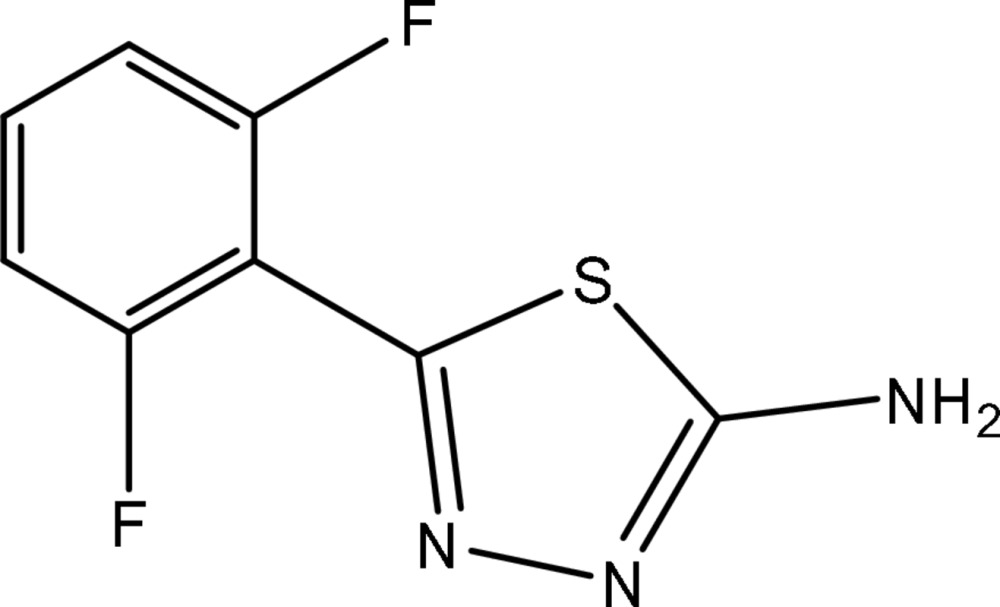
Experimental
Crystal data
C8H5F2N3S
M r = 213.21
Monoclinic,

a = 9.0920 (18) Å
b = 8.7400 (17) Å
c = 10.936 (2) Å
β = 95.85 (3)°
V = 864.5 (3) Å3
Z = 4
Mo Kα radiation
μ = 0.37 mm−1
T = 293 K
0.20 × 0.10 × 0.10 mm
Data collection
Enraf–Nonius CAD-4 diffractometer
Absorption correction: ψ scan (North et al., 1968 ▶) T min = 0.931, T max = 0.964
1670 measured reflections
1568 independent reflections
1189 reflections with I > 2σ(I)
R int = 0.018
3 standard reflections every 200 reflections intensity decay: 1%
Refinement
R[F 2 > 2σ(F 2)] = 0.042
wR(F 2) = 0.109
S = 1.01
1568 reflections
127 parameters
H-atom parameters constrained
Δρmax = 0.26 e Å−3
Δρmin = −0.28 e Å−3
Data collection: CAD-4 EXPRESS (Enraf–Nonius, 1989 ▶); cell refinement: CAD-4 EXPRESS; data reduction: XCAD4 (Harms & Wocadlo, 1995 ▶); program(s) used to solve structure: SHELXS97 (Sheldrick, 2008 ▶); program(s) used to refine structure: SHELXL97 (Sheldrick, 2008 ▶); molecular graphics: SHELXTL (Sheldrick, 2008 ▶); software used to prepare material for publication: SHELXL97.
Supplementary Material
Crystal structure: contains datablocks global, I. DOI: 10.1107/S1600536809047990/rn2058sup1.cif
Structure factors: contains datablocks I. DOI: 10.1107/S1600536809047990/rn2058Isup2.hkl
Additional supplementary materials: crystallographic information; 3D view; checkCIF report
Table 1. Hydrogen-bond geometry (Å, °).
| D—H⋯A | D—H | H⋯A | D⋯A | D—H⋯A |
|---|---|---|---|---|
| N3—H3A⋯N2i | 0.86 | 2.17 | 3.017 (4) | 166 |
| N3—H3B⋯N1ii | 0.86 | 2.30 | 3.088 (3) | 152 |
Symmetry codes: (i)  ; (ii)
  .
.
Acknowledgments
The authors gratefully acknowledge Professor Hua-Qin Wang of the Analysis Center, Nanjing University, for providing the Enraf–Nonius CAD-4 diffractometer for this research project.
supplementary crystallographic information
Comment
1,3,4-Thiadiazole derivatives represent a class of biologically important compounds, which often exhibit insecticidal, fungicidal and other biological activities (Nakagawa et al., 1996; Wang et al., 1999). We report here the crystal structure of the title compound, (I).
The molecular structure of (I) is shown in Fig.1, in which the bond lengths and angles are generally within normal ranges (Allen et al., 1987). The dihedral angle between the thiadiazole and phenyl ring is 35.19 (14)°. In the crystal structure, intermolecular N—H···N hydrogen bonds (Fig. 2) form chains along the b and c axes. There are also intermolecular N-H···S contacts between the molecules, which may further stabilize the structure.
Experimental
2,6-difluorobenzoic acid (2 mmol) and thiosemicarbazide (5 mmol) were mixed in a 25 ml flask, and kept in the oil bath at 90°C for 6 h. After cooling, the crude product (I) precipitated and was filtrated. Pure compound (I) was obtained by crystallization from ethanol (20 ml). Crystals of (I) suitable for X-ray diffraction were obtained by slow evaporation of an acetone solution.
Refinement
All H atoms bonded to the C atoms were placed geometrically at distances of 0.93–0.97 Å and included in the refinement in riding motion approximation with Uiso(H) = 1.2 or 1.5Ueq of the carrier atom.
Figures
Fig. 1.

A view of the molecular structure of (I). Displacement ellipsoids are drawn at the 50% probability level.
Fig. 2.

Partial packing view showing the hydrogen-bonded network. Dashed lines indicate intermolecular N—H···N hydrogen bonds and intermolecular N-H···S contacts between the molecules.
Crystal data
| C8H5F2N3S | Dx = 1.638 Mg m−3 |
| Mr = 213.21 | Melting point: 533 K |
| Monoclinic, P21/c | Mo Kα radiation, λ = 0.71073 Å |
| a = 9.0920 (18) Å | Cell parameters from 25 reflections |
| b = 8.7400 (17) Å | θ = 10–13° |
| c = 10.936 (2) Å | µ = 0.37 mm−1 |
| β = 95.85 (3)° | T = 293 K |
| V = 864.5 (3) Å3 | Block, colorless |
| Z = 4 | 0.20 × 0.10 × 0.10 mm |
| F(000) = 432 |
Data collection
| Enraf–Nonius CAD-4 diffractometer | 1189 reflections with I > 2σ(I) |
| Radiation source: fine-focus sealed tube | Rint = 0.018 |
| graphite | θmax = 25.3°, θmin = 2.3° |
| ω/2θ scans | h = 0→10 |
| Absorption correction: ψ scan (North et al., 1968) | k = 0→10 |
| Tmin = 0.931, Tmax = 0.964 | l = −13→13 |
| 1670 measured reflections | 3 standard reflections every 200 reflections |
| 1568 independent reflections | intensity decay: 1% |
Refinement
| Refinement on F2 | Primary atom site location: structure-invariant direct methods |
| Least-squares matrix: full | Secondary atom site location: difference Fourier map |
| R[F2 > 2σ(F2)] = 0.042 | Hydrogen site location: inferred from neighbouring sites |
| wR(F2) = 0.109 | H-atom parameters constrained |
| S = 1.01 | w = 1/[σ2(Fo2) + (0.060P)2 + 0.150P] where P = (Fo2 + 2Fc2)/3 |
| 1568 reflections | (Δ/σ)max < 0.001 |
| 127 parameters | Δρmax = 0.26 e Å−3 |
| 0 restraints | Δρmin = −0.28 e Å−3 |
Special details
| Geometry. All e.s.d.'s (except the e.s.d. in the dihedral angle between two l.s. planes) are estimated using the full covariance matrix. The cell e.s.d.'s are taken into account individually in the estimation of e.s.d.'s in distances, angles and torsion angles; correlations between e.s.d.'s in cell parameters are only used when they are defined by crystal symmetry. An approximate (isotropic) treatment of cell e.s.d.'s is used for estimating e.s.d.'s involving l.s. planes. |
| Refinement. Refinement of F2 against ALL reflections. The weighted R-factor wR and goodness of fit S are based on F2, conventional R-factors R are based on F, with F set to zero for negative F2. The threshold expression of F2 > σ(F2) is used only for calculating R-factors(gt) etc. and is not relevant to the choice of reflections for refinement. R-factors based on F2 are statistically about twice as large as those based on F, and R- factors based on ALL data will be even larger. |
Fractional atomic coordinates and isotropic or equivalent isotropic displacement parameters (Å2)
| x | y | z | Uiso*/Ueq | ||
| S | 0.68075 (8) | 0.14488 (8) | 0.16016 (6) | 0.0423 (2) | |
| F1 | 0.8904 (2) | 0.0561 (2) | −0.18276 (16) | 0.0637 (5) | |
| N1 | 0.6608 (3) | 0.1922 (3) | −0.07123 (19) | 0.0475 (6) | |
| C1 | 0.8942 (4) | −0.3350 (4) | −0.0851 (3) | 0.0606 (9) | |
| H1B | 0.9319 | −0.4285 | −0.1078 | 0.073* | |
| F2 | 0.6767 (2) | −0.1812 (2) | 0.14183 (15) | 0.0616 (5) | |
| N2 | 0.6037 (3) | 0.3224 (3) | −0.02139 (19) | 0.0495 (6) | |
| C2 | 0.9208 (3) | −0.2061 (4) | −0.1510 (3) | 0.0531 (8) | |
| H2B | 0.9784 | −0.2110 | −0.2165 | 0.064* | |
| N3 | 0.5574 (3) | 0.4251 (3) | 0.1678 (2) | 0.0521 (7) | |
| H3A | 0.5209 | 0.5074 | 0.1338 | 0.062* | |
| H3B | 0.5620 | 0.4143 | 0.2462 | 0.062* | |
| C3 | 0.8608 (3) | −0.0705 (3) | −0.1185 (2) | 0.0442 (7) | |
| C4 | 0.7736 (3) | −0.0549 (3) | −0.0209 (2) | 0.0362 (6) | |
| C5 | 0.7556 (3) | −0.1889 (3) | 0.0433 (3) | 0.0446 (7) | |
| C6 | 0.8123 (4) | −0.3281 (3) | 0.0144 (3) | 0.0571 (8) | |
| H6A | 0.7961 | −0.4149 | 0.0603 | 0.069* | |
| C7 | 0.7062 (3) | 0.0912 (3) | 0.0101 (2) | 0.0357 (6) | |
| C8 | 0.6070 (3) | 0.3142 (3) | 0.0986 (2) | 0.0376 (6) |
Atomic displacement parameters (Å2)
| U11 | U22 | U33 | U12 | U13 | U23 | |
| S | 0.0628 (5) | 0.0382 (4) | 0.0272 (3) | 0.0092 (3) | 0.0101 (3) | 0.0052 (3) |
| F1 | 0.0799 (13) | 0.0567 (11) | 0.0601 (11) | 0.0030 (10) | 0.0351 (10) | 0.0104 (9) |
| N1 | 0.0737 (17) | 0.0411 (12) | 0.0278 (11) | 0.0138 (12) | 0.0064 (11) | −0.0010 (9) |
| C1 | 0.069 (2) | 0.0476 (18) | 0.065 (2) | 0.0180 (16) | 0.0049 (17) | −0.0093 (16) |
| F2 | 0.0825 (13) | 0.0493 (10) | 0.0580 (11) | 0.0069 (9) | 0.0309 (10) | 0.0111 (8) |
| N2 | 0.0803 (18) | 0.0405 (13) | 0.0280 (11) | 0.0176 (12) | 0.0069 (11) | 0.0017 (10) |
| C2 | 0.0503 (18) | 0.063 (2) | 0.0471 (17) | 0.0128 (16) | 0.0116 (14) | −0.0065 (15) |
| N3 | 0.0824 (19) | 0.0448 (13) | 0.0304 (12) | 0.0185 (13) | 0.0128 (12) | 0.0008 (10) |
| C3 | 0.0479 (16) | 0.0468 (16) | 0.0384 (14) | 0.0020 (13) | 0.0071 (13) | 0.0000 (12) |
| C4 | 0.0389 (15) | 0.0363 (14) | 0.0333 (13) | 0.0015 (11) | 0.0035 (11) | −0.0016 (11) |
| C5 | 0.0468 (16) | 0.0446 (15) | 0.0430 (15) | 0.0022 (13) | 0.0077 (13) | 0.0015 (13) |
| C6 | 0.069 (2) | 0.0377 (16) | 0.065 (2) | 0.0041 (15) | 0.0080 (17) | 0.0039 (14) |
| C7 | 0.0432 (15) | 0.0362 (13) | 0.0279 (12) | 0.0014 (12) | 0.0051 (11) | 0.0011 (11) |
| C8 | 0.0481 (16) | 0.0348 (14) | 0.0298 (13) | 0.0042 (12) | 0.0042 (11) | 0.0032 (10) |
Geometric parameters (Å, °)
| S—C8 | 1.733 (3) | C2—C3 | 1.367 (4) |
| S—C7 | 1.745 (2) | C2—H2B | 0.9300 |
| F1—C3 | 1.352 (3) | N3—C8 | 1.336 (3) |
| N1—C7 | 1.291 (3) | N3—H3A | 0.8600 |
| N1—N2 | 1.385 (3) | N3—H3B | 0.8600 |
| C1—C2 | 1.372 (4) | C3—C4 | 1.400 (4) |
| C1—C6 | 1.382 (4) | C4—C5 | 1.384 (4) |
| C1—H1B | 0.9300 | C4—C7 | 1.471 (3) |
| F2—C5 | 1.356 (3) | C5—C6 | 1.370 (4) |
| N2—C8 | 1.311 (3) | C6—H6A | 0.9300 |
| C8—S—C7 | 86.98 (12) | C5—C4—C3 | 114.2 (2) |
| C7—N1—N2 | 113.4 (2) | C5—C4—C7 | 123.0 (2) |
| C2—C1—C6 | 121.1 (3) | C3—C4—C7 | 122.8 (2) |
| C2—C1—H1B | 119.5 | F2—C5—C6 | 118.0 (2) |
| C6—C1—H1B | 119.5 | F2—C5—C4 | 117.4 (2) |
| C8—N2—N1 | 112.2 (2) | C6—C5—C4 | 124.6 (3) |
| C3—C2—C1 | 118.6 (3) | C5—C6—C1 | 117.8 (3) |
| C3—C2—H2B | 120.7 | C5—C6—H6A | 121.1 |
| C1—C2—H2B | 120.7 | C1—C6—H6A | 121.1 |
| C8—N3—H3A | 120.0 | N1—C7—C4 | 123.1 (2) |
| C8—N3—H3B | 120.0 | N1—C7—S | 113.60 (19) |
| H3A—N3—H3B | 120.0 | C4—C7—S | 123.26 (18) |
| F1—C3—C2 | 117.9 (2) | N2—C8—N3 | 123.6 (2) |
| F1—C3—C4 | 118.4 (2) | N2—C8—S | 113.82 (19) |
| C2—C3—C4 | 123.7 (3) | N3—C8—S | 122.63 (19) |
| C7—N1—N2—C8 | −0.5 (4) | C2—C1—C6—C5 | 1.5 (5) |
| C6—C1—C2—C3 | −1.9 (5) | N2—N1—C7—C4 | −178.8 (2) |
| C1—C2—C3—F1 | 178.7 (3) | N2—N1—C7—S | 0.9 (3) |
| C1—C2—C3—C4 | 0.2 (5) | C5—C4—C7—N1 | −146.4 (3) |
| F1—C3—C4—C5 | −176.8 (2) | C3—C4—C7—N1 | 33.2 (4) |
| C2—C3—C4—C5 | 1.6 (4) | C5—C4—C7—S | 33.9 (4) |
| F1—C3—C4—C7 | 3.6 (4) | C3—C4—C7—S | −146.5 (2) |
| C2—C3—C4—C7 | −178.0 (3) | C8—S—C7—N1 | −0.7 (2) |
| C3—C4—C5—F2 | 177.7 (2) | C8—S—C7—C4 | 178.9 (2) |
| C7—C4—C5—F2 | −2.7 (4) | N1—N2—C8—N3 | −179.6 (3) |
| C3—C4—C5—C6 | −2.1 (4) | N1—N2—C8—S | −0.1 (3) |
| C7—C4—C5—C6 | 177.5 (3) | C7—S—C8—N2 | 0.4 (2) |
| F2—C5—C6—C1 | −179.2 (3) | C7—S—C8—N3 | −180.0 (3) |
| C4—C5—C6—C1 | 0.6 (5) |
Hydrogen-bond geometry (Å, °)
| D—H···A | D—H | H···A | D···A | D—H···A |
| N3—H3A···N2i | 0.86 | 2.17 | 3.017 (4) | 166 |
| N3—H3B···N1ii | 0.86 | 2.30 | 3.088 (3) | 152 |
Symmetry codes: (i) −x+1, −y+1, −z; (ii) x, −y+1/2, z+1/2.
Footnotes
Supplementary data and figures for this paper are available from the IUCr electronic archives (Reference: RN2058).
References
- Allen, F. H., Kennard, O., Watson, D. G., Brammer, L., Orpen, A. G. & Taylor, R. (1987). J. Chem. Soc. Perkin Trans. 2, pp. S1–19.
- Enraf–Nonius (1989). CAD-4 Software. Enraf–Nonius, Delft, The Netherlands.
- Harms, K. & Wocadlo, S. (1995). XCAD4. University of Marburg, Germany.
- Nakagawa, Y., Nishimura, K., Izumi, K., Kinoshita, K., Kimura, T. & Kurihara, N. (1996). J. Pesticide Sci, 21, 195–201.
- North, A. C. T., Phillips, D. C. & Mathews, F. S. (1968). Acta Cryst. A24, 351–359.
- Sheldrick, G. M. (2008). Acta Cryst. A64, 112–122. [DOI] [PubMed]
- Wang, Y. G., Cao, L., Yan, J., Ye, W. F., Zhou, Q. C. & Lu, B. X. (1999). Chem. J. Chin. Univ. 20, 1903–1905.
Associated Data
This section collects any data citations, data availability statements, or supplementary materials included in this article.
Supplementary Materials
Crystal structure: contains datablocks global, I. DOI: 10.1107/S1600536809047990/rn2058sup1.cif
Structure factors: contains datablocks I. DOI: 10.1107/S1600536809047990/rn2058Isup2.hkl
Additional supplementary materials: crystallographic information; 3D view; checkCIF report