Age-related macular degeneration is the leading cause of severe, irreversible vision loss in Canada
The main risk factors include increasing age, smoking, family history and white race.1 Specific polymorphisms in the complement factor H gene have been shown to be strongly associated with the disease.2 In large epidemiologic studies, sunlight, hypertension, alcohol use and hypercholesterolemia have not been validated as consistent risk factors.
The dry form of the disease is usually asymptomatic. Progression to the wet form may be indicated by sudden, severe vision loss or new onset of visual distortion (metamorphopsia)
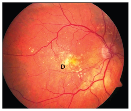
The dry form of the disease is characterized by macular drusen and alterations in the pigmented epithelium of the retina. Intermediate to severe cases of the dry form are characterized by larger drusen and geographic atrophy, which can cause severe vision loss if the fovea is involved (Figure 1). The wet form manifests clinically as macular hemorrhage and exudation from choroidal neo-vascularization (Appendix 1, available at www.cmaj.ca/cgi/content/full/cmaj.090378/DC1). It accounts for more than 80% of the vision loss associated with age-related macular degeneration.3
Figure 1.
Fundus of the right eye of a patient with the dry form of age-related macular degeneration, showing medium and large drusen (D).
Patients should undergo regular dilated funduscopic examination to detect subclinical disease progression4
Regular examinations are important to determine whether patients may benefit from certain interventions. For patients over age 55 with no risk factors, a comprehensive eye exam every one to two years is recommended.4 Patients with early-stage disease or a family history of the condition may require closer follow-up. Those with an intermediate or advanced case of the dry form of the disease should be advised to take a particular combination multivitamin recommended in the Age-Related Eye Disease Study. These supplements reduce the risk of progression to the wet form of the disease by 25%.5 However, patients with early-stage disease may not benefit from such supplementation. Smoking cessation is associated with a substantial reduction in the risk of progression to late-stage disease.6
Patients who describe a sudden change in vision should be referred for urgent ophthalmic evaluation
Self-monitoring with an Amsler grid (available at www.macular.org/chart.html) is critical and can help detect disease progression early. New onset of visual distortion noted on an Amsler grid, or any other sudden change in vision, may indicate progression to the wet form of age-related macular degeneration. In some cases, timely treatment can reduce the risk of permanent loss of vision.
If the wet form of the disease is detected, treatment with a vascular endothelial growth factor inhibitor may be indicated
The current standard of care for sub-foveal choroidal neovascular membranes secondary to age-related macular degeneration is intravitreal injection of an anti-vascular endothelial growth factor medication. With this treatment, 90% of patients experience stabilization in visual acuity, and up to 40% achieve substantial improvements in vision.7 Patients with extrafoveal choroidal neovascular membranes may benefit from argon laser photocoagulation.
Supplementary Material
Footnotes
Previously published at www.cmaj.ca
Competing interests: None declared.
For references, please see Appendix 2 (available at www.cmaj.ca/cgi/content/full/cmaj.090378/DC1).
This article has been peer reviewed.
Associated Data
This section collects any data citations, data availability statements, or supplementary materials included in this article.


