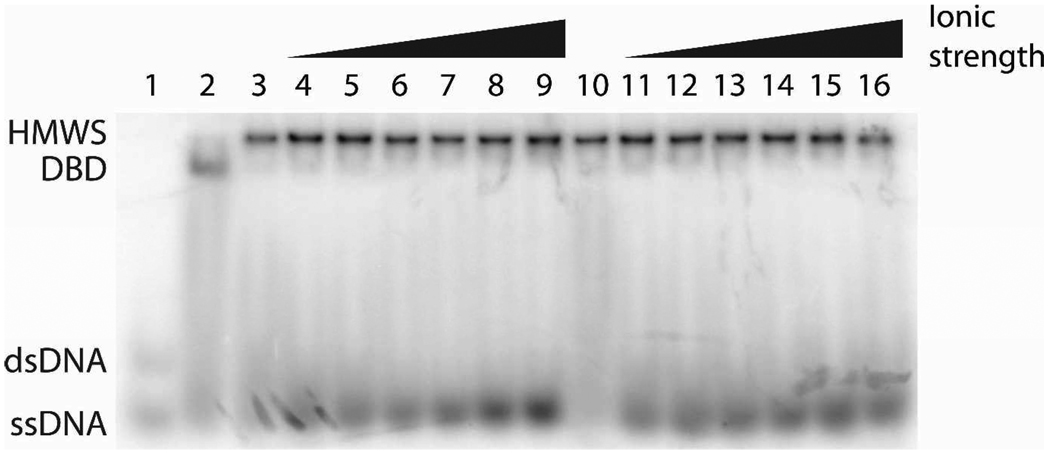
Figure 5.
Native gel electrophoresis of samples in binding buffer containing KOAc or NaCl concentrations from 0 – 300 mM. All samples contain 1 nM 32P-18-1 DNA, 1 µM SgrAI, and 1 µM PC DNA with the exception of that loaded in lane 1, which contains only the 32P-18-1, and lane 2, which contains 32P-18-1 and SgrAI. All samples were incubated in buffer containing 20 mM Tris-OAc (pH 8.0), 10 mM Ca(OAc)2, 10% glycerol and 1 mM DTT and varied concentrations of KOAc (lanes 4-9, with 50, 100, 150, 200, 250, 300 mM respectively) or NaCl (lanes 11–16, with 50, 100, 150, 200, 250, 300 mM respectively). Samples loaded in lanes 1–3 and 10 contain no KOAc or NaCl.

