
Figure 3.
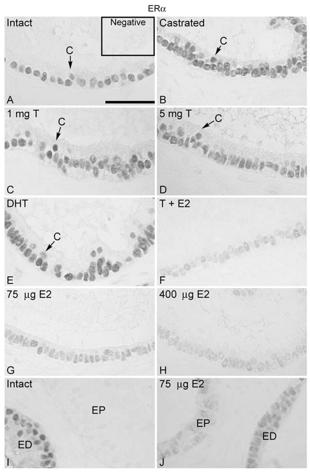
Regulation of ERα in the efferent ductule of the rat. (A) ERα was expressed in the nuclei of the ciliated (C) and nonciliated cells of efferent ductule epithelium in intact control rats. Insert represents the negative control. Castration (B) or castration followed by androgen replacement (C, D, E) did not affect ERα expression. After replacement with 400 μg estradiol (E2) associated with 5 mg testosterone (T) (F), or with estradiol (75 μg and 400 μg) alone (G, H) there was a remarkable decrease in ERα expression. The higher dose of estradiol induced a greater decrease in ERα (F, H). (I) The initial segment of the epididymis (EP) was negative for ERα in control animals. ED, distal efferent ductules. (J) After replacement with estradiol (75 μg and 400 μg) a slight staining for ERα was found in the initial segment epithelium (EP). The results from efferent ductules of sham-operated, unilateral or bilateral ligated and unilateral castrated rats were similar to those of intact controls (not shown). Bar = 100 μm.