Abstract
Background
Epithelial ovarian cancer (EOC) constitutes more than 90% of ovarian cancers and is associated with high mortality. EOC comprises a heterogeneous group of tumours, and the causes and molecular pathology are essentially unknown. Improved insight into the molecular characteristics of the different subgroups of EOC is urgently needed, and should eventually lead to earlier diagnosis as well as more individualized and effective treatments. Previously, we reported a limited number of mRNAs strongly upregulated in human osteosarcomas and other malignancies, and six were selected to be tested for a possible association with three subgroups of ovarian carcinomas and clinical parameters.
Methodology/Principal Findings
The six selected mRNAs were quantified by RT-qPCR in biopsies from eleven poorly differentiated serous carcinomas (PDSC, stage III–IV), twelve moderately differentiated serous carcinomas (MDSC, stage III–IV) and eight clear cell carcinomas (CCC, stage I–IV) of the ovary. Superficial scrapings from six normal ovaries (SNO), as well as biopsies from three normal ovaries (BNO) and three benign ovarian cysts (BBOC) were analyzed for comparison. The gene expression level was related to the histological and clinical parameters of human ovarian carcinoma samples. One of the mRNAs, DNA polymerase delta 2 small subunit (POLD2), was increased in average 2.5- to almost 20-fold in MDSC and PDSC, respectively, paralleling the degree of dedifferentiation and concordant with a poor prognosis. Except for POLD2, the serous carcinomas showed a similar transcription profile, being clearly different from CCC. Another mRNA, Killer-specific secretory protein of 37 kDa (KSP37) showed six- to eight-fold higher levels in CCC stage I compared with the more advanced staged carcinomas, and correlated positively with an improved clinical outcome.
Conclusions/Significance
We have identified two biomarkers which are markedly upregulated in two subgroups of ovarian carcinomas and are also associated with stage and outcome. The results suggest that POLD2 and KSP37 might be potential prognostic biomarkers.
Introduction
In Norway and the United States, ovarian cancer is the fourth and fifth most frequent cause of cancer death in women, respectively [1], [2]. At the time of diagnosis, almost 70% of the patients have distant spread of disease (stage III–IV), and their 5-year relative survival rate is only about 30% [1], [2]. The cause(s) and mode of progression are poorly understood, and the patients are treated similarly in spite of tumour heterogeneity [3]–[6].
EOC comprises several subtypes of histopathologically different tumours [7]. There is growing evidence for the existence of at least two distinct tumourigenetic pathways, corresponding to the development of type I and type II tumours [3], [6], [8]–[10]. Type I tumours include highly differentiated serous carcinomas, mucinous carcinomas, endometroid carcinomas, clear cell carcinomas and malignant Brenner tumours. They are thought to arise from precursor lesions such as cystadenomas, borderline tumours or endometriosis and suggested to be a result of mutations in e.g. KRAS, BRAF, CTNNB1 or PTEN genes [4], [6], [8], [9]. Type II carcinomas include moderately and poorly differentiated serous carcinomas, carcinosarcomas and undifferentiated carcinomas, and appear to originate de novo from as yet no known identified precursor lesions, possibly resulting from mutations in e.g. TP53 [4], [6], [8], [9], [11]. Thus, ovarian carcinogenesis appears to be associated with abnormalities in multiple gene families. How these genetic alterations are reflected in changes in transcriptional activity and carcinogenesis are not understood.
Previously, we reported a limited number of mRNAs strongly upregulated in human osteosarcomas and several other malignancies [12]. Further analyses on various types of human malignant cell lines and normal tissues showed that six mRNAs were highly expressed: KSP37, C9orf89, PRAT4A, NOLA2, ANT2 and POLD2 (Table 1). Apart from C9orf89 and PRAT4A (unknown at project start), these mRNAs code for proteins known to be associated with malignancy [13]–[16]. We hypothesized that these mRNAs might as well be associated with ovarian cancer. In the present study, we quantified these mRNAs by RT-qPCR in biopsies from eleven PDSC (stage III–IV), twelve MDSC (stage III–IV) and eight CCC (stage I–IV) as well as control tissue representing six SNO, three BNO and three BBOC. The expression levels were related to histological, clinical and laboratory parameters. We found that two of the mRNAs were markedly upregulated in two subgroups of ovarian carcinomas and also associated with stage and outcome.
Table 1. Title and assumed function of six selected mRNAs [12].
| Title | Assumed function |
| Killer-specific secretory protein of 37 kDa; KSP37 | Cytotoxic lymphocyte-mediated immunity [13] |
| Chromosome 9 open reading frame 89; C9orf89 | CARD binding region* [29] |
| Protein associated with TLR4,A; PRAT4A | TLR4 associated* [30] |
| Nucleolar protein family A, member 2; NOLA2 | Associated with telomerase and snoRNPs [14] |
| Adenine nucleotide translocator 2; ANT2 | ADP/ATP exchange [15] |
| DNA polymerase delta 2 small subunit; POLD2 | DNA replication and repair [16] |
Unknown at project start. CARD: Caspase Recruitment Domain. TLR: Toll-like receptor. SnoRNPs: small nucleolar ribonucleoproteins. KSP37 is synonymous with fibroblast growth factor binding protein 2; FGFBP2 (www.ncbi.nlm.nih.gov/genebank).
Results
Mean expression levels of six selected mRNAs in three subgroups of ovarian carcinomas compared with three different control groups
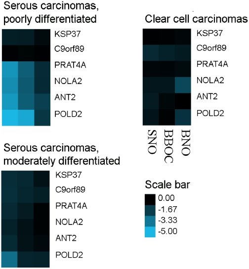
Expression levels of the six selected mRNAs in PDSC, MDSC and CCC are presented in Figures 1,2,3. Figure 1 shows heat-maps of log10 transformed p-values (t-test) comparing the mean expression levels as ΔCq (delta quantification cycles) values in PDSC, MDSC and CCC with SNO, BNO and BBOC. P-values less than 0.05 were used as cut-off value for significance. When comparing PDSC with SNO and BBOC, respectively, the following mRNAs were significantly differentially expressed: PRAT4A (p = 8.1×10⁻⁵ and 2.6×10⁻³), NOLA2 (p = 1.3×10⁻⁴ and 3.5×10⁻³), ANT2 (p = 6.3×10⁻⁵ and 2.6×10⁻³) and POLD2 (p = 3.4×10⁻⁸ and 2.4×10⁻⁵), whereas comparing these carcinomas with BNO, ANT2 (p = 1.9×10⁻²) and POLD2 (p = 3.1×10⁻²) showed a differential expression. For MDSC, POLD2 (p = 9.1×10⁻⁴) showed differential transcription when compared with SNO. NOLA2 (p = 1.1×10⁻²) and POLD2 (p = 4.3×10⁻²) were differentially expressed when CCC were compared with BNO. These significantly differentially expressed mRNAs were all upregulated in PDSC and MDSC, while downregulated in CCC (data not shown). Thus, several of the six previously shown upregulated mRNAs in osteosarcomas were also differentially expressed in the ovarian carcinomas. Furthermore, the overall transcriptional activity of these genes was similar when comparing BBOC with SNO and BNO, while PRAT4A and POLD2 showed significant differential expression (p<0.05) when BNO and SNO were compared (data not shown).
Figure 1. Mean differential expression levels of six selected mRNAs (horizontal) in three subgroups of ovarian carcinomas compared with three different control tissues (vertical).
Log10 p-values of the T-test of delta Cq values are shown as heat-maps, where the smaller the p-value, the brighter the blue colour (scale bar). P<0.05 represents significant differential expression. SNO: superficial scrapings from normal ovaries. BBOC: biopsies from benign ovarian cysts. BNO: biopsies from normal ovaries.
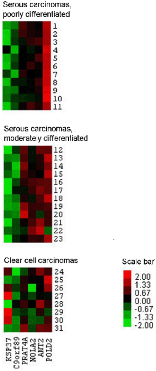
Figure 2. Differential expression levels of six selected mRNAs (vertical) in 31 individual tissue samples (horizontal) of three subgroups of ovarian carcinomas compared with superficial scrapings from normal ovaries.
Normalized log2 transformed original FC values (Z-scores) are shown as heat-maps, where the higher/lower the FC value, the brighter the red/green colour, respectively (scale bar). Black colour illustrates no difference in FC values of cancer tissue and control tissue.
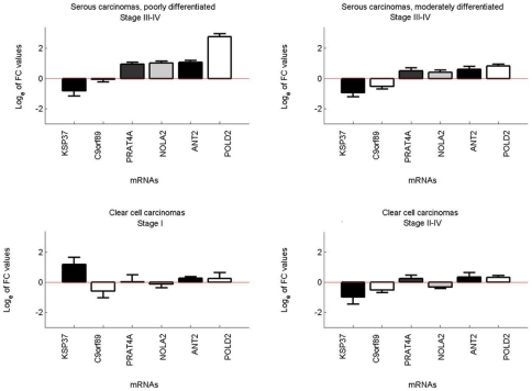
Figure 3. Mean expression levels of six selected mRNAs in moderately and poorly differentiated serous carcinomas (stage III–IV) and clear cell carcinomas (stage I and II–IV) compared with superficial scrapings from normal ovaries.
Loge transformed original FC values with standard deviation are shown as bar plots.
Individual expression levels of six selected mRNAs in three subgroups of ovarian carcinomas compared with SNO controls
Figure 2 shows mRNA expression profiles of all 31 carcinomas employing SNO as a control group, depicted as heat-maps of normalized log2 transformed original fold change (FC) values. Higher mRNA levels were detected in PDSC and MDSC for PRAT4A, NOLA2, ANT2 and POLD2. PRAT4A, NOLA2 and ANT2 showed a similar mRNA expression in PDSC and MDSC in contrast to POLD2, being clearly more upregulated in PDSC compared with MDSC. The mRNA levels were reduced for KSP37 and C9orf89 in both PDSC and MDSC. Furthermore, except for C9orf89, a distinct mRNA expression pattern of the mRNAs was present in CCC. The heat-maps looked almost identical when BBOC were used as the control group, but differed slightly when BNO were used (Figure S1).
Mean expression levels of six selected mRNAs in three subgroups of ovarian carcinomas compared with SNO controls
Figure 3 shows bar plots of mean mRNA expression (loge transformed original FC values) in PDSC, MDSC and different stages of CCC, using SNO for comparison. In PDSC, POLD2 was almost 20-fold upregulated (FC 19.4), whereas C9orf89, PRAT4A, NOLA2 and ANT2 were only moderately upregulated (FC 1.2–3.1) and KSP37 slightly downregulated (FC 0.7). In MDSC, transcription levels of KSP37 and C9orf89 were reduced (FC 0.5 and 0.7 respectively), while the other mRNAs showed moderate upregulations (FC 1.8–2.5). In CCC stage I, KSP37 was markedly upregulated (FC 4.3), but downregulated in the more advanced stages of CCC (FC 0.5). In both stage I and stages II–IV of CCC, PRAT4A, ANT2 and POLD2 were slightly upregulated (FC 1.4–1.7), whereas C9orf89 and NOLA2 were slightly downregulated (FC 0.6–0.96). Thus, when comparing KSP37 expression levels in CCC stage I with the more advanced stages of CCC, MDSC and PDSC, a six- to eight-fold difference was detected. Further analyses of the FC values in Figure 3 are shown in Table 2. The mean mRNA profiles were almost identical when BBOC were used as control tissue, but differed more when BNO were used (Figure S2).
Table 2. Statistical analyses of the FC values shown in Figure 3.
| KSP37 | C9orf89 | PRAT4A | NOLA2 | ANT2 | POLD2 | |
| PDSC, stage III–IV | ||||||
| Average | 0.70 | 1.17 | 2.77 | 3.03 | 3.12 | 19.42 |
| Stdev | 0.62 | 0.93 | 1.06 | 1.43 | 1.11 | 14.79 |
| Min | 0.07 | 0.50 | 1.24 | 1.36 | 1.51 | 5.90 |
| Max | 1.64 | 3.27 | 4.82 | 6.73 | 4.92 | 59.30 |
| MDSC, stage III–IV | ||||||
| Average | 0.52 | 0.69 | 1.98 | 1.78 | 2.40 | 2.50 |
| Stdev | 0.39 | 0.46 | 1.15 | 1.42 | 2.36 | 1.13 |
| Min | 0.06 | 0.29 | 0.60 | 0.74 | 0.77 | 1.38 |
| Max | 1.42 | 1.79 | 3.97 | 6.06 | 9.42 | 4.66 |
| CCC, stage I | ||||||
| Average | 4.28 | 0.69 | 1.42 | 0.96 | 1.35 | 1.66 |
| Stdev | 3.11 | 0.46 | 1.32 | 0.50 | 0.28 | 1.54 |
| Min | 0.95 | 0.17 | 0.39 | 0.57 | 1.17 | 0.67 |
| Max | 8.40 | 1.29 | 3.34 | 1.68 | 1.77 | 3.94 |
| CCC, stage II–IV | ||||||
| Average | 0.49 | 0.64 | 1.38 | 0.73 | 1.60 | 1.43 |
| Stdev | 0.40 | 0.23 | 0.65 | 0.12 | 0.91 | 0.35 |
| Min | 0.12 | 0.36 | 0.90 | 0.56 | 0.82 | 1.11 |
| Max | 1.04 | 0.90 | 2.31 | 0.83 | 2.76 | 1.75 |
PDSC: Poorly differentiated serous carcinomas. MDSC: Moderately differentiated serous carcinomas. CCC: Clear cell carcinomas. Stdev: Standard deviation. Min: minimal value. Max: Maximal value. mRNA description is given in Table 1.
The mean mRNA expressions, given as loge transformed original FC values, in the different ovarian carcinoma subgroups were also compared (t-test). P-values less than 0.001 were used as cut-off value for significance. POLD2 mRNA levels were significantly higher in PDSC compared with both MDSC (FC 19.4 vs. 2.5; p = 1.7×10⁻⁸) and CCC (FC 19.4 vs. 1.5; p = 5.6×10⁻⁸), whereas transcription levels of NOLA2 and ANT2 were significantly higher in PDSC compared with CCC (FC 3.0 vs. 0.8; p = 3.0×10⁻⁶ and FC 3.1 vs. 1.5; p = 5.7×10⁻⁴, respectively). The results were similar irrespectively of the control tissue used (data not shown).
Correlation of mRNA expression to clinical, laboratory and histological parameters
In a single-factor linear regression model, normalized FC values of the six mRNAs, employing SNO as controls, were correlated with clinical, laboratory and histological parameters. The parameters shown in Table S1 as well as histological subgroups were included in the regression analysis. The significant positive correlations (p<0.05) between mRNA expression levels and these parameters are shown in Table 3. Only KSP37 was significantly associated with several clinical parameters, being positively associated with favourable prognostic factors such as localized disease, long progression-free survival (>18 months) and long overall survival (>36 months). Furthermore, it was negatively associated with unfavourable prognostic factors such as more advanced disease, short progression-free survival and short overall survival (data not shown). When correlating the FC values with histological subgroups, KSP37 expression was positively associated with CCC, whereas PRAT4A, NOLA2 and POLD2 were positively associated with PDSC. The transcriptional levels of C9orf89 and ANT2 did not correlate with any of the parameters.
Table 3. Results of single-factor regression analysis.
| KSP37 | PRAT4A | NOLA2 | POLD2 | |
| Clinical parameters | ||||
| FIGO stage I (all CCC) | 7.9×10⁻⁷ | |||
| Progression-free survival ≥18 months | 1.6×10⁻² | |||
| Overall survival ≥36 months | 3.3×10⁻² | |||
| Status at last follow-up: Alive, no relapse of EOC | 8.0×10⁻⁵ | |||
| Status at last follow-up: Alive, relapse of EOC | 1.2×10⁻² | |||
| Histological parameters | ||||
| PDSC | 1.8×10⁻² | 2.1×10⁻³ | 1.2×10⁻⁵ | |
| CCC | 6.8×10⁻³ |
Discussion
A major finding in this study was the strong upregulation of POLD2 in PDSC compared to control tissues and other histological subgroups of ovarian carcinomas examined. POLD2 is a subunit of the DNA polymerase delta complex, encoding a protein involved in DNA replication and repair [16]. It is downregulated by the PTEN tumour suppressor gene [17], already known to be involved in ovarian carcinogenesis [4], [6], [8], [9]. In gliomas, a consistent pattern of chromosomal alterations were found involving altered regions which harboured seven “landscape genes” associated with patient survival, among these POLD2 [18].
KSP37 mRNA levels were clearly and distinctly regulated in early stage of CCC, another histological subgroup of ovarian cancer. KSP37 is identified as FGFBP2, a member of the fibroblast growth factor binding protein 2 family (www.ncbi.nlm.nih.gov/genebank). It is expressed in cytotoxic T lymphocytes and natural killer cells, and is suggested to have a “cytotoxic potential” which so far has not been identified [13]. Yamanaka et al. found that a high KSP37 expression in high-grade gliomas was positively correlated with survival. Furthermore, KSP37 was more closely correlated with survival than histological grade [19], while in the present study, a positive correlation with histological type, clinical stage as well as good prognosis was observed.
A challenge related to the understanding of molecular portraits of ovarian cancer has been the lack of representative control tissue. Histologically, EOC is thought to originate from the single layer of ovarian surface epithelium (OSE) [5], [7], [20]–[22], which therefore should be the most representative control tissue. Because the OSE represents only a small fraction of the total ovary, the availability of OSE RNA is limited. Zorn et al [23] compared the gene expression profiles of OSE brushings, whole ovary samples, cultures of normal OSE and immortalized OSE cell lines. The transcriptional profiles were markedly distinct, but it was concluded that OSE brushings were most representative as control material, since it is not exposed to in vitro manipulations and does not contain stromal components. In the present study, OSE, as represented by six superficial scrapings from normal ovaries (SNO) was used as reference material. Furthermore, three biopsies from normal ovaries (BNO) and three biopsies from benign ovarian cysts (BBOC) were included for additional comparisons. Our results showed that the investigated six mRNAs were similarly expressed in SNO and BBOC, but differed more in BNO (data not shown). Furthermore, the mRNA levels of the carcinomas were similar both when compared to SNO and BBOC, but different when compared to BNO (Figures 1,2,3 and Figure S1, S2). Apparently, SNO and BBOC showed comparable transcriptional activity for these six mRNAs. The findings are not unexpected, since the benign ovarian cysts used for control tissue are believed to originate from OSE, whereas BNO mainly consist of stromal tissue [7]. Thus, for study purposes, benign cysts originating from OSE, being simpler to obtain than OSE, and superficial scrapings of normal ovaries appear to be alternative choices as control tissue for EOC.
Except for the marked upregulation of POLD2 in PDSC, the expression levels of the other mRNAs in PDSC and MDSC were similar, in agreement with a common tumourigenetic pathway for moderately and poorly differentiated serous carcinomas as previously suggested [10]. Thus, the fact that POLD2 mRNA expression paralleled the dedifferentiation of MDSC to PDSC, increasing from 2.5-fold in MDSC to almost 20-fold in PDSC, underscores the uniqueness of this transcript. Since patients with PDSC generally have a worse clinical outcome than patients with MDSC, the significantly higher POLD2 expression in PDSC compared with MDSC could have a bearing on a poor prognosis, possibly through a replication advantage in cells overexpressing POLD2.
The marked upregulation of KSP37 confined to CCC stage I, as well as its positive association with clinical variables of good prognosis, suggest also a possible predictive role of this transcript. Even though these results are very much in concordance with overall results from studies on other malignancies, the present results are novel related to ovarian carcinomas and need to be confirmed. The different transcriptional profiles for clear cell carcinomas and serous carcinomas are in agreement with distinct tumourigenetic pathways for these carcinomas and also consistent with other studies [24], [25]. Although the present study is based on a limited patient cohort of only three subgroups of ovarian carcinomas, the strong association of two of the mRNAs with histology, stage and outcome suggest that they may have potential as cancer markers.
Materials and Methods
Patients and tissue material
The study was approved by the Regional Committee of Medical and Health Research Ethics (REK) in Eastern Norway and all participating women signed informed consent. Tissue specimens as well as clinical and laboratory information were obtained from women primarily operated for gynecological tumours at Oslo University Hospital, Ulleval, in the period 2003 to 2008. All tissue samples were snap-frozen in liquid nitrogen, except SNO, which were transferred to 500 µl TRIzol solution (Invitrogen.com) immediately after harvesting in order to avoid mRNA degradation. The samples were stored in a biobank at −80°C until processed.
The expression of the six selected mRNAs was studied in a total of 31 epithelial ovarian carcinomas and twelve benign samples. The carcinomas included twelve MDSC (stage III–IV), eleven PDSC (stage III–IV) and eight CCC (stage I–IV). Six SNO, three BNO and three BBOC were used for comparison. SNO were taken from the surface of normal ovaries by scraping the ovaries with a scalpel, as cervical pap smear brushings yielded too little material (data not shown). By this method, the vast majority of harvested cells were immunologically verified as epithelial (data not shown). The three benign cysts were cystadenofibromas, containing both epithelial and stromal cells. BNO consisted almost exclusively of stromal cells as confirmed by histology. In accordance with the literature [23], we used OSE, represented by SNO, as reference material. The histological diagnoses of all samples were confirmed by an experienced pathologist.
Clinical and laboratory information was collected from hospital records and additional preoperative patient interviews, shown in Table S1. All patients and controls were of Western European descent, postmenopausal (apart from two being perimenopausal) and had no diseases influencing survival other than the ovarian cancer. All patients but four (two with MDSC and two with PDSC) were primarily operated by at least a total hysterectomy or a uterus amputation, a bilateral salpingo-oophorectomy and an omentectomy. No patients received neoadjuvant chemotherapy, whereas all patients but three (one in each histological group) received adjuvant chemotherapy. The effect of treatment was evaluated by clinical examinations and serum CA125 measurements at minimum.
Selected mRNAs
Six mRNAs were selected from a subtraction cDNA library of human osteosarcoma [12]. They represented interesting candidate genes, being strongly upregulated in several osteosarcoma and other malignant human cell lines, and showed a differential expression between human cancers and normal tissues. Except for C9orf89 and PRAT4A, whose identities and functions were unknown at project start, these mRNAs code for proteins possibly associated with malignancies. The titles and assumed protein functions of the selected candidate mRNAs are shown in Table 1.
Primer sequences
PCR primers (Table S2) were designed by using the Invitrogen database and tested for homology with other sequences at the NCBI gene website (www.ncbi.nlm.nih.gov). All primers were intron spanning to avoid co-amplification of genomic DNA.
RNA isolation
Tissue specimens were either crushed frozen or homogenized directly for 2×2 minutes in 750 µl TRIzol using a Tissuelyzer (Qiagen.com). Total RNA was extracted using the TRIzol method according to the manufacturer's instructions. Isolated total RNA was quantified (Nano Drop spectrophotometer, Saveen Werner AB) and quality controlled by the RNA Nano 6000 assay on the Bioanalyzer 2100 system (Agilent). RNA integrity number (RIN) and 28S/18S ratios were calculated to ensure a satisfactory RNA quality and integrity of the samples. To remove genomic DNA, total RNA was treated using RNase-free DNase I (Roche.com). Total RNA was further purified on RNeasy MinElute clean up spin columns (Qiagen.com), eluted with RNase free water, aliquoted and stored at −80°C until analyzed.
Quantitative reverse transcription-polymerase chain reaction (RT-qPCR)
One µg of total RNA from each sample was reversely transcribed using 2.5 U/µl Omniscript enzyme (Qiagen.com), 1 X RT-buffer, 1 mM dNTPs, 2.5 µM oligo-d(T)-primer and 1 U/µl RNase inhibitor (final concentrations) in a total volume of 20 µl for one hour at 37°C. For all samples, a negative RT-control without Reverse Transcriptase enzyme was included. cDNA was PCR-amplified with primers from the six specific mRNAs and two endogenous reference genes (β-actin and GAPDH) in replicate sets of two to six, with a coefficient of variation of less than 1.6 percent. The samples were analyzed on a real-time fluorescence LightCycler instrument (Roche.com) according to the manufacturer's instructions in a final volume of 20 µl using a LightCycler Fast start SYBR Green kit. PCR conditions essentially contained 2 µl cDNA, 25 mM MgCl2 and 0.5 µM of forward and reverse primers. The following cycle conditions were used: 10 min denaturation at 95°C before 45 cycles at 95°C for 0 s, 56°C for 10 s and 70°C for 5 s.
Gene expression patterns for the six selected mRNAs were calculated using the comparative crossing threshold method of relative quantification (ΔΔCq method) [26], and presented as relative (ΔCq) and fold change (FC) values. All expression levels were normalized to the reference genes separately, giving overall similar results. β-actin quantification was most linear over a wide dilution range and preferred as reference gene. ΔCq was designated as the mean quantification cycle of an mRNA in a tissue subtracted with the mean quantification cycle of a reference RNA in the same tissue. ΔΔCq was calculated as mean ΔCq of each of the three different control groups subtracted by ΔCq of each cancer tissue sample (mean of replicates), whereas FC was 2ΔΔCq.
Statistical analysis
Mean ΔCq values of each histological subgroup of ovarian carcinomas were compared to mean ΔCq values of each control group by performing a two-tailed t-test, presented in heat-maps by log10 transformed p-values (Figure 1). Log2 transformed original FC values of each individual sample (n = 31) were normalized (Z-scores) and shown as heat-maps by applying a two-way clustering method [27] (Figure 2 and Figure S1). Mean original FC values of the three ovarian carcinoma subgroups were presented by loge transformed bar plots (Figure 3 and Figure S2). Finally, a linear regression model [28], testing the correlation of histological, clinical and laboratory parameters with mRNA expression levels given as normalized FC values, was used (Table 3).
Supporting Information
Differential expression levels of six selected mRNAs (vertical) in 31 individual tissue samples (horizontal) of three subgroups of ovarian carcinomas compared with biopsies from benign ovarian cysts (a) and biopsies from normal ovaries (b). Normalized log2 transformed original FC values (Z-scores) are shown as heat-maps, where the higher/lower the FC value, the brighter the red/green color, respectively (scale bar). Black color illustrates no difference in FC values of cancer tissue and control tissue.
(1.92 MB TIF)
Mean expression levels of six selected mRNAs in moderately and poorly differentiated serous carcinomas (stage III–IV) and clear cell carcinomas (stage I and II–IV) compared with biopsies from benign ovarian cysts (a) and biopsies from normal ovaries (b). Loge transformed original FC values with standard deviation are shown as bar plots.
(2.74 MB TIF)
Clinical and laboratory information for patients included.
(0.05 MB DOC)
Primer sequences of six selected mRNAs.
(0.04 MB DOC)
Acknowledgments
We thank Anne-Marie Siebke Trøseid for technical assistance as well as Elin Ødegaard and Lise Levy for patient recruitment and tissue sampling.
Footnotes
Competing Interests: The authors have declared that no competing interests exist.
Funding: The authors acknowledge former Eastern Norway Regional Health Authority for economical support as well as Organon (currently part of Merck, Sharp and Dohme) for providing a research grant through the Norwegian Society for Gynecology and Obstetrics. The funders had no role in study design, data collection and analysis, decision to publish, or preparation of the manuscript.
References
- 1.Bray F. Oslo: Cancer in Norway 2008 - Cancer incidence, mortality, survival and prevalence in Norway; 2009. Cancer Registry of Norway. [Google Scholar]
- 2.Jemal A, Siegel R, Ward E, Hao Y, Xu J, et al. Cancer statistics, 2009. CA Cancer J Clin. 2009;59:225–249. doi: 10.3322/caac.20006. [DOI] [PubMed] [Google Scholar]
- 3.Farley J, Ozbun LL, Birrer MJ. Genomic analysis of epithelial ovarian cancer. Cell Res. 2008;18:538–548. doi: 10.1038/cr.2008.52. [DOI] [PMC free article] [PubMed] [Google Scholar]
- 4.Naora H. The heterogeneity of epithelial ovarian cancers: reconciling old and new paradigms. Expert Rev Mol Med. 2007;9:1–12. doi: 10.1017/S1462399407000324. [DOI] [PubMed] [Google Scholar]
- 5.Auersperg N, Wong AS, Choi KC, Kang SK, Leung PC. Ovarian surface epithelium: biology, endocrinology, and pathology. Endocr Rev. 2001;22:255–288. doi: 10.1210/edrv.22.2.0422. [DOI] [PubMed] [Google Scholar]
- 6.Landen CN, Jr, Birrer MJ, Sood AK. Early events in the pathogenesis of epithelial ovarian cancer. J Clin Oncol. 2008;26:995–1005. doi: 10.1200/JCO.2006.07.9970. [DOI] [PubMed] [Google Scholar]
- 7.Prat J. Ovarian serous and mucinous epithelial-stromal tumors. Robboy's pathology of the female reproductive tract. 2009.
- 8.Shih I, Kurman RJ. Ovarian tumorigenesis: a proposed model based on morphological and molecular genetic analysis. Am J Pathol. 2004;164:1511–1518. doi: 10.1016/s0002-9440(10)63708-x. [DOI] [PMC free article] [PubMed] [Google Scholar]
- 9.Bell DA. Origins and molecular pathology of ovarian cancer. Mod Pathol. 2005;18(Suppl 2):S19–S32. doi: 10.1038/modpathol.3800306. [DOI] [PubMed] [Google Scholar]
- 10.Meinhold-Heerlein I, Bauerschlag D, Hilpert F, Dimitrov P, Sapinoso LM, et al. Molecular and prognostic distinction between serous ovarian carcinomas of varying grade and malignant potential. Oncogene. 2005;24:1053–1065. doi: 10.1038/sj.onc.1208298. [DOI] [PubMed] [Google Scholar]
- 11.Chan WY, Cheung KK, Schorge JO, Huang LW, Welch WR, et al. Bcl-2 and p53 protein expression, apoptosis, and p53 mutation in human epithelial ovarian cancers. Am J Pathol. 2000;156:409–417. doi: 10.1016/S0002-9440(10)64744-X. [DOI] [PMC free article] [PubMed] [Google Scholar]
- 12.Olstad OK, Gautvik VT, Reppe S, Rian E, Jemtland R, et al. Molecular heterogeneity in human osteosarcoma demonstrated by enriched mRNAs isolated by directional tag PCR subtraction cloning. Anticancer Res. 2003;23:2201–2216. [PubMed] [Google Scholar]
- 13.Ogawa K, Tanaka K, Ishii A, Nakamura Y, Kondo S, et al. A novel serum protein that is selectively produced by cytotoxic lymphocytes. J Immunol. 2001;166:6404–6412. doi: 10.4049/jimmunol.166.10.6404. [DOI] [PubMed] [Google Scholar]
- 14.Pogacic V, Dragon F, Filipowicz W. Human H/ACA small nucleolar RNPs and telomerase share evolutionarily conserved proteins NHP2 and NOP10. Mol Cell Biol. 2000;20:9028–9040. doi: 10.1128/mcb.20.23.9028-9040.2000. [DOI] [PMC free article] [PubMed] [Google Scholar]
- 15.Battini R, Ferrari S, Kaczmarek L, Calabretta B, Chen ST, et al. Molecular cloning of a cDNA for a human ADP/ATP carrier which is growth-regulated. J Biol Chem. 1987;262:4355–4359. [PubMed] [Google Scholar]
- 16.Perez A, Leon A, Lee MY. Characterization of the 5′-flanking region of the gene encoding the 50 kDa subunit of human DNA polymerase delta. Biochim Biophys Acta. 2000;1493:231–236. doi: 10.1016/s0167-4781(00)00153-6. [DOI] [PubMed] [Google Scholar]
- 17.Matsushima-Nishiu M, Unoki M, Ono K, Tsunoda T, Minaguchi T, et al. Growth and gene expression profile analyses of endometrial cancer cells expressing exogenous PTEN. Cancer Res. 2001;61:3741–3749. [PubMed] [Google Scholar]
- 18.Bredel M, Scholtens DM, Harsh GR, Bredel C, Chandler JP, et al. A network model of a cooperative genetic landscape in brain tumors. JAMA. 2009;302:261–275. doi: 10.1001/jama.2009.997. [DOI] [PMC free article] [PubMed] [Google Scholar]
- 19.Yamanaka R, Arao T, Yajima N, Tsuchiya N, Homma J, et al. Identification of expressed genes characterizing long-term survival in malignant glioma patients. Oncogene. 2006;25:5994–6002. doi: 10.1038/sj.onc.1209585. [DOI] [PubMed] [Google Scholar]
- 20.Orsulic S, Li Y, Soslow RA, Vitale-Cross LA, Gutkind JS, et al. Induction of ovarian cancer by defined multiple genetic changes in a mouse model system. Cancer Cell. 2002;1:53–62. doi: 10.1016/s1535-6108(01)00002-2. [DOI] [PMC free article] [PubMed] [Google Scholar]
- 21.Sasaki R, Narisawa-Saito M, Yugawa T, Fujita M, Tashiro H, et al. Oncogenic transformation of human ovarian surface epithelial cells with defined cellular oncogenes. Carcinogenesis. 2009;30:423–431. doi: 10.1093/carcin/bgp007. [DOI] [PubMed] [Google Scholar]
- 22.Okamura H, Katabuchi H. Pathophysiological dynamics of human ovarian surface epithelial cells in epithelial ovarian carcinogenesis. Int Rev Cytol. 2005;242:1–54. doi: 10.1016/S0074-7696(04)42001-4. [DOI] [PubMed] [Google Scholar]
- 23.Zorn KK, Jazaeri AA, Awtrey CS, Gardner GJ, Mok SC, et al. Choice of normal ovarian control influences determination of differentially expressed genes in ovarian cancer expression profiling studies. Clin Cancer Res. 2003;9:4811–4818. [PubMed] [Google Scholar]
- 24.Schwartz DR, Kardia SL, Shedden KA, Kuick R, Michailidis G, et al. Gene expression in ovarian cancer reflects both morphology and biological behavior, distinguishing clear cell from other poor-prognosis ovarian carcinomas. Cancer Res. 2002;62:4722–4729. [PubMed] [Google Scholar]
- 25.Shimizu M, Nikaido T, Toki T, Shiozawa T, Fujii S. Clear cell carcinoma has an expression pattern of cell cycle regulatory molecules that is unique among ovarian adenocarcinomas. Cancer. 1999;85:669–677. doi: 10.1002/(sici)1097-0142(19990201)85:3<669::aid-cncr17>3.0.co;2-f. [DOI] [PubMed] [Google Scholar]
- 26.Livak KJ, Schmittgen TD. Analysis of relative gene expression data using real-time quantitative PCR and the 2(-Delta Delta C(T)) Method. Methods. 2001;25:402–408. doi: 10.1006/meth.2001.1262. [DOI] [PubMed] [Google Scholar]
- 27.Wang J, Delabie J, Aasheim H, Smeland E, Myklebost O. Clustering of the SOM easily reveals distinct gene expression patterns: results of a reanalysis of lymphoma study. BMC Bioinformatics. 2002;3:36. doi: 10.1186/1471-2105-3-36. [DOI] [PMC free article] [PubMed] [Google Scholar]
- 28.Wang J. A new framework for identifying combinatorial regulation of transcription factors: a case study of the yeast cell cycle. J Biomed Inform. 2007;40:707–725. doi: 10.1016/j.jbi.2007.02.003. [DOI] [PubMed] [Google Scholar]
- 29.Woo HN, Hong GS, Jun JI, Cho DH, Choi HW, et al. Inhibition of Bcl10-mediated activation of NF-kappa B by BinCARD, a Bcl10-interacting CARD protein. FEBS Lett. 2004;578:239–244. doi: 10.1016/j.febslet.2004.10.094. [DOI] [PubMed] [Google Scholar]
- 30.Wakabayashi Y, Kobayashi M, Akashi-Takamura S, Tanimura N, Konno K, et al. A protein associated with toll-like receptor 4 (PRAT4A) regulates cell surface expression of TLR4. J Immunol. 2006;177:1772–1779. doi: 10.4049/jimmunol.177.3.1772. [DOI] [PubMed] [Google Scholar]
Associated Data
This section collects any data citations, data availability statements, or supplementary materials included in this article.
Supplementary Materials
Differential expression levels of six selected mRNAs (vertical) in 31 individual tissue samples (horizontal) of three subgroups of ovarian carcinomas compared with biopsies from benign ovarian cysts (a) and biopsies from normal ovaries (b). Normalized log2 transformed original FC values (Z-scores) are shown as heat-maps, where the higher/lower the FC value, the brighter the red/green color, respectively (scale bar). Black color illustrates no difference in FC values of cancer tissue and control tissue.
(1.92 MB TIF)
Mean expression levels of six selected mRNAs in moderately and poorly differentiated serous carcinomas (stage III–IV) and clear cell carcinomas (stage I and II–IV) compared with biopsies from benign ovarian cysts (a) and biopsies from normal ovaries (b). Loge transformed original FC values with standard deviation are shown as bar plots.
(2.74 MB TIF)
Clinical and laboratory information for patients included.
(0.05 MB DOC)
Primer sequences of six selected mRNAs.
(0.04 MB DOC)


