Abstract
Objective
We assessed the outcomes of a simplified technique for the percutaneous placement of a hepatic artery port-catheter system for chemotherapy infusion in advanced hepatocellular carcinoma with portal vein invasion.
Materials and Methods
From February 2003 to February 2008, percutaneous hepatic artery port-catheter insertion was performed in 122 patients who had hepatocellular carcinoma with portal vein invasion. The arterial access route was the common femoral artery. The tip of the catheter was wedged into the right gastroepiploic artery without an additional fixation device. A side hole was positioned at the distal common hepatic artery to allow the delivery of chemotherapeutic agents into the hepatic arteries. Coil embolization was performed only to redistribute to the hepatic arteries or to prevent the inadvertent delivery of chemotherapeutic agents into extrahepatic arteries. The port chamber was created at either the supra-inguinal or infra-inguinal region.
Results
Technical success was achieved in all patients. Proper positioning of the side hole was checked before each scheduled chemotherapy session by port angiography. Catheter-related complications occurred in 19 patients (16%). Revision was achieved in 15 of 18 patients (83%).
Conclusion
This simplified method demonstrates excellent technical feasibility, an acceptable range of complications, and is hence recommended for the management of advanced hepatocellular carcinoma with portal vein thrombosis.
Keywords: Liver cancer, Hepatic arterial catheterization, Percutaneous catheter placement
Hepatic arterial infusion chemotherapy is an alternative treatment for patients with unresectable liver tumors (1, 2). Repeated hepatic arterial delivery of chemotherapeutic agents to the liver by a percutaneously implanted port-catheter system has been widely used to treat advanced unresectable liver cancer (3, 4). Instead of laparotomy, percutaneous implantation of port-catheter systems for intra-arterial use in various target organs, particularly in regional chemotherapy of the liver, has been successfully performed by interventional radiologists under local anesthesia (5-11). Among the various interventional techniques, the classic fixed-catheter-tip method has some advantages, such as increased stability of the indwelling catheter and a decreased rate of hepatic arterial occlusion (12, 13). However, this method, which uses microcoils alone or n-butyl cyanoacrylate, is a lengthy procedure, and removal of the implanted port-catheter system can be difficult. At our institute, a new, simplified fixed-catheter-tip method without microcoils or n-butyl cyanoacrylate was adapted for the indwelling catheter in hepatic arterial infusion chemotherapy. The purpose of this study was to assess the patency and complications of the new fixed-catheter-tip method.
MATERIALS AND METHODS
Patients
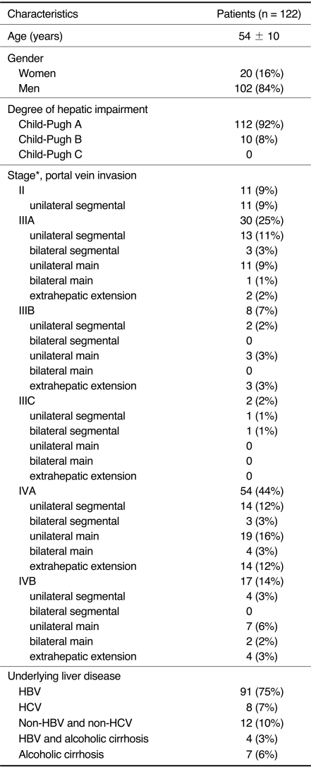
Between February 2003 and February 2008, a port-catheter system was implanted percutaneously by interventional radiologists in 122 consecutive patients who had unresectable hepatocellular carcinoma (HCC) with portal vein invasion. Indications for hepatic arterial infusion chemotherapy were as follows: 1) unresectable liver tumors (n = 103), 2) hepatic lobectomy status with recurrent HCC combined portal vein invasion (n = 7), 3) size reduction of HCC before surgery (n = 1), 4) other concomitant malignancy (n = 1), 5) refusal of both surgery and repeated transarterial chemoembolization (n = 9), and 6) unsuitable general condition for surgery (n = 1). The age of the patients ranged from 29 to 76 years (54±10 years, mean ± standard deviation [SD]). Clinical characteristics of the 122 patients are shown in Table 1.
Table 1.
Clinical Characteristics of 122 Patients with Advanced Hepatocellular Carcinoma with Portal Vein Thrombosis Who Underwent Percutaneous Hepatic Artery Port-Catheter Insertion
Note.-Data are presented as mean ± standard deviation or number (percent).
*= American Joint Committee on Cancer (AJCC) in 2010 (29).
HBV = hepatitis B virus, HCV = hepatitis C virus
Before the procedure, written informed consent was obtained from all the patients. Institutional review board approval was also obtained for this retrospective study.
Techniques
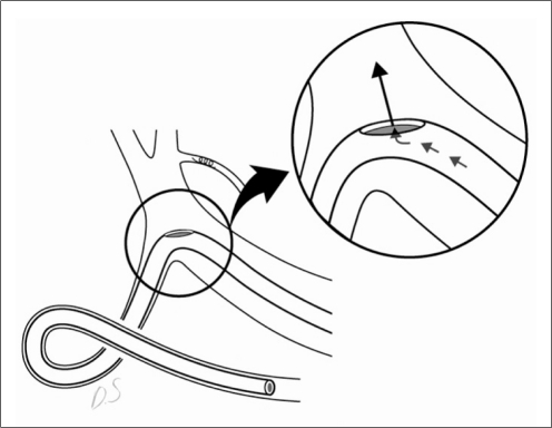
All procedures were performed percutaneously in angiographic suites by interventional radiologists. Before catheter placement, all patients underwent transfemoral angiography for arterial road mapping and, if necessary, arterial redistribution to convert multiple hepatic arteries into a single arterial supply (14, 15). If necessary, extrahepatic arterial branches arising from the hepatic artery were occluded to prevent inadvertent drug delivery using stainless steel microcoils (3-6 mm; Tornado embolization coils; Cook Medical Inc., Bloomington, IN). Catheter placement was performed under local anesthesia immediately after angiography. The right or left common femoral artery was chosen as the access route. The catheter tip was placed in the right gastroepiploic artery without a distal fixation device such as a coil or n-butyl cyanoacrylate (Fig. 1). The procedure was performed as follows:
Fig. 1.
Illustration of our simplified fixed-catheter-tip method with no additional fixation material.
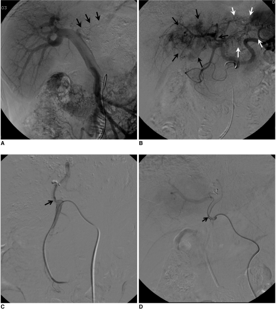
A 5-Fr catheter (RH-R; Cook Medical Inc., Bloomington, IN) was inserted under local anesthesia from the common femoral artery and was advanced to the superior mesenteric artery, where the angiography was performed (Fig. 2A). The catheter tip was repositioned in the common hepatic artery via the celiac trunk, and a common hepatic artery arteriography was performed (Fig. 2B). For accurate anatomical evaluation, 5 µg of Lipo-PGE1 (lipidencapsulated preparation of prostaglandin E1, Eglandin; WelFide Korea Co., Ltd., Seoul, Korea) was administered directly into the superior mesenteric artery immediately before angiography.
A microcatheter was inserted coaxially, and coil embolization was performed to prevent extrahepatic drug distribution or arterial redistribution.
The catheter tip location was determined by celiac arteriography, and was placed at the right gastroepiploic artery about 3-5 cm from the ostium of the right gastroepiploic artery.
After determining the catheter tip location, a side hole was made in the catheter using small scissors, and was adjusted at the distal common hepatic artery. The distance between the side hole and the distal end of the indwelling catheter was determined from the results of the celiac arteriography. Before implantation of the port-catheter system, a test injection using sterile saline was performed through the catheter to confirm the absence of flow through the distal end of the catheter. The port chamber was created after povidone-iodine soaking of the low abdominal wall or upper anterior thigh, and then subcutaneous tunneling was performed.
Using the catheter-exchange method, an indwelling catheter with one side hole (5-Fr polyurethane catheter [Healthport LP set; Baxter Healthcare Co., Deerfield, IL] or 5.8-Fr polyurethane catheter [PolyFlow; Deltec Inc., St. Paul, MN]) was inserted via the right or left common femoral artery. The tip of the catheter was wedged into the right gastroepiploic artery. No additional distal-tip-fixation material, such as a coil or n-butyl cyanoacrylate, was used. The distal tip of the catheter was not occluded. The side hole was placed into the distal common hepatic artery, and its location and patency were confirmed by a test injection of contrast media (Fig. 2C).
The proximal segment of the indwelling catheter was embedded in the subcutaneous tunnel, and the proximal end of the catheter was cut and connected to a port (Healthport LP [Baxter] or Port-A-Cath [Deltec Inc., St. Paul, MN]), which was implanted in the subcutaneous space. Port-catheter angiography was performed to confirm the location of the side hole. The subcutaneous tissue was flushed with antibiotic solution and sutured aseptically (the skin was closed with staples) and 2000 IU of heparin was inserted into the port. To prevent thrombosis of the port-catheter system, the port was flushed with saline and heparin (2000 IU) after each administration of chemotherapeutic drugs.
Fig. 2.
Hepatocellular carcinoma treatment using implantable port-catheter system with our simplified fixed-catheter-tip method. 72-year-old man with advanced hepatocellular carcinoma and left portal vein invasion. Transarterial chemoembolization was performed 1-week before procedure.
A. Superior mesenteric artery angiography was performed using 5-Fr catheter with nonvisualization of left portal vein (arrows) because of tumor invasion.
B. Celiac angiography revealed extensive tumor staining (black arrows) involving left hepatic lobe. Previously treated region of hepatocellular carcinoma involving left lateral segment was not stained (white arrows).
C. 5-Fr indwelling catheter with side hole was inserted in right common femoral artery. Tip of catheter was wedged into proximal portion of right gastroepiploic artery. No additional distal tip-fixation material, such as coil or n-butyl cyanoacrylate, was used. Location of side hole was confirmed by test injection of contrast media, and proper hepatic artery (arrow) was opacified.
D. 6-month follow-up angiography revealed that location of indwelling catheter was not changed, and hepatic artery (arrow) was patent.
Follow-Up
Before chemotherapy, all patients underwent digital subtraction angiography after infusion of the contrast material via the port to confirm the patency of the hepatic artery and indwelling catheter (Fig. 2D). Follow-up intervals depended on the clinical characteristics of the patients.
Port-catheter system removal was performed by pulling gently on the catheter under fluoroscopic guidance in angiographic suites by radiologists, and under local anesthesia, at the physician's request. The duration of this procedure was only a few minutes.
RESULTS
The port-catheter systems were implanted successfully, and hepatic arterial infusion chemotherapy was initiated in all 122 patients, with a mean follow-up duration of 145±133.6 days (mean ± SD; range, 1-861 days; median, 101 days). The number of arterial infusion chemotherapy sessions using the port-catheter system was 3.9 ± 2.4 (mean ± SD; range, 1-15; median, 3).
In 115 of 122 patients, the right common femoral artery was chosen as the puncture site for implantation of the port-catheter system. For the other seven patients, the left common femoral artery was chosen as the puncture site, because of right iliac artery occlusion (n = 1), severe fibrosis at the right inguinal region caused by previous repeated transarterial chemoembolization (n = 1), or the patient's preference for the chamber site of the port-catheter system (n = 5). In 30 of 122 patients (25%, n = 32), coil embolization was performed to prevent extrahepatic drug distribution or arterial redistribution. The distribution of the embolized arteries was as follows: right gastric artery (n = 15), superior pancreaticoduodenal artery (n = 7), left gastric artery (n = 6), replaced left hepatic artery (n = 2), and replaced right hepatic artery (n = 2).
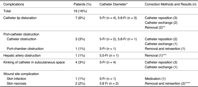
In total, 30 cases with complications were related to the port-catheter system (n = 19, 16%) (Table 2), and to chemotherapy (i.e., acute gastroduodenal mucosal lesions; n = 11, 9%). On scheduled digital subtraction angiographies that were performed before chemotherapy, 106 of 122 patients demonstrated patent hepatic arterial flow with no complication. For the remaining 16 patients, complications related to the port-catheter system were observed. Wound infection cases (n = 3) were detected by a physician during a routine physical examinations. Port-catheter system correction was attempted in 18 of 19 patients. In one case, local infection was controlled by medical treatment and did not require correction of the port-catheter system. Correction was possible and chemotherapy was continued in 15 of 18 patients (83%). Among the complications, catheter tip dislocation occurred in seven patients; in five of the seven cases, successful correction was performed, but in the other two cases, the correction failed. Detailed correction methods and results are presented in Table 2. If the correction of the catheter position was necessary, repositioning of the catheter was first attempted using a hydrophilic guidewire. However, if the catheter repositioning failed, then catheter exchange was attempted. In three cases, correction of the port-catheter system could not be accomplished because of proximal segment obstruction of the proper hepatic artery in one patient, celiac trunk stenosis in another patient, and gastroduodenal artery obstruction in another patient. No correlation between the difference of the distal catheter tip location and the complication rate was observed. Acute gastroduodenal mucosal lesions occurred in 11 patients (9%) who underwent hepatic arterial infusion chemotherapy using an indwelling catheter, which was confirmed by endoscopy. All of these patients recovered with medical treatment. No other complication associated with this procedure was observed.
Table 2.
Complications in 122 Patients after Percutaneous Hepatic Artery Port-Catheter Insertion
Note.-*= Diameter of indwelling catheter. **= Correction of port-catheter system could not be accomplished because gastroduodenal artery obstruction occurred in one patient and celiac stenosis in another. Port-catheter systems were removed and treatment method was switched. ***= Correction of port-catheter system could not be accomplished because of hepatic artery obstruction. Port-catheter systems were removed and treatment method was switched. ****= Reinsertion was performed via contralateral common femoral artery.
Combined radiation therapy was performed in 50 of the 122 patients (41%) with hepatic arterial infusion chemotherapy for the treatment of hepatocellular carcinoma.
During follow-up, 49 of 122 patients were referred for removal of the port-catheter system due to a switch in treatment method; from hepatic arterial infusion chemotherapy to other methods (n = 47) and local infection (n = 2). The removal was performed successfully without complications in 48 of 49 patients. In one case, the port-catheter system could not be removed: when the operator tried to pull it out, the catheter did not move to the outside. Celiac angiography revealed patent hepatic arterial flow, but the indwelling catheter was fixed to the adjacent celiac trunk. We considered that adhesion had occurred between the indwelling catheter and the adjacent celiac trunk wall, and it was left alone. The remaining 73 patients continued with hepatic arterial infusion chemotherapy and were followed as outpatients, or were scheduled for catheter removal.
DISCUSSION
Among the various interventional techniques to achieve implantation of port-catheter systems (6-11, 16-22), the fixed-catheter-tip method (6, 17-22) is advantageous because it prevents catheter dislocation and hepatic artery obstruction (6, 18-22). A review of large published studies describing percutaneous port-catheter system placement for hepatic arterial infusion chemotherapy using the fixed-catheter-tip method, revealed that the rate of indwelling catheter dislocation was 2-8% (16, 18, 19, 21-25), and the rate of hepatic artery obstruction was 3-20% (19, 21-25). However, several important problems exist with this method such as the difficulty in catheter removal (19), the long procedure duration for catheter-tip fixation using microcoils alone or with n-butyl cyanoacrylate, and the migration of the coil or cast of n-butyl cyanoacrylate. Yamagami et al. (19) described one case (1%) of migration of the cast of n-butyl cyanoacrylate occurring during the procedure, and three cases (3%) of removal failure. Another report described the withdrawal method of the port-catheter system implanted using the fixed-catheter-tip method (26). According to that report, 14 cases of catheter withdrawal were performed because of catheter dislocation (n = 6), infection (n = 3), brain infarction (n = 2), catheter obstruction (n = 2), and hepatic artery obstruction (n = 1). In that report, 14 of 15 cases of port-catheter system removal were successful. However, meticulous care and special devices were used to avoid dislocation of the microcoils or n-butyl cyanoacrylate mixture that had been used to fix the catheter tip into the hepatic arteries or aorta: the leading edge of the indwelling catheter was slowly moved while being carefully observed under real-time fluoroscopy (26). Additionally, 10 of 15 patients underwent reinsertion of the port-catheter system in that study, but the new location of the distal catheter tip was the splenic artery (n = 5) and a branch of the hepatic artery (n = 5), but not the gastroduodenal artery. To resolve these problems, Tajima et al. (27) developed a new catheter system, termed the self-retaining indwelling catheter system. This method was feasible and safe, but required a special device.
A clinical trial using the simplified technique for the implantable port-catheter system without tip-fixation materials, such as coils, was previously performed (28). However, the total number of patients was small (n = 43), and a detailed evaluation of the catheter patency and complication rate was required.
In this study, we performed a detailed evaluation of a simplified fixed-catheter-tip method that did not require additional special devices. We chose the right or left common femoral artery as the access route for implantation of the port-catheter system following angiography. We used a non-tapered indwelling catheter, such as a 5-Fr or 5.8-Fr polyurethane catheter, where a tip-fixation effect may be expected, and no coil insertion or n-butyl cyanoacrylate embolization was performed for additional catheter-tip fixation. In our study, catheter dislocation occurred more frequently than in previous reports using the classic fixed-catheter-tip method (18, 19, 21-25). However, among 18 of the 19 cases with complications that required correction of the implantable port-catheter system, correction was possible in the majority of cases with the distal catheter tip at the gastroepiploic or gastroduodenal artery.
Seki et al. (12) reported that hepatic artery patency was significantly higher in patients with port-catheter systems implanted with the fixed-catheter-tip method using microcoils alone or with n-butyl cyanoacrylate, than in patients in whom the catheter tip had been placed in the proper or common hepatic artery without fixation. Additionally, the catheter tip location was different between the two groups: gastroduodenal artery in the former and proper or common hepatic artery in the latter, and this difference could result in a bias. Hepatic arterial occlusion resulted in mechanical stimulation to the vascular wall by the catheter tip (16). Thus, the important factor in hepatic arterial occlusion is not distal catheter tip fixation, but the distal catheter tip location. In our study, the hepatic artery obstruction rate was 1% without the fixed-catheter-tip method, and this was a very low rate compared with previous studies using the fixed-catheter-tip method (16, 19, 22, 27, 28). Thus, we conclude that distal catheter tip fixation did not correlate with the hepatic arterial obstruction rate. Additionally, our very low hepatic arterial obstruction rate was at least in part due to the relatively few chemotherapy sessions. Our result indicated that the mean number of arterial infusion chemotherapy sessions with the portcatheter system was 3.9 ± 2.4 (mean ± SD), and may consequently affect the patency of the hepatic artery.
Catheter dislocation is a frustrating problem associated with our method, but its occurrence was compensated for by the ability to correct the problem. Additionally, hepatic arterial occlusion was prevented by placing the indwelling catheter tip in the gastroduodenal artery with no additional fixation devices such as a coil or n-butyl cyanoacrylate. Difficulty in removal is one important problem of the conventional fixed-catheter-tip method. In our study, removal failure occurred in only one case, and the duration of catheterization in this case was 722 days. In the present study, we did not measure the precise procedure duration, but, according to our method, catheter-tip fixation is not necessary and it will reduce the procedure duration compared with the conventional fixed-catheter-tip method.
The incidence of acute gastroduodenal mucosal lesions in our study was 9%, which was higher than reported for the conventional fixed-catheter-tip method, at 2-6% (15, 16, 22-24). This high incidence was partially caused by the influx of anticancer agents into the arteries originating from the hepatic arteries and supplying the gastroduodenal region. However, about 40% of our patients underwent combined computer-controlled radiation therapy during chemotherapy, and all of the patients with acute gastroduodenal mucosal lesions underwent this therapy. Thus, this radiation therapy could have contributed to the high incidence of gastroduodenal mucosal lesions.
Taken together, in our study, catheter tip dislocation occurred slightly more frequently, but within an acceptable range, compared with the conventional fixed-catheter-tip method. Additionally, the occurrence of hepatic arterial occlusion was low, difficulty in removal was rare, and our method may reduce the procedure duration and medical expenses. In conclusion, this simplified method demonstrates excellent technical feasibility and an acceptable range of problems. Furthermore, we recommend it for the management of patients with advanced hepatocellular carcinoma with portal vein thrombosis.
References
- 1.Arai Y, Inaba Y, Takeuchi Y, Ariyoshi Y. Intermittent hepatic arterial infusion of high-dose 5FU on a weekly schedule for liver metastases from colorectal cancer. Cancer Chemother Pharmacol. 1997;40:526–530. doi: 10.1007/s002800050697. [DOI] [PubMed] [Google Scholar]
- 2.Sadahiro S, Suzuki T, Ishikawa K, Yasuda S, Tajima T, Makuuchi H, et al. Prophylactic hepatic arterial infusion chemotherapy for the prevention of liver metastasis in patients with colon carcinoma: a randomized control trial. Cancer. 2004;100:590–597. doi: 10.1002/cncr.11945. [DOI] [PubMed] [Google Scholar]
- 3.Lorenz M, Müller HH. Randomized, multicenter trial of fluorouracil plus leucovorin administered either via hepatic arterial or intravenous infusion versus fluorodeoxyuridine administered via hepatic arterial infusion in patients with nonresectable liver metastases from colorectal carcinoma. J Clin Oncol. 2000;18:243–254. doi: 10.1200/JCO.2000.18.2.243. [DOI] [PubMed] [Google Scholar]
- 4.Allen-Mersh TG, Earlam S, Fordy C, Abrams K, Houghton J. Quality of life and survival with continuous hepatic-artery floxuridine infusion for colorectal liver metastases. Lancet. 1994;344:1255–1260. doi: 10.1016/s0140-6736(94)90750-1. [DOI] [PubMed] [Google Scholar]
- 5.Arai K, Kitamura M, Iwasaki Y. Extra-arterial dislocation of indwelling catheter in hepatic arterial infusion chemotherapy--special reference to tissue destruction by 5-fluorouracil. Gan To Kagaku Ryoho. 1997;24:1832–1834. [Japanese] [PubMed] [Google Scholar]
- 6.Yamagami T, Nakamura T, Yamazaki T, Iida S, Kato T, Nishimura T. Catheter-tip fixation of a percutaneously implanted port-catheter system to prevent dislocation. Eur Radiol. 2002;12:443–449. doi: 10.1007/s003300100943. [DOI] [PubMed] [Google Scholar]
- 7.Wacker FK, Boese-Landgraf J, Wagner A, Albrecht D, Wolf KJ, Fobbe F. Minimally invasive catheter implantation for regional chemotherapy of the liver: a new percutaneous transsubclavian approach. Cardiovasc Intervent Radiol. 1997;20:128–132. doi: 10.1007/s002709900120. [DOI] [PubMed] [Google Scholar]
- 8.Clouse ME, Ahmed R, Ryan RB, Oberfield RA, McCaffrey JA. Complications of long term transbrachial hepatic arterial infusion chemotherapy. AJR Am J Roentgenol. 1977;129:799–803. doi: 10.2214/ajr.129.5.799. [DOI] [PubMed] [Google Scholar]
- 9.Oberfield RA, McCaffrey JA, Polio J, Clouse ME, Hamilton T. Prolonged and continuous percutaneous intra-arterial hepatic infusion chemotherapy in advanced metastatic liver adenocarcinoma from colorectal primary. Cancer. 1979;44:414–423. doi: 10.1002/1097-0142(197908)44:2<414::aid-cncr2820440207>3.0.co;2-3. [DOI] [PubMed] [Google Scholar]
- 10.Herrmann KA, Waggershauser T, Sittek H, Reiser MF. Liver intraarterial chemotherapy: use of the femoral artery for percutaneous implantation of catheter-port systems. Radiology. 2000;215:294–299. doi: 10.1148/radiology.215.1.r00ap14294. [DOI] [PubMed] [Google Scholar]
- 11.Strecker EP, Boos IB, Ostheim-Dzerowycz W, Heber R, Vetter SC. Percutaneously implantable catheter-port system: preliminary technical results. Radiology. 1997;202:574–577. doi: 10.1148/radiology.202.2.9015093. [DOI] [PubMed] [Google Scholar]
- 12.Seki H, Kimura M, Yoshimura N, Yamamoto S, Ozaki T, Sakai K. Hepatic arterial infusion chemotherapy using percutaneous catheter placement with an implantable port: assessment of factors affecting patency of the hepatic artery. Clin Radiol. 1999;54:221–227. doi: 10.1016/s0009-9260(99)91155-8. [DOI] [PubMed] [Google Scholar]
- 13.Seki H, Shiina M. Placement of a long tapered side-hole catheter in the hepatic artery: technical advantages, catheter stability, and arterial patency. AJR Am J Roentgenol. 2006;187:1312–1320. doi: 10.2214/AJR.05.0741. [DOI] [PubMed] [Google Scholar]
- 14.Chuang VP, Wallace S. Hepatic arterial redistribution for intraarterial infusion of hepatic neoplasms. Radiology. 1980;135:295–299. doi: 10.1148/radiology.135.2.7367615. [DOI] [PubMed] [Google Scholar]
- 15.Inaba Y, Arai Y, Matsueda K, Takeuchi Y, Aramaki T. Right gastric artery embolization to prevent acute gastric mucosal lesions in patients undergoing repeat hepatic arterial infusion chemotherapy. J Vasc Interv Radiol. 2001;12:957–963. doi: 10.1016/s1051-0443(07)61576-0. [DOI] [PubMed] [Google Scholar]
- 16.Chen Y, He X, Chen W, Lu W, Mei Q, Zeng Q, et al. Percutaneous implantation of a Port-Catheter System using the left subclavian artery. Cardiovasc Intervent Radiol. 2000;23:22–25. doi: 10.1007/s002709910004. [DOI] [PubMed] [Google Scholar]
- 17.Irie T. Intraarterial chemotherapy of liver metastases: implantation of a microcatheter-port system with use of modified fixed catheter tip technique. J Vasc Interv Radiol. 2001;12:1215–1218. doi: 10.1016/s1051-0443(07)61682-0. [DOI] [PubMed] [Google Scholar]
- 18.Tanaka T, Arai Y, Inaba Y, Matsueda K, Aramaki T, Takeuchi Y, et al. Radiologic placement of side-hole catheter with tip fixation for hepatic arterial infusion chemotherapy. J Vasc Interv Radiol. 2003;14:63–68. doi: 10.1097/01.rvi.0000052292.26939.59. [DOI] [PubMed] [Google Scholar]
- 19.Yamagami T, Iida S, Kato T, Tanaka O, Hirota T, Nakamura T, et al. Using n-butyl cyanoacrylate and the fixed-catheter-tip technique in percutaneous implantation of a port-catheter system in patients undergoing repeated hepatic arterial chemotherapy. AJR Am J Roentgenol. 2002;179:1611–1617. doi: 10.2214/ajr.179.6.1791611. [DOI] [PubMed] [Google Scholar]
- 20.Shindoh N, Ozaki Y, Kyogoku S, Yamana D, Sumi Y, Katayama H. Stabilization of a percutaneously implanted port catheter system for hepatic artery chemotherapy infusion. Cardiovasc Intervent Radiol. 1999;22:344–347. doi: 10.1007/s002709900404. [DOI] [PubMed] [Google Scholar]
- 21.Yamagami T, Kato T, Hirota T, Yoshimatsu R, Matsumoto T, White RI, Jr, et al. Value of Micronester coils in port-catheter implantation for continuous hepatic arterial infusion chemotherapy with fixed catheter tip method. Eur Radiol. 2008;18:152–157. doi: 10.1007/s00330-007-0706-y. [DOI] [PubMed] [Google Scholar]
- 22.Yamagami T, Kato T, Iida S, Tanaka O, Nishimura T. Value of transcatheter arterial embolization with coils and n-butyl cyanoacrylate for long-term hepatic arterial infusion chemotherapy. Radiology. 2004;230:792–802. doi: 10.1148/radiol.2303021564. [DOI] [PubMed] [Google Scholar]
- 23.Yamagami T, Kato T, Iida S, Hirota T, Nishimura T. Management of end hole in placement of port-catheter system for continuous hepatic arterial infusion chemotherapy using the fixed catheter tip method. AJR Am J Roentgenol. 2005;184:1332–1339. doi: 10.2214/ajr.184.4.01841332. [DOI] [PubMed] [Google Scholar]
- 24.Seki H, Ozaki T, Shiina M. Side-hole catheter placement for hepatic arterial infusion chemotherapy in patients with liver metastases from colorectal cancer: long-term treatment and survival benefit. AJR Am J Roentgenol. 2008;190:111–120. doi: 10.2214/ajr.07.2038. [DOI] [PubMed] [Google Scholar]
- 25.Yamagami T, Terayama K, Yoshimatsu R, Matsumoto T, Miura H, Nishimura T. Use of n-butyl cyanoacrylate in implantation of a port-catheter system for hepatic arterial infusion chemotherapy with the fixed-catheter-tip method: is it necessary? AJR Am J Roentgenol. 2008;191:1523–1529. doi: 10.2214/AJR.07.3717. [DOI] [PubMed] [Google Scholar]
- 26.Yamagami T, Kato T, Hirota T, Yoshimatsu R, Matsumoto T, Nishimura T. Withdrawal of port-catheter system for hepatic arterial infusion chemotherapy implanted with fixed catheter tip method. J Vasc Interv Radiol. 2006;17:651–656. doi: 10.1097/01.RVI.0000208981.37434.5E. [DOI] [PubMed] [Google Scholar]
- 27.Tajima T, Yoshimitsu K, Kuroiwa T, Ishibashi T, Irie H, Aibe H, et al. Percutaneous femoral catheter placement for long-term chemotherapy infusions: preliminary technical results. AJR Am J Roentgenol. 2005;184:906–914. doi: 10.2214/ajr.184.3.01840906. [DOI] [PubMed] [Google Scholar]
- 28.Hashimoto M, Watanabe O, Takahashi S, Watarai J, Sato T, Yamamoto Y. Efficacy and safety of hepatic artery infusion catheter placement without fixation in the right gastroepiploic artery. J Vasc Interv Radiol. 2005;16:465–470. doi: 10.1097/01.RVI.0000147075.70631.DE. [DOI] [PubMed] [Google Scholar]
- 29.Greene FL. American Joint Committee on Cancer, American Cancer Society. AJCC cancer staging manual. 7th ed. New York: Springer-Verlag; 2010. [Google Scholar]


