Abstract
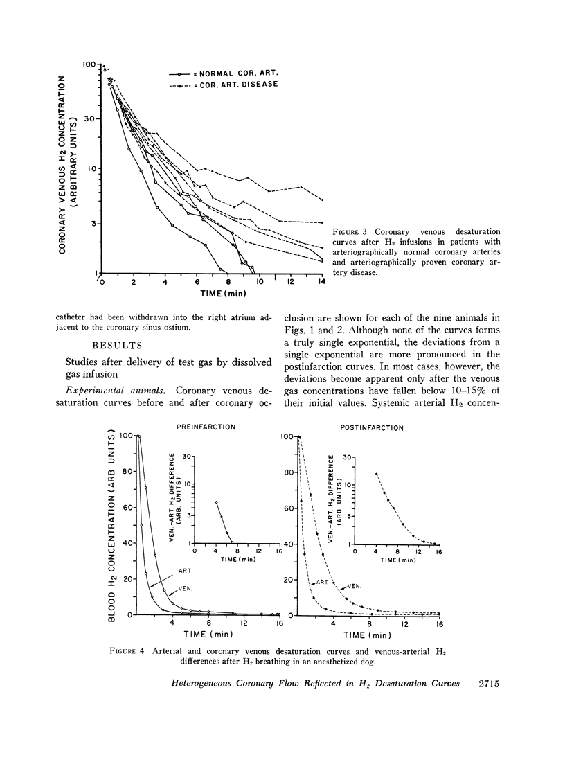
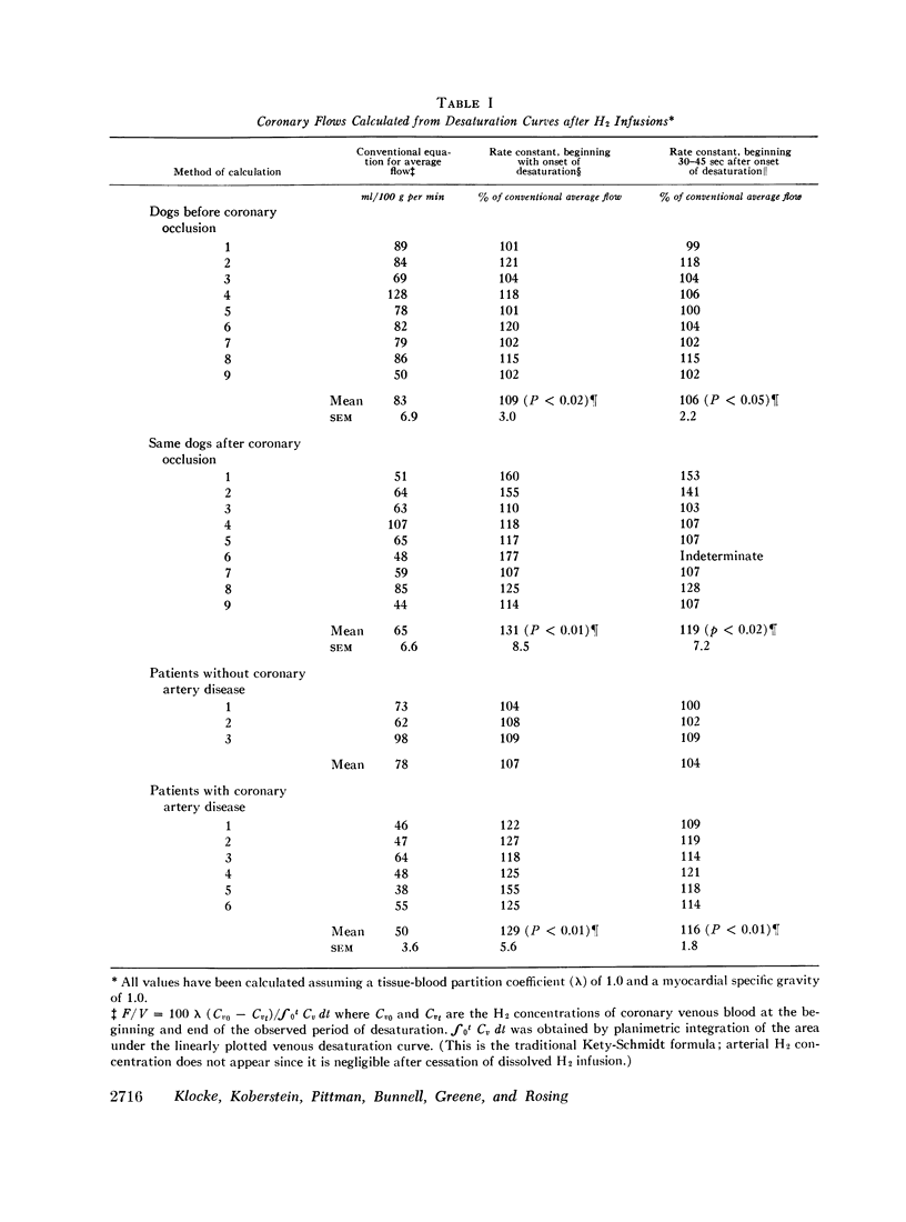
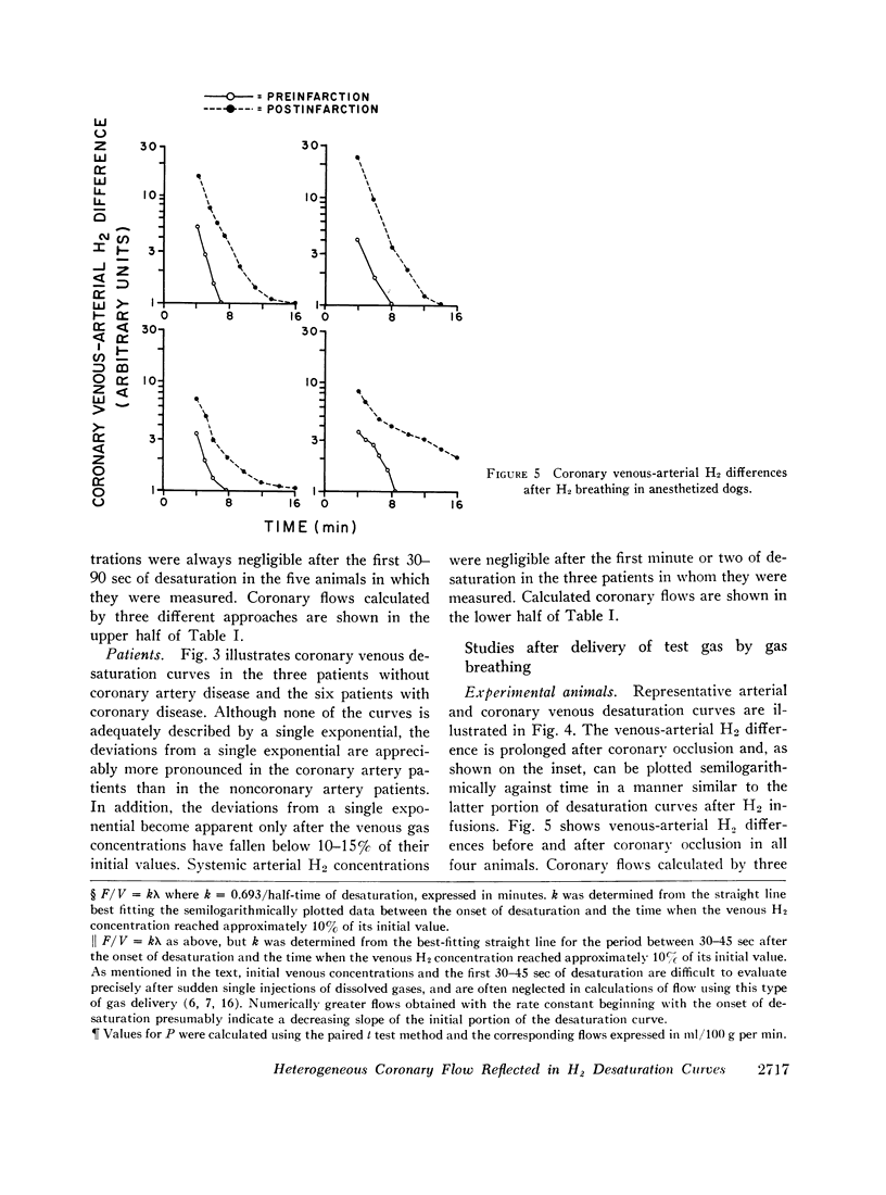
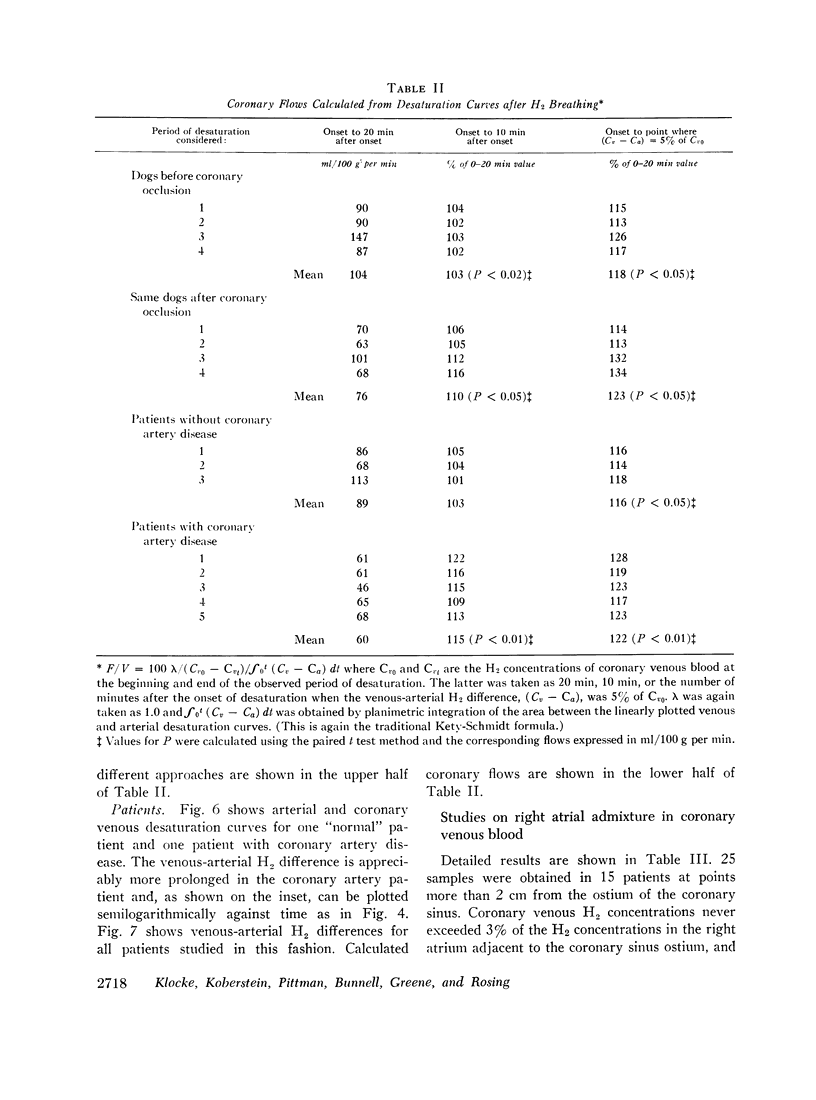
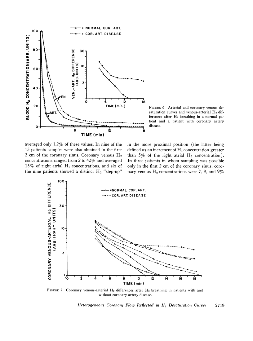
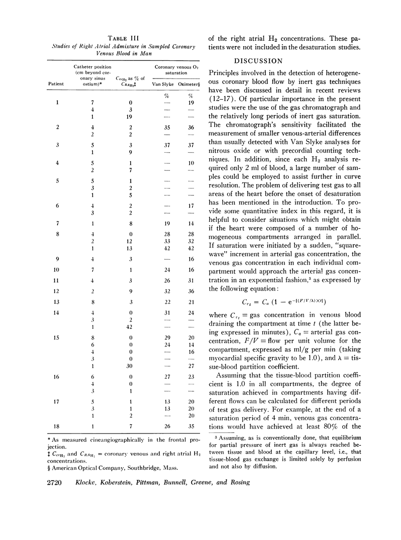
The present investigation was intended to evaluate myocardial inert gas desaturation curves for manifestations of heterogeneous coronary perfusion. The test gas was hydrogen (H2) and blood H2 analyses were performed with a gas chromatograph capable of detecting small but prolonged venous-arterial H2 differences produced by areas of reduced flow. Curves were initially obtained after 4-min left ventricular infusions of H2-saturated saline in six patients with arteriographically proven coronary artery disease, three patients with normal coronary arteries, and nine closed-chest dogs. The dogs were studied before and after embolic occlusion of a portion of the left coronary artery. Although the slopes of their semilogarithmically plotted venous desaturation curves varied with time before embolization, they showed more distinct deviations from single exponentials after embolization (after H2 concentrations had fallen below 15% of their initial values). The human curves divided similarly, those from coronary artery patients deviating appreciably from single exponentials. A similar separation was also evident in studies of coronary venous-arterial H2 differences after 20 min of breathing 2% H2: data were obtained in four dogs before and after coronary embolization, and in three normal patients, and five patients with coronary artery disease. Additional data indicated that the findings were not the result of right atrial admixture in sampled coronary venous blood, although admixture occurred frequently when blood was sampled in the first 2 cm of the coronary sinus (as seen in the frontal projection). Finally, average coronary flows calculated from a given set of data varied significantly with different methods of calculation. Areas of below-average flow seemed likely to be overlooked when single rate constants of desaturation, relatively insensitive analytical techniques, or relatively short periods of saturation and (or) desaturation are employed.
Full text
PDF
Selected References
These references are in PubMed. This may not be the complete list of references from this article.
- Brandi G., Fam W. M., McGregor M. Measurement of coronary flow in local areas of myocardium using xenon 133. J Appl Physiol. 1968 Mar;24(3):446–450. doi: 10.1152/jappl.1968.24.3.446. [DOI] [PubMed] [Google Scholar]
- COHEN L. S., ELLIOTT W. C., GORLIN R. MEASUREMENT OF MYOCARDIAL BLOOD FLOW USING KRYPTON 85. Am J Physiol. 1964 May;206:997–999. doi: 10.1152/ajplegacy.1964.206.5.997. [DOI] [PubMed] [Google Scholar]
- Farhi L. E. Elimination of inert gas by the lung. Respir Physiol. 1967 Aug;3(1):1–11. doi: 10.1016/0034-5687(67)90018-7. [DOI] [PubMed] [Google Scholar]
- GOODALE W. T., HACKEL D. B. Measurement of coronary blood flow in dogs and man from rate of myocardial nitrous oxide desaturation. Circ Res. 1953 Nov;1(6):502–508. doi: 10.1161/01.res.1.6.502. [DOI] [PubMed] [Google Scholar]
- KETY S. S. The theory and applications of the exchange of inert gas at the lungs and tissues. Pharmacol Rev. 1951 Mar;3(1):1–41. [PubMed] [Google Scholar]
- KLOCKE R. A., FARHI L. E. SIMPLE METHOD FOR DETERMINATION OF PERFUSION AND VENTILATION-PERFUSION RATIO OF THE UNDERVENTILATED ELEMENTS (THE SLOW COMPARTMENT) OF THE LUNG. J Clin Invest. 1964 Dec;43:2227–2232. doi: 10.1172/JCI105096. [DOI] [PMC free article] [PubMed] [Google Scholar]
- Klocke F. J., Greene D. G., Koberstein R. C. Indicator-dilution measurement of cardiac output with dissolved hydrogen. Circ Res. 1968 Jun;22(6):841–853. doi: 10.1161/01.res.22.6.841. [DOI] [PubMed] [Google Scholar]
- ROSS R. S., UEDA K., LICHTLEN P. R., REES J. R. MEASUREMENT OF MYOCARDIAL BLOOD FLOW IN ANIMALS AND MAN BY SELECTIVE INJECTION OF RADIOACTIVE INERT GAS INTO THE CORONARY ARTERIES. Circ Res. 1964 Jul;15:28–41. doi: 10.1161/01.res.15.1.28. [DOI] [PubMed] [Google Scholar]
- ROWE G. G., CASTILLO C. A., AFONSO S., CRUMPTON C. W. CORONARY FLOW MEASURED BY THE NITROUS-OXIDE METHOD. Am Heart J. 1964 Apr;67:457–468. doi: 10.1016/0002-8703(64)90092-4. [DOI] [PubMed] [Google Scholar]
- ROWE G. G. The nitrous-oxide method for determining coronary blood flow in man. Am Heart J. 1959 Aug;58(2):268–281. doi: 10.1016/0002-8703(59)90344-8. [DOI] [PubMed] [Google Scholar]
- SAPIRSTEIN L. A., OGDEN E. Theoretic limitations of the nitrous oxide method for the determination of regional blood flow. Circ Res. 1956 May;4(3):245–249. doi: 10.1161/01.res.4.3.245. [DOI] [PubMed] [Google Scholar]
- Sullivan J. M., Taylor W. J., Elliott W. C., Gorlin R. Regional myocardial blood flow. J Clin Invest. 1967 Sep;46(9):1402–1412. doi: 10.1172/JCI105632. [DOI] [PMC free article] [PubMed] [Google Scholar]
- Van Liew H. D. Semilogarithmic Plots of Data Which Reflect a Continuum of Exponential Processes. Science. 1962 Nov 9;138(3541):682–683. doi: 10.1126/science.138.3541.682. [DOI] [PubMed] [Google Scholar]
- ZIERLER K. L. EQUATIONS FOR MEASURING BLOOD FLOW BY EXTERNAL MONITORING OF RADIOISOTOPES. Circ Res. 1965 Apr;16:309–321. doi: 10.1161/01.res.16.4.309. [DOI] [PubMed] [Google Scholar]


