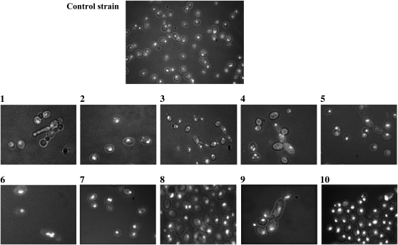
Figure 6.—
Microphotography of DAPI-stained control strain cells and SUSU translocants. (1–10) SUSU1–SUSU10 translocant cells. Various abnormal cell morphologies and karyokinetic defects are shown: germination tube formation (1), pseudohyphal growth of cells (4, 5, and 9), and multi-budded cells or cells with elongated buds (3 and 4). Nuclear fragmentation and/or defects in nuclear segregation are evident in some of the cells shown in each field (2, 4, 5, 6, 7, 8, and 10).

