Despite the growing literature on the treatment of arterial injuries, comparable information on the care of great venous injuries has been slow to accumulate. Prior to 1961, there was only one report of the successful treatment of more than a single case of injury to the inferior vena cava.5 Since that time, large series of vena caval injuries have been reported from Houston3 and from Chicago.6
The purpose of the present report is to re-emphasize certain clinical features of inferior vena caval perforations, and to outline steps for the effective treatment of this potentially lethal injury. The 12 cases used for illustration of various problems have been previously tabulated6 and Cases 1–4 and 8 have been previously described in detail.5, 6
CASE REPORTS
Case 1
J.M.H., Miami, 246987 (previously reported in detail5).
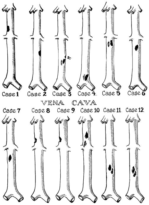
A 27 year old Negro man was shot in the abdomen with a .22 caliber pistol on September 8, 1956, and was admitted to the hospital in shock The bullet had passed through the second part of the duodenum and the head of the pancreas and had avulsed the right lateral wall of the inferior vena cava above the renal veins (Fig. 1). A large retropancreatic hematoma was present, but there was no active intraperitoneal bleeding. Massive hemorrhage, which occurred when the plane of the hematoma was entered by a Kocher maneuver (Fig. 2, C), was controlled with pressure and with the use of vascular clamps. The caval perforation was closed with continuous 5–0 arterial silk. The duodenal perforations were closed and the retroperitoneal space was extensively drained. Transfusions of 5000 cc. of plasma or blood were administered. Convalescence was uncomplicated, and there were no late untoward sequelae.
Fig. 1.
The location and extent of the injuries to the inferior vena cava. The diagrams are in the same order as the case reports. (From Surgery 51: 195, 1962.6 By permission of the publishers.)
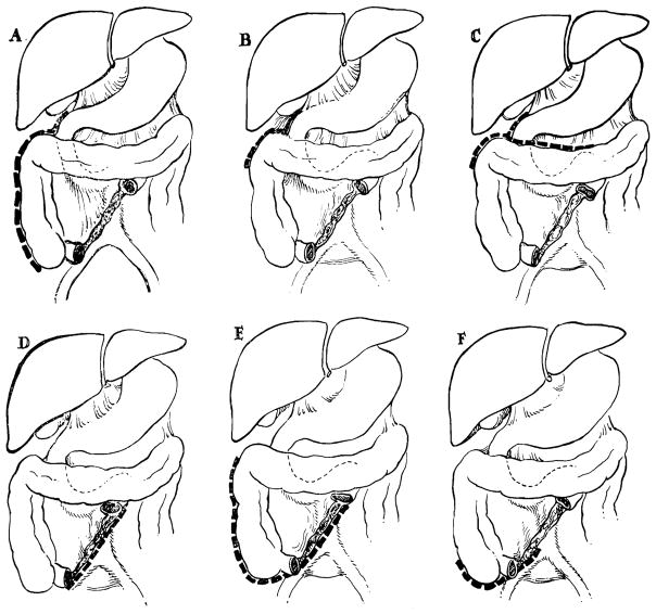
Fig. 2.
Different posterior peritoneal incisions used for retroperitoneal exploration.
Case 2
J.M.H., Miami, 252952 (previously reported in detail5).
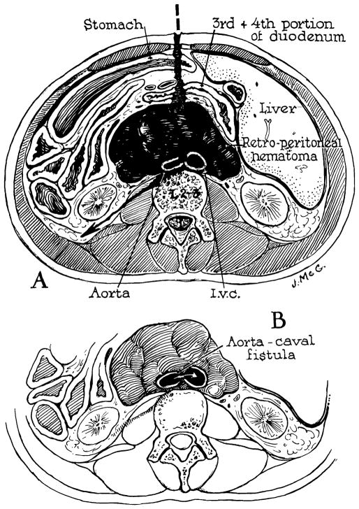
A 38 year old man was admitted in shock on October 19, 1956, after being shot in the abdomen with a .32 caliber pistol. At operation, active intraperitoneal bleeding had ceased, and a large retroperitoneal hematoma was found beneath the transverse colon and the mesentery of the small bowel. The bullet had passed through the stomach, the third portion of the duodenum, and between the aorta and vena cava approximately 2 inches below the renal vessels (Fig. 1), causing injury to the contiguous aortic and caval wall (Fig. 3, A). The vessels were approached through an incision in the base of the small bowel mesentery (Fig. 2, D). Abrupt hemorrhage occurred upon opening the hematoma. This was controlled with proximal and distal compression and both the aorta and vena cava were repaired with 5–0 arterial silk. The gastrointestinal perforations were closed and the retroperitoneal space drained. Transfusions of 4000 cc. of plasma or blood were given. Convalescence was rapid and follow-up revealed no late complications.
Fig. 3.
A, The injury seen in Case 2, with laceration of the contiguous aorta and inferior vena cava.
B, Possible result if vascular injuries had not been repaired, with formation of aorta-caval fistula. Nine such cases have been reported.
Case 3
J.M.H., Miami, 266747 (previously reported in detail5).
A 25 year old Puerto Rican male was admitted on January 4, 1957, after being shot in the back with a .357 magnum police pistol. He was normotensive and did not appear to be critically injured. At exploration, free intraperitoneal bleeding had ceased. The bullet had passed through the jejunum and third part of the duodenum. A small hematoma elevated the ligament of Treitz. On entering the hematoma, at operation, massive venous bleeding occurred. Broad exposure of the retroperitoneal space was obtained as shown in Figure 2, E. Application of Potts clamps to both iliac veins and to the vena cava above the site of injury was ineffective in controlling the hemorrhage owing to intercostal influx, and local pressure was necessary. The missile had passed through the vena cava 2 inches above the bifurcation causing 1.5 cm. lacerations of both anterior and posterior walls and avulsing a lumbar vein (Fig. 1). The caval rents were repaired with continuous 5–0 arterial silk. After suturing the intestinal perforations, the abdomen was closed without drainage. Transfusions of 5000 cc. of plasma or blood were required. Recovery was complicated by left iliofemoral thrombosis which was thought to be secondary to operative trauma to the left iliac vein. Two months after discharge, there was no evidence of venous stasis in either leg.
Case 4
U.S.V.A.R., 13588 (previously reported in detail6).
A 27 year old male Negro was admitted on November 2, 1956, to the U.S.V.A. Research Hospital, Chicago, three hours after having been shot in the upper midabdomen. The bullet had passed through the transverse colon, two loops of small bowel and the inferior vena cava (Fig. 1). At operation, active bleeding had ceased, but a large retroperitoneal hematoma was present.
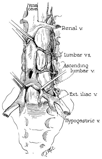
Despite preliminary occlusion of the cava and iliac veins with tapes (Fig. 4), massive hemorrhage occurred when the retroperitoneal space was entered, owing to venous return from the intervening lumbar veins. This was eventually controlled by pressure and packing on each side of the injured vessel. An anterior caval laceration was closed with 5–0 arterial silk, and the posterior wall rolled over and closed in a similar manner. The colon and small bowel lacerations were closed in two layers, and the abdomen closed without drainage. Transfusions totaled 3500 cc. Recovery was uneventful except for a superficial wound infection.
Fig. 4.
Large collaterals which connect different segments of inferior vena cava. Note that tapes placed at a distance from vascular injury fail to control hemorrhage due to collateral inflow. (From Surgery, 51: 195, 1962.6 By permission of the publishers.)
Case 5
C.C.H., 56–65477.
A 26 year old male Negro was admitted to Cook County Hospital on October 21, 1956, six hours after being shot in the lower chest. He was unconscious from the time of injury until after admission, and his last recollection was that of leaning over an automobile hood prior to injury. He had been admitted to another hospital in shock, treated with 500 cc. plasma and Levarterenol and transferred to Cook County Hospital. Blood pressure was 90/64 and pulse 124. There was a wound of entry in the right eighth intercostal space, just lateral to the midclavicular line. The bullet, subsequently shown to be .32 caliber, could be palpated subcutaneously in the left flank, opposite the third lumbar vertebra. He had generalized peritonitis. No blood was present in the urine, gastric aspirate, or feces. Hematocrit was 35.
After transfusion, operation was performed eight hours after the original injury, under ether anesthesia. A long right paramedian incision was used. A large quantity of free peritoneal blood was removed, and a large retroperitoneal hematoma was noted, but active bleeding had ceased. The bullet had passed downward and to the left from the point of entry, passing through the right lobe of the liver and the anterior and posterior walls of the lower second part of the duodenum, and had entered the retroperitoneal space at the level of the second lumbar vertebra.
The retroperitoneal space was entered by mobilization of the right colon medially and with a Kocher maneuver (Fig. 2, A). The perforations in the duodenum were closed in two layers. The hematoma, at a somewhat lower level, was then explored further. The field was immediately flooded with blood. The hemorrhage was controlled by pressure with a stick sponge over a through-and-through perforation of the vena cava just below the renal veins (Fig. 1). Ligatures were placed above and below the lacerations, and the vena cava doubly ligated with silk. Two Penrose drains and a Chaffin tube were used to drain the retroperitoneal and subhepatic spaces and the abdomen was closed. Transfusions totaled 3000 cc. of blood and 500 cc. of plasma.
The wounds healed primarily. Full diet was resumed by the fourth postoperative day. Drains were removed and antibiotics discontinued on the tenth day. The principal postoperative complaint was pain and swelling in both legs, particularly the left. The patient was outfitted with elastic stockings and discharged on the twenty-sixth day after operation. In June, 1960, the patient was well and without evidence of leg edema.
Case 6
C.C.H., 58–88026.
A 36 year old male Negro was admitted to Cook County Hospital on December 26, 1958, after being stabbed with a large butcher knife. He was drowsy and could be aroused only with effort. Blood pressure was 84/60, pulse 84, and respirations 20. There were two stab wounds in the right upper quadrant just below the costal margin, both lateral to the rectus muscle. He had peritonitis. No gross blood was present in the urine, gastric aspirate, or feces. After a 1500 cc. transfusion, he was anesthetized with ether and the abdomen explored through a right subcostal incision.
There was a small amount of blood in the peritoneal cavity and no active bleeding. The knife had passed sharply downward through the right lobe of the liver and the head of the pancreas. The third part of the duodenum and the mesentery of the small bowel were elevated by a large retroperitoneal hematoma. It was feared that retroperitoneal injury of the third portion of the duodenum had occurred. The retroperitoneal space was exposed with a Kocher maneuver and by mobilization of the hepatic flexure (Fig. 2, B). Massive hemorrhage occurred when the hematoma was entered. This was quickly controlled by stick sponge pressure immediately above and below the site of caval injury.
A 1.5 cm. laceration of the anterior wall of the inferior vena cava was demonstrated, 2 or 3 cm. below the renal veins (Fig. 1). This was repaired with 5–0 arterial silk. The posterior wall of the cava had not been damaged. Three Penrose drains and a Chaffin tube were placed about the head of the pancreas and the abdomen closed. A total of 5000 cc. of blood was administered.
The early postoperative course was complicated by right middle lobe pneumonitis, which cleared by the tenth day. Two weeks postoperatively, a pancreatic fistula developed. This closed spontaneously after wide incision and drainage of the subcostal incision on January 19, 1959. The patient was discharged in good condition on February 10, 1959. He was last seen on June 30, 1959, and was without complaints.
Case 7
C.C.H., 59–07122.
A 36 year old male Negro was admitted to Cook County Hospital on January 28, 1959, four hours after a knife fight, after having been previously seen in another hospital. He did not appear to be gravely wounded. Blood pressure was 104/60 and pulse 72. There were superficial lacerations of the supraclavicular area and of both hands. A stab wound was present half-way between the xiphoid and umbilicus through which protruded a loop of small bowel. He had generalized peritonitis. Hematocrit was 46.
Under Pentothal and ether anesthesia, abdominal exploration was carried out five and a half hours after injury, through an upper right paramedian incision. Only a few cubic centimeters of free blood was in the peritoneal cavity. The pathway of the knife had been in a postero-inferior direction. Through-and-through perforations of the jejunum, ileum and third portion of the duodenum were identified. A moderate sized hematoma elevated the third part of the duodenum.
In an effort to visualize the posterior duodenal perforation adequately, the retroperitoneal space was entered by a Kocher maneuver with extension of the peritoneal incision along the right colon (Fig. 2, A). When the place of the hematoma was entered, massive hemorrhage ensued. The bleeding was rapidly controlled with finger compression above and below the focus of bleeding. A 1.5 cm. laceration, 1 inch below the renal veins, was identified on the anterolateral surface of the vena cava (Fig. 1). There was no injury to the posterior vessel wall. The rent was trapped with a partially occluding Satinsky clamp and closed with continuous 5–0 arterial silk.
The multiple small bowel perforations were closed in two layers and, after bringing a Penrose drain from the retroperitoneal space out of the right flank, the abdomen was closed. Altogether, 3500 cc. of blood were given.
Convalescence was satisfactory. The patient was not febrile after the second postoperative day. The drain was removed on the seventh day, and he was discharged 11 days after operation. On February 16, 1960, he was asymptomatic and working.
Case 8
C.C.H., 60–20934 (previously reported in detail6).
A 23 year old male Negro was treated on March 20, 1960, for a gunshot wound of the abdomen. The bullet had passed through the right lobe of the liver, through the second part of the duodenum just below the entrance of the common duct, and through the pancreas, portal vein and inferior vena cava. The head of the pancreas was elevated by a large retroperitoneal hematoma, but free hemorrhage from the caval and portal lacerations had ceased.
The retroperitoneal space was exposed by medial mobilization of duodenum and pancreas (Kocher maneuver) and by moblilzation of the right colon (Fig. 2, B). When the plane of the hematoma was entered, massive hemorrhage occurred with the sudden loss of 1500 cc. of blood. This was rapidly controlled with pressure and with the tangential application of several straight Potts clamps to the general focus of bleeding. The vena caval and portal venous lacerations were identified and repaired with continuous arterial silk. The duodenal perforations were closed, and the area extensively drained. During the preoperative, operative and immediate postoperative periods, the patient received 750 cc. of plasma and 7500 cc. of blood.
He was discharged on May 3, 1960, six weeks after operation. On June 18, 1960, he was asymptomatic, without evidence of caval or portal thrombosis, and had returned to work.
Case 9
C.C.H., 60–11580.
A 20 year old white woman was admitted to Cook County Hospital on February 11, 1960, forty-two days after operative treatment elsewhere of an abdominal wound which was self-inflicted with a .38 caliber pistol. Information from the primary hospitalization indicated that the patient was not in shock on arrival. She was 4½ months pregnant. At operation, active bleeding had ceased. The bullet had passed from the midepigastrium posteriorly to the first lumbar vertebra, causing an avulsing laceration of the vena cava just above the renal veins (Fig. 1). A moderate-sized hematoma was present. There were concomitant perforations of the transverse colon, small bowel, pancreas, and branches of the superior mesenteric vein. Exposure was obtained as in Figure 2, C. The visceral perforations were closed and the cava repaired with arterial suture. The pancreas was drained. Postoperatively, a subphrenic abscess required posterolateral drainage, during which the right chest was accidentally entered.
Upon admission to Cook County Hospital, the patient was profoundly ill with pulse of 180 and temperatures as high as 108°. Two days after admission, she delivered a 2 pound, 8 ounce stillborn fetus. A large empyema on the right was treated with multiple aspirations and antibiotics. Pulmonary decortication of the empyema cavity was performed on March 7, 1960. A small residual empyema was treated with tube drainage on April 25, 1960. She was discharged on May 3, 1960. At the time of last examination on July 2, 1960, her condition was excellent, with no evidence of vena caval occlusion.
Case 10
C.C.H., 60–23320.
An intoxicated 27 year old Negro woman was admitted to Cook County Hospital on March 29, 1960. She had been shot three times at close range with a .38 caliber pistol four hours previously, and treated with 1000 cc. of blood and plasma at another hospital. She did not appear to be mortally wounded. Blood pressure was 140/90 and pulse 96. A superficial wound was in the left posterior axillary fold with adjacent entrance and exit punctures. Two other wounds of entry were found, one anteriorly in the fifth intercostal space just to the right of the midline, and the other posteriorly in the right flank. There was generalized peritonitis. No blood was found in the stool, gastric aspirate or urine. Abdominal x-rays revealed one bullet just below the diaphragm posteriorly, and another in the upper anterior abdominal wall.
Because of blood pressure instability, an additional 1500 cc. of blood were given through an arm cut-down. Exploration was carried out two hours after arrival through an upper right paramedian incision, with Pentothal-cyclopropane-ether anesthesia. 1500 cc. of blood were removed from the abdomen. Active bleeding had ceased. The only visceral injury was a perforating wound of the right lobe of the liver, just to the right of the midline. A small hematoma of no more than 5 or 10 cc. was seen in the posterior wall of the foramen of Winslow. No other disruption of the posterior peritoneum could be found. It was thought that the flank bullet had passed around the circumference of the body in the soft tissues, and that the only abdominal penetration had been through the inferiorly directed anterior chest wound.
During digital examination of the hematoma, massive hemorrhage occurred, which was controlled with pressure. The hepatic flexure and duodenum were mobilized (Fig. 2, C), and a Potts clamp placed on the vena cava above the renals, distal to the hematoma. Profuse hemorrhage continued. The incision was extended into the right chest and, after retraction of the liver to the left and superiorly, the cava was occluded above the site of injury with considerable reduction of bleeding. The bullet had passed through the vessel in a postero-inferior direction, passing through the anterior wall in the floor of the foramen of Winslow, and the posterior wall at a lower level just above the entrance of the right renal vein (Fig. 1). The anterior laceration was closed with 5–0 arterial silk. In rolling the vena cava to expose the posterior rent, the right renal vein was partially avulsed. This was repaired, in addition to the posterior bullet hole, with 5–0 arterial silk. The patient received 14,000 cc. of blood during the operation.
During closure, oozing was noted from all raw surfaces, suggesting a hemorrhagic diathesis. Postoperatively, blood drainage erupted from the multiple abdominal drains and from the chest tube. The patient died 18 hours after operation. At autopsy, massive hemothorax and hemoperitoneum were found. The vascular wounds appeared to be well closed.
Case 11
C.C.H., 60–67350.
A 13 year old male Negro was admitted to Cook County Hospital on September 10, 1960, four hours after being shot in the abdomen with a .22 caliber rifle. Blood pressure was 100/60, pulse 80, and respirations 24. There was a wound of entry in the right upper abdomen, 2 inches above and lateral to the umbilicus. He had generalized peritonitis. Gastric contents, feces and urine did not contain blood. Hematocrit was 39. X-rays showed the bullet to the right of the fourth lumbar vertebra. A major vascular injury was not suspected.
At operation a hemoperitoneum of 750 cc. was evacuated. Active bleeding had ceased. The bullet had perforated the jejunum in six places, and had caused a through-and-through perforation at the junction of the second and third portions of the duodenum. A small retroperitoneal hematoma was found below the third portion of the duodenum. The right colon and duodenum were reflected medially (Fig. 2, A). Upon entering the plane of the hematoma, severe bleeding occurred which was immediately controlled by local pressure. Anterior and posterior perforations of the vena cava were encountered (Fig. 1) below the renal veins. These were closed with continuous 5–0 arterial silk. The duodenal perforations were closed in two layers, and because this resulted in constriction of the lumen, a gastroenterostomy was performed. The multiple jejunal perforations were close together and were removed by a 14 cm. resection. The retroperitoneal space was drained. A total of 1000 cc. of blood was given. The patient was discharged nine days after operation. Two months after the accident, there were no complaints.
Case 12
C.C.H., 60–70998.
A 28 year old male Negro was admitted to Cook County Hospital on September 23, 1960, after having been shot in the abdomen with a .32 caliber pistol. Blood pressure was unobtainable initially, but after 1500 cc. of blood it stabilized at 160/60. A wound of entry was found in the midline halfway between the xiphoid process and umbilicus. The patient had generalized peritonitis. Abdominal x-rays showed a chip fracture of the fourth lumbar vertebra and the bullet near the right iliac crest. A major vascular injury was suspected.
Exploration was performed through a right paramedian incision. Approximately 1500 cc. of free blood and bile were in the abdomen. Active bleeding had ceased. A large retroperitoneal hematoma elevated the transverse mesocolon and the base of the small bowel mesentery. The bullet had projected postero-inferiorly, passing through the right lobe of the liver, the gallbladder and the head of the pancreas. The ascending colon and duodenum were reflected medially (Fig. 2, A). Upon entering the plane of the hematoma, massive hemorrhage occurred. This was controlled by stick sponge pressure above and below a through-and-through perforation of the vena cava (Fig. 1). The anterior wound was closed with continuous 5–0 arterial silk. With some difficulty, the cava was rolled over and the posterior laceration also was closed. Cholecystectomy, common duct exploration, and insertion of a T-tube were carried out. The retroperitoneal space was drained extensively. Total transfusion before, during and after surgery was 5500 cc.
Convalescence was complicated. The patient had a temporary partial paraplegia probably due to concussion injury of the cauda equina. Renal shutdown developed postoperatively with a rise of the blood urea nitrogen to 204, before diuresis started on the fifteenth postoperative day. After 2½ weeks, the T-tube was removed. A bile fistula developed which closed spontaneously three weeks later. The patient was discharged in good condition 6½ weeks after operation. There was no evidence of venous stasis in the lower extremities.
DISCUSSION
The high incidence of inferior vena caval injury with penetrating wounds of the abdomen has not been generally appreciated. It is probable that caval laceration occurs approximately once in every 50 cases of civilian gunshot wounds of the abdomen2, 4, 6, 7 and once in every 300 knife wounds.6
Despite the difference in the weapons of assault, the location of the wounds, and the magnitude of associated injuries, the problems encountered at operation were remarkably similar in all the cases. There was, for example, no active bleeding from the injured vessel at the time of operation in any case, despite the fact that six of the 12 patients entered the hospital in shock. Although varying degrees of hemoperitoneum were present at the time of exploration, spontaneous tamponade had invariably occurred, with the formation of a retroperitoneal hematoma. Only when the hematoma was disturbed did bleeding recur, often massively to the surprise of the unsuspecting surgeon.
This feature of hematoma formation and self-tamponade, which is so characteristic of the inferior vena caval laceration, has important implications relating both to the judgment involved in exploring the retroperitoneal space and the method to be followed if retroperitoneal exploration is decided upon. Ochsner, Crawford and DeBakey3 have cautioned against disturbing retroperitoneal hematomas if circumstances permit. This policy may be advisable where the only injury appears to be to the vena cava. Commonly, however, retroperitoneal exploration is mandatory to treat or rule out injuries to other retroperitoneal structures which, if neglected, will lead to death. This was the situation in 11 of the 12 cases in the present series. It is interesting to speculate on the fate of the patient in Case 2, had the aortic injury not been detected and repaired. Although a fatal hemorrhage would have been the most likely outcome, it is possible that an aortocaval fistula might have developed as depicted in Figure 3, B. Nine such cases following penetrating trauma have been described in the world literature.
If retroperitoneal exploration is decided upon in the presence of a retroperitoneal hematoma, preliminary precautionary measures can be exercised in an unhurried and orderly manner. Exposure and lighting can be improved. Additional assistance can be called. Vascular clamps can be prepared. A large-bore needle or cut-down can be placed in the arm, since forced transfusions from the lower extremities may only aggravate the situation by flooding the field with citrated blood through the caval rent. An adequate supply of blood should be brought to the operating room. Plans can be made for the maximum exposure of the retroperitoneal space, according to the location of the injury (Fig. 2). Isolation of the great vessels above and below the site of the hematoma may reduce the ultimate extent of blood loss.
When at last the retroperitoneal space is exposed, the dissection should proceed rapidly or exsanguination may occur before the site of hemorrhage is controlled. The most effective means for immediate control of hemorrhage has been finger or stick sponge pressure upon or near the site of vascular injury, or the direct application of vascular clamps. Attempts at occlusion at any distance above and below the bleeding site do not achieve hemostasis (Case 4). The abundant lumbar collaterals1 interconnect the different levels of the vena cava so effectively (Fig. 4) that hemorrhage may be almost as severe as with the cava open. The difficulties with hemorrhage can be appreciated by the fact that an average of 5200 cc. of blood or plasma was given to these 12 cases.
Suture repair of the caval rent was performed in all except Case 5, in which ligation was done. Subsequent complications attributable to the venous repair occurred only in Case 3, in which ileofemoral thrombosis occurred. In the two knife wounds (Cases 6 and 7), the blade lacerated only the anterior wall. With gunshot wounds there was either a double perforation (Cases 3, 5, 10, 11, 12) or a lateral avulsion (Fig. 1) with loss of tissue (Cases 1, 2, 4, 8, 9). In the cases with injury above the renal veins (Fig. 1), suture repair is mandatory since caval ligation at this level leads to death.8
In the cases presented, vena caval laceration never occurred as an isolated injury, a minimum of one, a maximum of four and an average of 2.4 other organs being involved. These included associated injuries to the liver, pancreas, stomach, duodenum, jejunum, ileum, colon, gallbladder, aorta, portal vein and superior mesenteric vein. These injuries necessitated extensive use of intra- or extraperitoneal drains in all but two cases, and contributed to serious complications which included pancreatic fistula, subphrenic abscess, empyema, biliary fistula, and acute renal failure. The high survival rate of 11 of 12 patients was undoubtedly partly due to the fact that all the patients were less than 38 years old and in previous good health.
SUMMARY
Clinical summaries are presented of 12 patients who sustained penetrating wounds of the inferior vena cava in addition to other visceral injuries. The wounds were inflicted by gunshot in ten cases and by knife lacerations in two. Eleven of the 12 patients survived.
Injury to the inferior vena cava as a result of penetrating wounds of the abdomen is more common than is generally appreciated. Characteristically, hemorrhage from the venous injury spontaneously ceases as the result of retroperitoneal tamponade. Exsanguinating hemorrhage is frequently provoked by exploring the retroperitoneal hematoma at the time of operation.
If a retroperitoneal hematoma is found at operation in the locality of the great vessels, precautions should be taken before the retroperitoneal space is explored. These include provisions for rapid transfusion, adequate or extended exposure, and instruments for vascular reconstruction.
Acknowledgments
Supported by grants A-3176 and A-6283 from the United States Public Health Service.
References
- 1.Davis RA, Milloy FJ, Anson BJ. Lumbar, renal, and associated parietal and visceral veins based upon a study of 100 specimens. Surg Gynec & Obst. 1958;107:1. [PubMed] [Google Scholar]
- 2.Elkin DC, Ward WC. Gunshot wounds of the abdomen. Ann Surg. 1943;118:780. doi: 10.1097/00000658-194311850-00002. [DOI] [PMC free article] [PubMed] [Google Scholar]
- 3.Ochsner JL, Crawford ES, DeBakey ME. Injuries of the vena cava caused by external trauma. Surgery. 1961;49:397. [PubMed] [Google Scholar]
- 4.Oberhelman HA, LeCount ER. Peacetime bullet wounds of the abdomen. Arch Surg. 1936;32:373. [Google Scholar]
- 5.Starzl TE, Broadaway RK, Dever RL, Reams GB. Management of penetrating wounds of the inferior vena cava. Am Surgeon. 1957;23:455. [PubMed] [Google Scholar]
- 6.Starzl TE, Kaupp HA, Jr, Beheler EM, Freeark RJ. Treatment of penetrating wounds of the inferior vena cava. Surgery. 1962;52:195. [PubMed] [Google Scholar]
- 7.Walker WH. Penetrating abdominal wounds. Ann Surg. 1950;60:55. [PubMed] [Google Scholar]
- 8.Whittenberger JT, Huggins C. Ligation of the inferior vena cava. Arch Surg. 1940;41:1334. [Google Scholar]


