Abstract
Several vectors that facilitate stable fluorescent labeling of Burkholderia pseudomallei and Burkholderia thailandensis were constructed. These vectors combined the effectiveness of the mini-Tn7 site-specific transposition system with fluorescent proteins optimized for Burkholderia spp., enabling bacterial tracking during cellular infection.
Burkholderia pseudomallei is a highly infectious Gram-negative bacterium and a facultative intracellular pathogen. The ability to observe infectious processes of this bacterium at various stages is critical to understand its pathogenesis. Fluorescent proteins facilitate bacterial tagging and have been powerful investigative tools in deciphering biological processes (3, 23, 24, 29, 31, 32). However, the lack of optimized fluorescent constructs used to label B. pseudomallei for visualization necessitates further development. Although there are many fluorescent tools available besides the green fluorescent protein (GFP), they are not optimized for use in B. pseudomallei or the less pathogenic model species Burkholderia thailandensis. Commercially available fluorescent proteins are optimized for eukaryotic expression or, at best, for bacteria with low-G/C-content genomes, and hence codon preference may cause problems (13, 19) during protein expression in Burkholderia spp. Also, there is usually an ineffective promoter driving transcription in Burkholderia spp., and available constructs usually replicate plasmids that require selective maintenance. The restricted use of antibiotic markers in select agents (e.g., B. pseudomallei) adds another level of complexity to the genetic manipulation of these species (27). Hence, these obstacles have limited the applications of fluorescent proteins in pathogenesis studies of Burkholderia spp.
The well-established mini-Tn7 system (7, 14, 18) inserts itself at a unique neutral site(s) in the bacterial genome with the aid of a nonreplicating helper plasmid encoding the transposase (1). The B. pseudomallei chromosome contains three insertion sites downstream of three glmS genes, whereas B. thailandensis contains two insertion sites downstream of two glmS genes (8). Mini-Tn7 inserted in bacterial genomes is quite stable (7, 20), and selective maintenance is not required. In this study, we constructed and demonstrated the use of fluorescent proteins encoded on mini-Tn7-based site-specific transposition vectors for fluorescent tagging of B. pseudomallei and B. thailandensis. We optimized for B. pseudomallei the fluorescent protein genes (cyan, red, and yellow), which were synthesized through Genscript Corporation based on the amino acid sequences from Evrogen, by driving their transcription with a PS12 promoter (36) and changing the codons to those preferred by B. pseudomallei. Since enhanced GFP (eGFP) is sufficiently bright in Burkholderia spp., we utilized the gfp gene driven by the PS12 promoter (Table 1). The four new fluorescent proteins (green, cyan, red, and yellow fluorescent proteins [GFP, CYP, RFP, and YFP, respectively]) were combined with two mini-Tn7 backbones to produce eight fluorescent tagging vectors (Fig. 1 and Table 1). The first series of four vectors encode the nonantibiotic selectable marker gat (resistance to glyphosate) (5, 21), and the other series of vectors encode kanamycin resistance (Kanr) (Fig. 1). Both are currently approved selectable markers for B. pseudomallei (8, 21). Nevertheless, prior CDC/USDA approval for each laboratory to use these markers is necessary and must be sought. All manipulations of B. pseudomallei were carried out in a CDC/USDA-approved biosafety level 3 laboratory by following the guidelines presented in Biosafety in Microbiological and Biomedical Laboratories, 5th ed. (34).
TABLE 1.
Bacterial strains and plasmids used in this studya
| Strain or plasmid | Lab IDb | GenBank ID no. | Relevant properties | Source or reference(s) |
|---|---|---|---|---|
| Strains | ||||
| E. coli | ||||
| EPMax10B-pir116 Δasd Δtrp::Gmrmob-Kanr | E1354 | Gmr Kanr F− λ−mcrAΔ(mrr-hsdRMS-mcrBC) φ80dlacZΔM15 ΔlacX74 deoR recA1 endA1 araD139 Δ(ara leu)7697 galU galK rpsL nupG Tn-pir116-FRT2 Δasd::wFRT Δtrp::Gmr-FRT5 mob[recA::RP4-2 Tc::Mu-Kanr] | Available lab strain | |
| EPMax10B-lacIqpir | E1869 | F− λ−mcrA Δ(mrr-hsdRMS-mcrBC) φ80dlacZΔM15 ΔlacX74 deoR recA1 endA1 araD139 Δ(ara leu)7697 galU galK rpsL nupG lacIq-FRT8 pir-FRT4 | Available lab strain | |
| B. pseudomallei | ||||
| 1026b | B0004 | Clinical melioidosis isolate | 10 | |
| 1026b/att-Tn7-gat-cfp | B0036 | GSr, 1026b with mini-Tn7-gat-cfp inserted | This study | |
| 1026b/att-Tn7-gat-gfp | B0037 | GSr, 1026b with mini-Tn7-gat-gfp inserted | This study | |
| 1026b/att-Tn7-gat-rfp | B0038 | GSr, 1026b with mini-Tn7-gat-rfp inserted | This study | |
| 1026b/att-Tn7-gat-yfp | B0039 | GSr, 1026b with mini-Tn7-gat-yfp inserted | This study | |
| B. thailandensis | ||||
| E264 | E1298 | Prototroph, environmental isolate | 2 | |
| E264/att-Tn7-kan-cfp | E2552 | Kanr, E264 with mini-Tn7-kan-cfp inserted | This study | |
| E264/att-Tn7-kan-gfp | E2491 | Kanr, E264 with mini-Tn7-kan-gfp inserted | This study | |
| E264/att-Tn7-kan-rfp | E2553 | Kanr, E264 with mini-Tn7-kan-rfp inserted | This study | |
| E264/att-Tn7-kan-yfp | E2554 | Kanr, E264 with mini-Tn7-kan-yfp inserted | This study | |
| Plasmidsc | ||||
| mini-Tn7-gat | E1981 | GSr, mini-Tn7 integration vector based on gat | 21 | |
| mini-Tn7-gat-cfp | E2460 | HM150704 | GSr, mini-Tn7-gat harboring cfp | This study |
| mini-Tn7-gat-gfp | E2462 | HM150705 | GSr, mini-Tn7-gat harboring gfp | This study |
| mini-Tn7-gat-rfp | E2326 | HM150706 | GSr, mini-Tn7-gat harboring rfp | This study |
| mini-Tn7-gat-yfp | E2468 | HM150707 | GSr, mini-Tn7-gat harboring yfp | This study |
| mini-Tn7-kan-cfp | E2480 | HM150708 | Kanr, mini-Tn7-kan harboring cfp | This study |
| mini-Tn7-kan-gfp | E2482 | HM150709 | Kanr, mini-Tn7-kan harboring gfp | This study |
| mini-Tn7-kan-rfp | E2486 | HM150710 | Kanr, mini-Tn7-kan harboring rfp | This study |
| mini-Tn7-kan-yfp | E2488 | HM150711 | Kanr, mini-Tn7-kan harboring yfp | This study |
| pPS747 | E0042 | Apr, plasmid harboring eGFP | 9, 15 | |
| pRK2013 | E1358 | Kanr, helper plasmid encoding conjugative proteins | 11 | |
| pTNS3-asdEc | E2237 | Suicidal helper plasmid containing E. coli asd and transposase for the Tn7 site-specific transposition system | 17 | |
| pUC57-PS12-cfp | E1739 | HM150701 | Apr, plasmid harboring B. pseudomallei codon-optimized cfp driven by PS12 | This study |
| pUC57-PS12-rfp | E1735 | HM150702 | Apr, plasmid harboring B. pseudomallei codon-optimized rfp driven by PS12 | This study |
| pUC57-PS12-yfp | E1738 | HM150703 | Apr, plasmid harboring B. pseudomallei codon-optimized yfp driven by PS12 | This study |
Abbreviations: Apr, ampicillin resistant; asdEc, E. coli asd gene; gat, gene encoding glyphosate acetyltransferase; Gmr, gentamicin resistant; GSr, glyphosate resistant; kan, gene encoding kanamycin resistance; Kanr, kanamycin resistant; PS12, rpsL promoter from B. pseudomallei; w, wild type.
Please use the laboratory identification number (Lab ID) when requesting strains and plasmids.
The mini-Tn7 plasmid backbone is based on pUC18R6KT-mini-Tn7T (7). Fluorescent protein genes (cfp, rfp, yfp) were synthesized by GeneScript and cloned into the EcoRV site of pUC57. These genes were removed by digestion with KpnI and PstI and then subcloned into mini-Tn7-gat cut with the same enzymes. Two-step PCR was performed to incorporate the PS12 promoter upstream of the gfp gene, which was digested with KpnI and PstI and cloned into mini-Tn7-gat that had been digested with the same enzymes. The mini-Tn7-kan constructs were created by replacing the gat cassette with the kan cassette.
FIG. 1.
Maps of mini-Tn7-gat-cfp and mini-Tn7-kan-cfp (A), mini-Tn7-gat-gfp and mini-Tn7-kan-gfp (B), mini-Tn7-gat-rfp and mini-Tn7-kan-rfp (C), and mini-Tn7-gat-yfp and mini-Tn7-kan-yfp (D). These constructs allow for site-specific transposition of fluorescent protein genes (cfp, gfp, rfp, and yfp), using the nonantibiotic glyphosate resistance marker (gat) or the kanamycin resistance marker (kan) assisted by the helper plasmid pTNS3-asdEc. Differences in plasmid size are indicated in parenthesis. The PS12 promoter drives all fluorescent proteins. The gat or kan cassette is driven by the rpsL promoter of B. cenocepacia (PCS12) on all constructs. These selectable markers are flanked by FRT sequences for Flp protein-mediated excision. Abbreviations: oriT, RP4 conjugal origin of transfer; PCS12, rpsL promoter of B. cenocepacia; PS12, rpsL promoter of B. pseudomallei; R6Kγori, π protein-dependent R6K origin of replication (γ indicates subtype of the origin); Tn7L and Tn7R, left and right transposase recognition sequences; T0T1, transcriptional terminator.
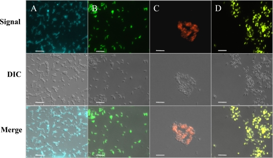
The four vectors based on gat were used to tag B. pseudomallei 1026b (Fig. 1). To introduce the fluorescent tags, triparental matings were conducted using an Escherichia coli donor (E1354) (Table 1) carrying a helper plasmid (pTNS3-asdEc), B. pseudomallei 1026b, and one of the four fluorescent vectors shown in Fig. 1, as previously described (21). B. pseudomallei containing the inserted transposon was selected for on 1× M9 minimal glucose medium containing 0.3% (vol/vol) glyphosate, as previously described (21). Colonies appeared ∼2 days later and were purified on the same medium. Insertion at one of the three glmS sites in the chromosome was verified by PCR as described previously (8, 17, 21). B. pseudomallei could be labeled, and all four colors could be observed as shown in Fig. 2. To visualize fluorescently labeled bacteria, all samples were fixed in 1% paraformaldehyde based on previously published protocols for 30 min (6, 25). Fluorescent microscopy was carried out using the suggested filter cube sets shown in Table 2. As our laboratory has not applied for approval to introduce Kanr genes into B. pseudomallei, we used the gat-based constructs to tag B. pseudomallei for the rest of our experiments. Regardless, the four other fluorescent vectors based on Kanr (Fig. 1) were constructed for use in B. pseudomallei by those laboratories with appropriate USDA/CDC approval, and we have validated proper transposition and fluorescence in B. thailandensis, as well as in Burkholderia cenocepacia strain K56-2 and Pseudomonas aeruginosa strain PAO1 (data not shown).
FIG. 2.
Fluorescence microscopy of B. pseudomallei labeled at the att-Tn7 site with gat-cfp (A), gat-gfp (B), gat-rfp (C), and gat-yfp (D). Fluorescent signals were obtained using the respective filter cube sets on a Zeiss AxioObserver D1 microscope and AxioCam MRc 5 monochrome camera. Pseudocolor was applied to the signal intensity at the time of capture using Zeiss AxioVision software. The middle row is comprised of differential interference contrast (DIC) images from the respective samples. At the time of capture, Zeiss AxioVision software was used to superimpose the fluorescent signal and DIC images, displayed in the bottom row. When comparing the overlay in the bottom row to the DIC image in the middle, it can be seen that almost all bacteria are fluorescing at one exposure time or at a set fluorescent intensity. Differing levels of expression of fluorescent protein genes and extended fixation can result in slightly different fluorescent intensities among a population of bacteria, since extended paraformaldehyde fixation could damage the fluorescent proteins (12). The total magnification is ×630, and scale bars equal 10 μm.
TABLE 2.
Fluorescent-protein characteristicsa
| Fluorescent protein (original source) | Excitation maximum (nm) | Emission maximum (nm) | Recommended fluorescence microscopy filter set(s) | Speed of maturation at 37°C |
|---|---|---|---|---|
| Tag CFP (Aequorea macrodactyla) | 458 | 480 | Omega Optical set XF114-2 or XF130-2 | Fast |
| eGFP (Aequorea victoria) | 488 | 509 | Omega Optical set XF116-2 or Chroma Technology set 41017 | Fast |
| Turbo YFP (Phialidium sp.) | 525 | 538 | Omega Optica set XF104-3 or Chroma Technology set 42003 | Superfast |
| Turbo RFP (Entacmaea quadricolor) | 553 | 574 | Omega Optical sets QMAX-Yellow, XF108-2, XF101-2, and XF111-2 | Superfast |
These characteristics were found on the Evrogen website.
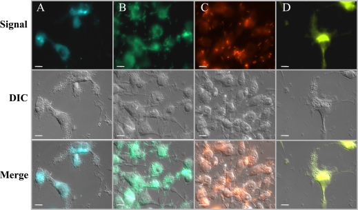
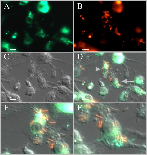
The ability to tag Burkholderia spp. with different colors could facilitate studies where one or more strains expressing different colors could be located or tracked. To demonstrate this, we took advantage of the intracellular replication of B. pseudomallei by using two of the fluorescent strains engineered above (e.g., green and red) to infect the murine macrophage cell line RAW 264.7 in a modified aminoglycoside protection assay (16). Briefly, the respective B. pseudomallei cultures were grown overnight and used to infect the RAW 264.7 cell monolayers at a multiplicity of infection (MOI) of 10:1 for 1 h (4, 26, 28, 30, 33). Afterwards, the extracellular bacteria were removed and the monolayers were washed twice with 1× phosphate-buffered saline (PBS). Fresh Dulbecco's modified Eagle's medium (DMEM) containing 750 μg/ml amikacin and 750 μg/ml kanamycin was then added to inhibit extracellular bacterial replication. At 7 h postinfection, the monolayers were fixed with 1% (wt/vol) paraformaldehyde for 30 min and visualized using fluorescence microscopy (6, 25). As Fig. 3 indicates, B. pseudomallei cells tagged with different colors were easily distinguishable within the murine macrophage monolayers and can be seen inside host cells as the bacteria replicate. When both green and red fluorescent B. pseudomallei cells are mixed together and used to infect a murine macrophage monolayer at a total MOI of 10:1, differently colored bacteria can be distinguished and neighboring bacteria of either color can be differentiated from one another (Fig. 4). To observe the different infectious stages with fluorescently tagged B. pseudomallei, RAW 264.7 macrophages were infected at an MOI of 1:5 (1 bacterium per 5 host cells). In Fig. 5A and B, the host cells were infected with RFP-tagged B. pseudomallei, fixed, permeabilized, and then stained with the far-red lipophilic styryl dye FM 4-64-FX (Molecular Probes). This stains all lipid bilayers far-red, including vacuoles, leaving the slightly orange color of the RFP-tagged B. pseudomallei visible (i) in the macrophage vesicle (Fig. 5A) and (ii) during vesicular escape (Fig. 5B). Alternatively, B. pseudomallei could be labeled with GFP for visualization (Fig. 5C and D). By infecting the macrophages at an MOI of 1:5 with GFP-tagged B. pseudomallei and then staining host cell actin far-red, one can visualize bacterial replication in the cytoplasm (Fig. 5C) and the formation of actin tails during protrusion from the host cell (Fig. 5D).
FIG. 3.
Fluorescence microscopy of B. pseudomallei labeled at the att-Tn7 site with gat-cfp (A), gat-gfp (B), gat-rfp (C), and gat-yfp (D) to infect the murine macrophage-like cell line RAW 264.7. Cell monolayers were seeded overnight onto poly-l-lysine-coated coverslips at the bottom of a 6-well plate and infected with fluorescently tagged B. pseudomallei. Images were obtained as described in the legend of Fig. 2. The total magnification is ×630, and scale bars equal 10 μm.
FIG. 4.
Dual infection of RAW 264.7 macrophages by differentially labeled (green and red) B. pseudomallei bacteria. Infections were carried out identically to those described in the legend of Fig. 3. (A) The green fluorescent signal indicates where gfp-tagged B. pseudomallei bacteria are replicating inside macrophages. (B) The red fluorescent signal was obtained from the same field and shows where rfp-tagged B. pseudomallei bacteria are replicating within macrophages. (C) A DIC image was then captured and is presented. (D) Overlay of images captured sequentially in panels A, B, and C. Images were superimposed at the time of capture using Zeiss AxioVision software. (E and F) Close-ups of the two macrophages indicated by arrows in panel D, where the two differently fluorescing B. pseudomallei strains are clearly visible and distinguishable within the macrophages and even within the same host cell. The total magnification in panels A to D is ×630, and all scale bars equal 10 μm.
FIG. 5.
Tracking of B. pseudomallei infectious stages. Infections were carried out as described in the legend of Fig. 3 except that B. pseudomallei bacteria were used to infect macrophages at an MOI of 1:5 to enable isolated bacterial infection. (A) RAW 264.7 monolayers were infected with RFP-tagged B. pseudomallei. The infection was allowed to progress for 1 h, and then vesicles were stained far-red with the lipophilic styryl dye FM-4-64-FX (Molecular Probes). Phase-contrast microscopy in the red fluorescent channel captured an image of two RFP-tagged B. pseudomallei bacteria in a phagocytic vesicle. The image in panel B was obtained similarly, except that a single RFP-tagged B. pseudomallei bacterium is possibly escaping the far-red-stained phagocytic vesicle. (C) RAW 264.7 macrophages were infected with GFP-tagged B. pseudomallei for 2 h, after which the monolayers were fixed and permeabilized and host cell actin was stained far-red with phalloidin (Invitrogen). GFP-tagged B. pseudomallei can be seen polymerizing host cell actin, enabling observation of actin-based intracellular motility. (D) GFP-tagged B. pseudomallei bacteria were used to infect RAW 264.7 monolayers for 6 h. The bacteria are polymerizing host cell actin to infect neighboring host cells via membrane protrusions. The arrows indicate GFP-tagged B. pseudomallei bacteria at the tips of polymerized actin tails. The total magnification in all panels is ×1,000.
In summary, we have constructed and demonstrated the use of transposon vectors for site-specific stable fluorescent tagging of B. pseudomallei with four unique colors. These tools will be beneficial for microbiological studies involving the tracking or microscopy of B. pseudomallei during cellular infection. There are real needs for these vectors in the field, and several applications can be envisioned. Infection studies that require tracking more than one strain through the infectious process would benefit from these tagging vectors, which do not require plasmid maintenance. Although bioluminescent tools have been of value in in vivo and noninvasive imaging of B. pseudomallei animal infections (22), our fluorescent constructs are of similar value (35, 37). Fluorescence-activated cell sorting could also be used to enumerate host cells infected with a particular strain or strains of fluorescent B. pseudomallei or to monitor gene expression when engineered constructs are used (9, 32). We believe that these constructs will be beneficial to colleagues in this field and can be obtained upon request (Table 1).
Acknowledgments
This work was supported by National Institutes of Health grant R21-AI074608 to T.T.H. A graduate stipend for M.H.N. was provided by an NSF IGERT award (0549514) to B.A.W.
Footnotes
Published ahead of print on 17 September 2010.
REFERENCES
- 1.Biery, M. C., M. Lopata, and N. L. Craig. 2000. A minimal system for Tn7 transposition: the transposon-encoded proteins TnsA and TnsB can execute DNA breakage and joining reactions that generate circularized Tn7 species. J. Mol. Biol. 297:25-37. [DOI] [PubMed] [Google Scholar]
- 2.Brett, P. J., D. DeShazer, and D. E. Woods. 1998. Burkholderia thailandensis sp. nov., description of Burkholderia pseudomallei-like species. Int. J. Syst. Bacteriol. 48:317-320. [DOI] [PubMed] [Google Scholar]
- 3.Bumann, D. 2001. In vivo visualization of bacterial colonization, antigen expression, and specific T-cell induction following oral administration of live recombinant Salmonella enterica serovar Typhimurium. Infect. Immun. 69:4618-4626. [DOI] [PMC free article] [PubMed] [Google Scholar]
- 4.Burtnick, M. N., P. J. Brett, V. Nair, J. M. Warawa, D. E. Woods, and F. C. Gherardini. 2008. Burkholderia pseudomallei type III secretion system mutants exhibit delayed vacuolar escape phenotypes in RAW 264.7 murine macrophages. Infect. Immun. 76:2991-3000. [DOI] [PMC free article] [PubMed] [Google Scholar]
- 5.Castle, L. A., D. L. Siehl, R. Gorton, P. A. Patten, Y. H. Chen, S. Bertain, H. Cho, N. Duck, J. Wong, D. Liu, and M. W. Lassner. 2004. Discovery and directed evolution of a glyphosate tolerance gene. Science 304:1151-1154. [DOI] [PubMed] [Google Scholar]
- 6.Chanchamroen, S., C. Kewcharoenwong, W. Susaengrat, M. Ato, and G. Lertmemongkolchai. 2009. Human polymorphonuclear neutrophil responses to Burkholderia pseudomallei in healthy and diabetic subjects. Infect. Immun. 77:456-463. [DOI] [PMC free article] [PubMed] [Google Scholar]
- 7.Choi, K.-H., J. B. Gaynor, K. G. White, C. Lopez, C. M. Bosio, R. R. Karkhoff-Schweizer, and H. P. Schweizer. 2005. A Tn7-based broad-range bacterial cloning and expression system. Nat. Methods 2:443-448. [DOI] [PubMed] [Google Scholar]
- 8.Choi, K.-H., T. Mima, Y. Casart, D. Rholl, A. Kumar, I. R. Beacham, and H. P. Schweizer. 2008. Genetic tools for select-agent-compliant manipulation of Burkholderia pseudomallei. Appl. Environ. Microbiol. 74:1064-1075. [DOI] [PMC free article] [PubMed] [Google Scholar]
- 9.Cormack, B., R. Valdivia, and S. Falkow. 1996. FACS-optimized mutants of the green fluorescent protein (GFP). Gene 173:33-38. [DOI] [PubMed] [Google Scholar]
- 10.DeShazer, D., P. J. Brett, R. Carlyon, and D. E. Woods. 1997. Mutagenesis of Burkholderia pseudomallei with Tn5-OT182: isolation of motility mutant and molecular characterization of the flagellin structural gene. J. Bacteriol. 179:2116-2125. [DOI] [PMC free article] [PubMed] [Google Scholar]
- 11.Figurski, D. H., and D. R. Helinski. 1979. Replication of an origin-containing derivative of plasmid RK2 dependent on a plasmid function provided in trans. Proc. Natl. Acad. Sci. U. S. A. 76:1648-1652. [DOI] [PMC free article] [PubMed] [Google Scholar]
- 12.Fischer, A. H., K. A. Jacobson, J. Rose, and R. Zeller. 2006. Basic methods in microscopy: preparation of cells and tissues for fluorescence microscopy. Cold Spring Harbor Laboratory Press, Cold Spring Harbor, NY.
- 13.Gustafsson, C., S. Govindarajan, and J. Minshull. 2004. Codon bias and heterologous protein expression. Trends Biotechnol. 22:346-353. [DOI] [PubMed] [Google Scholar]
- 14.Hagemann, A. T., and N. L. Craig. 1993. Tn7 transposition creates a hotspot for homologous recombination at the transposon donor site. Genetics 133:9-16. [DOI] [PMC free article] [PubMed] [Google Scholar]
- 15.Hoang, T. T., R. R. Karkhoff-Schweizer, A. J. Kutchma, and H. P. Schweizer. 1998. A broad-host-range Flp-FRT recombination system for site-specific excision of chromosomally-located DNA sequences: application for isolation of unmarked Pseudomonas aeruginosa mutants. Gene 212:77-86. [DOI] [PubMed] [Google Scholar]
- 16.Jones, A. L., T. J. Beveridge, and D. E. Woods. 1996. Intracellular survival of Burkholderia pseudomallei. J. Bacteriol. 64:782-790. [DOI] [PMC free article] [PubMed] [Google Scholar]
- 17.Kang, Y., M. H. Norris, A. R. Barrett, B. A. Wilcox, and T. T. Hoang. 2009. Engineering of tellurite-resistant genetic tools for single-copy chromosomal analysis of Burkholderia spp. and characterization of the Burkholderia thailandensis betBA operon. Appl. Environ. Microbiol. 75:4015-4027. [DOI] [PMC free article] [PubMed] [Google Scholar]
- 18.Koch, B., L. E. Jensen, and O. Nybroe. 2001. A panel of Tn7-based vectors for insertion of the gfp marker gene or for delivery of cloned DNA into Gram-negative bacteria at a neutral chromosomal site. J. Microbiol. Methods 45:187-195. [DOI] [PubMed] [Google Scholar]
- 19.Kurland, C. G. 1991. Codon bias and gene expression. FEBS Lett. 285:165-169. [DOI] [PubMed] [Google Scholar]
- 20.LoVullo, E. D., C. R. Molins-Schneekloth, H. P. Schweizer, and M. S. Pavelka, Jr. 2009. Single-copy chromosomal integration systems for Francisella tularensis. Microbiology 155:1152-1163. [DOI] [PMC free article] [PubMed] [Google Scholar]
- 21.Norris, M. H., Y. Kang, D. Lu, B. A. Wilcox, and T. T. Hoang. 2009. Glyphosate resistance as a novel select-agent-compliant, non-antibiotic selectable-marker in chromosomal mutagenesis of the essential genes asd and dapB of Burkholderia pseudomallei. Appl. Environ. Microbiol. 75:6062-6075. [DOI] [PMC free article] [PubMed] [Google Scholar]
- 22.Owen, S., M. Batzloff, F. Chehrehasa, A. Meedeniya, Y. Casart, C. A. Logue, R. Hirst, I. R. Peak, A. Mackay-Sim, and I. R. Beacham. 2009. Nasal associated lymphoid tissue and olfactory epithelium as portals of entry for Burkholderia pseudomallei in murine melioidosis. J. Infect. Dis. 199:1761-1770. [DOI] [PubMed] [Google Scholar]
- 23.Parker, A. E., and L. E. Bermudez. 1997. Expression of the green fluorescent protein (GFP) in Mycobacterium aviumas: a tool to study the interaction between mycobacteria and host cells. Microb. Pathog. 22:193-198. [DOI] [PubMed] [Google Scholar]
- 24.Poschet, J. F., J. C. Boucher, L. Tatterson, J. Skidmore, R. W. Van Dyke, and V. Deretic. 2001. Molecular basis for defective glycosylation and Pseudomonas pathogenesis in cystic fibrosis lung. Proc. Natl. Acad. Sci. U. S. A. 98:13972-13977. [DOI] [PMC free article] [PubMed] [Google Scholar]
- 25.Reckseidler, S. L., D. DeShazer, P. A. Sokol, and D. E. Woods. 2001. Detection of bacterial virulence genes by subtractive hybridization: identification of capsular polysaccharide of Burkholderia pseudomallei as a major virulence determinant. Infect. Immun. 69:34-44. [DOI] [PMC free article] [PubMed] [Google Scholar]
- 26.Ribot, W. J., and R. L. Ulrich. 2006. The animal pathogen-like type III secretion system is required for the intracellular survival of Burkholderia mallei within J774.2 macrophages. Infect. Immun. 74:4349-4353. [DOI] [PMC free article] [PubMed] [Google Scholar]
- 27.Schweizer, H. P., and S. J. Peacock. 2008. Antimicrobial drug-selection markers for Burkholderia pseudomallei and B. mallei. Emerg. Infect. Dis. 14:1689-1692. [DOI] [PMC free article] [PubMed] [Google Scholar]
- 28.Stevens, J. M., R. L. Ulrich, L. A. Taylor, M. W. Wood, D. Deshazer, M. P. Stevens, and E. E. Galyov. 2005. Actin-binding proteins from Burkholderia mallei and Burkholderia thailandensis can functionally compensate for the actin-based motility defect of a Burkholderia pseudomallei bimA mutant. J. Bacteriol. 187:7857-7862. [DOI] [PMC free article] [PubMed] [Google Scholar]
- 29.Tan, M.-W., S. Mahajan-Miklos, and F. M. Ausubel. 1999. Killing of Caenorhabditis elegans by Pseudomonas aeruginosa used to model mammalian bacterial pathogenesis. Proc. Natl. Acad. Sci. U. S. A. 96:715-720. [DOI] [PMC free article] [PubMed] [Google Scholar]
- 30.Utaisincharoen, P., S. Arjcharoen, I. Lengwehasatit, K. Limposuwan, and S. Sirisinha. 2005. Burkholderia pseudomallei invasion and activation of epithelial cells requires activation of p38 mitogen-activated protein kinase. Microb. Pathog. 38:107-112. [DOI] [PubMed] [Google Scholar]
- 31.Valdivia, R. H., and S. Falkow. 1998. Flow cytometry and bacterial pathogenesis. Curr. Opin. Microbiol. 1:359-363. [DOI] [PubMed] [Google Scholar]
- 32.Valdivia, R. H., and S. Falkow. 1997. Fluorescence-based isolation of bacterial genes expressed within host cells. Science 277:2007-2011. [DOI] [PubMed] [Google Scholar]
- 33.van Schaik, E. J., M. Tom, and D. E. Woods. 2009. Burkholderia pseudomallei isocitrate lyase is a persistence factor in pulmonary melioidosis: implications for the development of isocitrate lyase inhibitors as novel antimicrobials. Infect. Immun. 77:4275-4283. [DOI] [PMC free article] [PubMed] [Google Scholar]
- 34.Wilson, D. E., and L. C. Chosewood (ed.). 2007. Biosafety in microbiological and biomedical laboratories, 5th ed. Centers for Disease Control and Prevention, Atlanta, GA.
- 35.Yang, M., E. Baranov, A. R. Moossa, S. Penman, and R. M. Hoffman. 2000. Visualizing gene expression by whole-body fluorescence imaging. Proc. Natl. Acad. Sci. U. S. A. 97:12278-12282. [DOI] [PMC free article] [PubMed] [Google Scholar]
- 36.Yu, M., and J. S. H. Tsang. 2006. Use of ribosomal promoters from Burkholderia cenocepacia and Burkholderia cepacia for improved expression of transporter protein in Escherichia coli. Protein Expr. Purif. 49:219-227. [DOI] [PubMed] [Google Scholar]
- 37.Zhao, M., M. Yang, E. Baranov, X. Wang, S. Penman, A. R. Moossa, and R. M. Hoffman. 2001. Spatial-temporal imaging of bacterial infection and antibiotic response in intact animals. Proc. Natl. Acad. Sci. U. S. A. 98:9814-9818. [DOI] [PMC free article] [PubMed] [Google Scholar]


