

Abstract
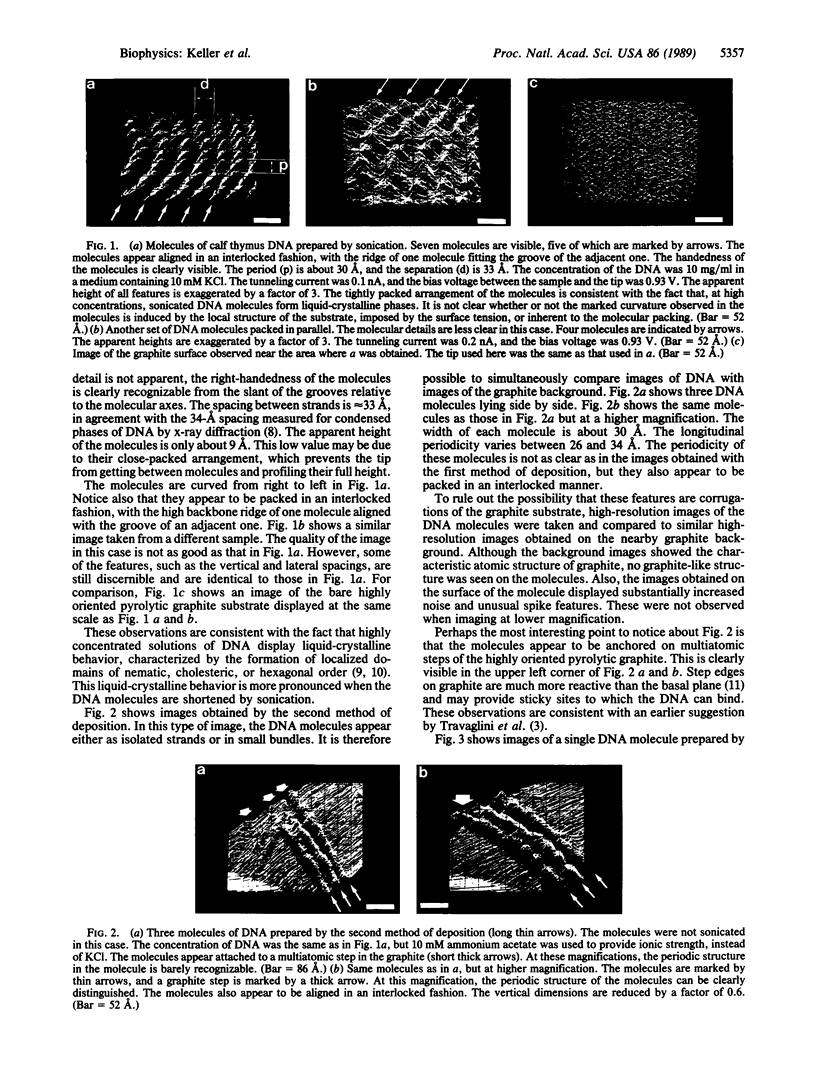
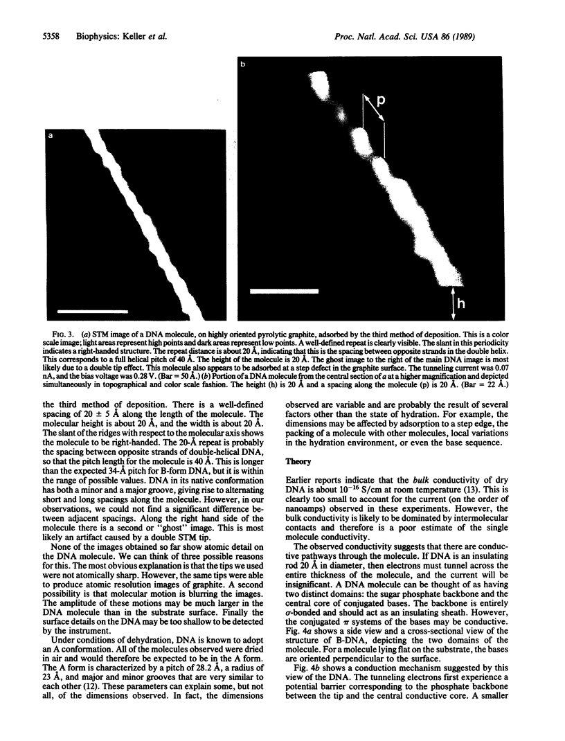
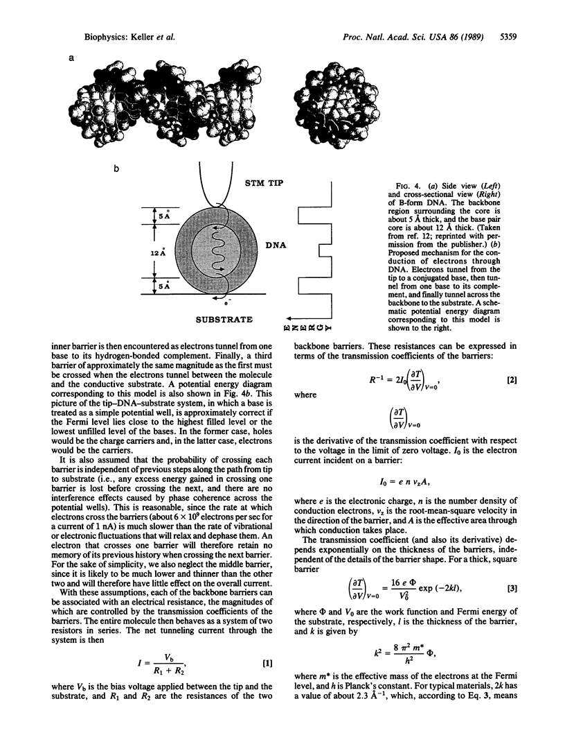
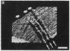
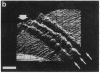
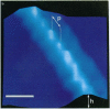
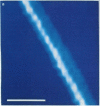
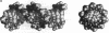
Scanning tunneling microscope images of DNA molecules absorbed onto highly oriented pyrolytic graphite have been obtained. Three methods of deposition and sample preparation have been utilized. In the first method, a highly concentrated solution of DNA is sonicated, and a drop is deposited on freshly cleaved graphite. Under these conditions, the molecules tend to align in a parallel fashion, forming liquid-crystalline phases. In the second method, a solution of DNA is deposited directly on the graphite surface without sonication. In this case, ammonium acetate, a volatile salt, is used to decrease the amount of the residual salt crystals left after drying. In the third method, a solution containing lysed phage particles and DNA is adsorbed onto a graphite surface. The molecules are seen either isolated or in small bundles. The values of height, periodicity, and thickness observed and the handedness of the molecules are consistent with those expected for DNA. In all cases, the molecules were identified by their characteristic periodic structure and because, at higher magnification, no graphite-like structure was detectable on the surface of the molecules. Often the DNA molecules appear to adsorb in areas of the graphite that have many steps and defects. A mechanism that explains the magnitude of the tunneling currents measured in DNA is proposed. This mechanism, in turn, suggests a general method by which large insulating molecules can be rendered conductive.
Full text
PDF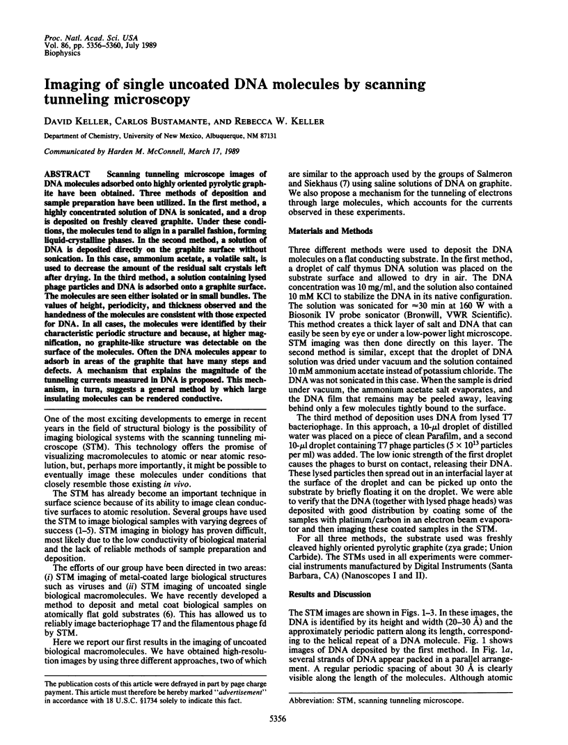
Images in this article
Selected References
These references are in PubMed. This may not be the complete list of references from this article.
- Baró A. M., Miranda R., Alamán J., García N., Binnig G., Rohrer H., Gerber C., Carrascosa J. L. Determination of surface topography of biological specimens at high resolution by scanning tunnelling microscopy. Nature. 1985 May 16;315(6016):253–254. doi: 10.1038/315253a0. [DOI] [PubMed] [Google Scholar]
- Beebe T. P., Jr, Wilson T. E., Ogletree D. F., Katz J. E., Balhorn R., Salmeron M. B., Siekhaus W. J. Direct observation of native DNA structures with the scanning tunneling microscope. Science. 1989 Jan 20;243(4889):370–372. doi: 10.1126/science.2911747. [DOI] [PubMed] [Google Scholar]
- Rill R. L. Liquid crystalline phases in concentrated aqueous solutions of Na+ DNA. Proc Natl Acad Sci U S A. 1986 Jan;83(2):342–346. doi: 10.1073/pnas.83.2.342. [DOI] [PMC free article] [PubMed] [Google Scholar]
- Zasadzinski J. A., Schneir J., Gurley J., Elings V., Hansma P. K. Scanning tunneling microscopy of freeze-fracture replicas of biomembranes. Science. 1988 Feb 26;239(4843):1013–1015. doi: 10.1126/science.3344420. [DOI] [PubMed] [Google Scholar]