Abstract
Highly pathogenic avian influenza (HPAI) is a striking disease in susceptible poultry, which leads to severe economic losses. Inactivated vaccines are the most widely used vaccines in avian influenza virus (AIV) vaccination programs. However, these vaccines interfere with the serological detection of wild-type AIV infections in immunized populations. The use of vaccines that allow differentiation between infected and vaccinated animals (DIVA strategy) would stop current stamping-out policies. Therefore, novel vaccination strategies are needed to allow improved protection of animals and humans against HPAI virus (HPAIV) infection. The presented study analyzed for the first time the immunogenic capacity of plant-expressed full-length hemagglutinin (rHA0) of HPAIV H5N1 in several vaccine formulations within the highly relevant host species chicken. We were able to express plant-expressed rHA0 at high levels and could show that, when administered with potent adjuvants, it is highly immunogenic and can fully protect chicken against lethal challenge infection. Real-time reverse transcription (RT)-PCR and serological tests demonstrated only marginally increased virus replication in animals vaccinated with plant-derived rHA0 compared to animals immunized with an inactivated reference vaccine. In addition, the use of plant-expressed rHA0 also allowed an easy serological differentiation of vaccinated from AIV-infected animals based on antibodies against the influenza virus NP protein.
Highly pathogenic avian influenza (AI) (HPAI) is a striking disease in susceptible poultry, which leads to severe economic losses (21). Since 2003, the H5N1 HPAI epidemic has claimed over 220 million poultry and other birds either through direct mortality from infection or from preemptive culling (22). The implementation of vaccination of poultry as a tool for the reduction of the viral load in the environment and, thus, for decreasing the risk of transmission within poultry—and, as a consequence, to humans—is still a discussed topic. Inactivated vaccines are the most widely used vaccines in AI vaccination programs. They are particularly addressed to protect adult chickens, turkeys, and other birds in emergency situations, e.g., when ring vaccination is used in an area of an HPAI virus (HPAIV) outbreak or when prophylactic vaccination is used in a region where H5 or H7 AI virus (AIV) infections are endemic. However, these vaccines limited the serological detection of wild-type AIV infections in immunized populations, as wild-type infection could be detectable only through higher antibody titers to nonstructural proteins or if the neuraminidase subtype of the vaccine differed from the subtype of the introduced wild-type virus (28).
The use of vaccines that fit in any case to the strategy of differentiating infected from vaccinated animals (DIVA) would make a strong case for turning away from current stamping-out policies in many countries. Vaccines that consist of only one major antigenic protein related to influenza A virus (e.g., hemagglutinin) would allow the identification of naturally infected herds by detecting seroconversion against further immunogenic proteins (e.g., nucleoprotein) and would therefore fit into the DIVA strategy (28). Furthermore, data generated for HPAIV could be used as a model for novel human vaccines, e.g., to protect against pandemic H1N1/2009 virus.
Transgenic plants have become attractive systems for the production of human and animal biopharmaceutical recombinant proteins. Plant-expressed proteins from infectious bursal disease virus and avian reovirus were previously tested successfully in chicken (29, 30, 31). The expression of influenza A virus antigen using plants was demonstrated recently, and protective efficacy has been investigated with mouse and ferret models (7, 19, 20).
Nevertheless, the presented study analyzed for the first time the immunogenic capacity of plant-expressed full-length recombinant hemagglutinin (rHA0) of HPAIV H5N1 in different vaccine formulations within the highly relevant host species chicken. Therefore, vaccine preparations containing different amounts of antigen and different adjuvants (Freund adjuvant, copolymer, and a cationic lipid-DNA complex) were compared.
MATERIALS AND METHODS
Design of constructs used for the expression of rHA0.
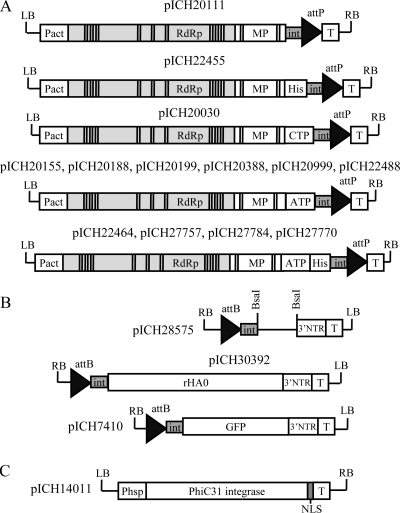
Viral vector modules (Fig. 1A) used in this study for the expression of full-length hemagglutinin (rHA0) are based on modules described previously (15). The T-DNA region of pICH20111, the 5′ provector for the cytosolic expression of the gene of interest, contains the Arabidopsis thaliana actin 2 (ACT2) gene promoter operably linked to the coding sequences of the RNA-dependent RNA polymerase (RdRp) and the movement protein (MP) genes of crucifer tobacco mosaic virus (crTMV) (8), followed by the CP subgenomic promoter (overlapping with the MP C-terminal coding sequences). The 5′ fragment of the intron sequence was derived from the third intron of the petunia (Petunia hybrida) Psk7 gene. The recombination site attP is recognized by the site-specific integrase phC31 and is followed by a nopaline synthase (nos) transcription termination sequence. The region coding for viral RNA was modified by inserting 15 plant-derived introns that improve intact viral RNA release into the cytoplasm (16). The entire fragment was cloned between the T-DNA borders of pICBV10, a pBIN19-derived binary vector. The 5′ provector construct for the cytosolic expression of His-tagged proteins, pICH22455, was identical to pICH20111 except for the presence of a His6 tag and enterokinase cleavage site coding sequences inserted between the MP gene and the 5′ fragment of the intron sequence. In the case of pICH20030, the DNA fragment contains the coding sequence for the synthetic (consensus) transit peptide of the small-subunit Rubisco gene for the transformation of dicotyledonous plants. Similarly, a set of 5′ provectors with different signal peptides (SPs) for apoplast targeting was designed. Vectors pICH20155 and pICH22488 contained the signal peptide of rice alpha-amylase (GenBank accession no. P27932), either with native codon usage or codon optimized for Nicotiana benthamiana, respectively; vector pICH20188 contained the signal peptide of Nicotiana plumbaginifolia calreticulin (GenBank accession no. Z71395.1); vector pICH20199 contained the signal peptide of the Phaseolus vulgaris endopolygalacturonase-inhibiting protein (PGIP) (GenBank accession no. P58823); vector pICH20388 contained the signal peptide of apple pectinase (GenBank accession no. P48978); and vector pICH2099 contained the signal peptide of barley alpha-amylase (GenBank accession no. CAX51374). Plasmids pICH22464, pICH27757, pICH27784, and pICH27770 contained the apoplast-targeting signal peptides of Nicotiana plumbaginifolia calreticulin, rice amylase, barley amylase, and apple pectinase, respectively, followed by the His6 tag and the enterokinase cleavage site.
FIG. 1.
Plasmid constructs. (A) TMV-based 5′ modules, including pICH20111 and pICH22455, for the cytosolic localization of proteins of interest, without and with a His6 tag and an enterokinase cleavage site, respectively; pICH20030, for chloroplast targeting, containing the consensus sequence of the transit peptide (CTP) for the small subunit of Rubisco (dicotyledonous plants); pICH20155 and pICH22488, for apoplast targeting, containing native and codon-optimized signal peptides (apoplast-targeting signal peptide [ATP]) from rice alpha-amylase, respectively; pICH20188, for apoplast targeting, containing a signal peptide from Nicotiana plumbaginifolia calreticulin; pICH20199, for apoplast targeting, containing a signal peptide of the endopolygalacturonase-inhibiting protein (PGIP) of Phaseolus vulgaris; and pICH20388 and pICH20999, for apoplast targeting, containing signal peptides of apple pectinase and barley alpha-amylase, respectively. Plasmids pICH22464, pICH27757, pICH27784, and pICH27770 contain signal peptides from Nicotiana plumbaginifolia calreticulin, rice alpha-amylase, barley alpha-amylase, and apple pectinase, respectively, followed by a His6 tag and an enterokinase cleavage site. (B) TMV-based 3′ provector modules, including cloning vector pICH28575 and 3′ modules for the expression of full-length hemagglutinin (rHA0) from NIBRG-14 influenza virus (pICH30392) and GFP (pICH7410). (C) Integrase module pICH14011 providing the site-specific integrase of phage C31 (PhiC31). LB and RB, left and right T-DNA borders, respectively; Pact, Arabidopsis actin 2 promoter; Phsp, promoter of the Arabidopsis gene encoding heat shock protein Hsp81.1; T, nos terminator; RdRp, RNA-dependent RNA polymerase; MP, movement protein; int, intron; AttP and AttB, recombination sites; his, His6 tag; BsaI, cloning sites recognized by class IIs restriction enzyme BsaI; 3′NTR, 3′ untranslated region of TMV; ATP, apoplast-targeting signal peptide; CTP, chloroplast-targeting transit peptide; NLS, nuclear localization signal.
The coding sequence for recombinant hemagglutinin was obtained by reverse transcription-PCR (RT-PCR) using purified viral RNA from the virus strain NIBRG-14 (catalog no. 05/204; National Institute for Biological Standards and Control [NIBSC], London, United Kingdom) that is a reverse genetic-derived 2:6 reassortant between A/Vietnam/1194/04 (H5N1) and A/Puerto Rico (PR)/8/23 viruses. Single-stranded cDNA was synthesized by using the SuperScript III first-strand synthesis system for RT-PCR (Invitrogen, Karlsruhe, Germany) and gene-specific primer nibf2 (5′-TTTGGTCTCAAGGTGATCAGATTTGCATTGGTTACCATGC-3′). The PCR amplification of the full-length hemagglutinin (rHA0) coding sequence lacking the native N-terminal signal peptide and flanked with BsaI restriction sites was performed by using primers nibf2 and nibr1 (5′-TTTGGTCTCAAAGCTTAAATGCAAATTCTGCATTGTAACG ACCCATTG-3′). The amplified gene fragment was cloned into the BsaI sites of the 3′ provector pICH28575, resulting in vector pICH30392 containing the full-length hemagglutinin gene lacking the signal peptide (Fig. 1B). Plasmids were transferred into Agrobacterium tumefaciens GV3101 competent cells by electroporation with a MicroPulser electroporation apparatus (Bio-Rad Laboratories, München, Germany) according to the manufacturer's instructions.
The design of a green fluorescent protein (GFP) open reading frame containing 3′ provector pICH7410 (Fig. 1b), used as an expression control, was described previously (15). Vector pICH14011 (Fig. 1C) was designed as described previously (11), providing the site-specific integrase phC31.
Agroinfiltration procedure.
Equal volumes of Agrobacterium cultures grown overnight (optical density at 600 nm [OD600] of 1.5 to 1.8) containing the desired provectors and integrase source were mixed and diluted with infiltration solution (10 mM MES [morpholineethanesulfonic acid] [pH 5.5], 10 mM MgSO4) to an OD600 of 0.05. For agroinfiltration, 6- to 7-week-old greenhouse-grown N. benthamiana plants were used. Infiltration of individual leaf sectors was performed by using a syringe without a needle. Alternatively, for inoculation of the whole plant, vacuum infiltration was performed as described previously (16).
SDS-PAGE and Western blotting.
Samples (100 mg) of N. benthamiana leaf tissue infiltrated with Agrobacterium were ground in liquid nitrogen and extracted with 10 min of heating at 95°C in 600 μl of Laemmli loading buffer (0.15 M Tris-HCl [pH 6.8], 2.3% SDS, 5% beta-mercaptoethanol, 10% glycerol, 20 μM bromphenol blue). The total soluble protein extracts were resolved on 12% polyacrylamide gels under reducing conditions by using the buffer system described previously by Laemmli (13), followed by Coomassie brilliant blue G-250 staining with PageBlue protein staining solution (Fermentas, Vilnius, Lithuania). For Western blot analysis, proteins from SDS-PAGE were electrophoretically transferred onto a Hybond P membrane (GE Healthcare, Munich, Germany) by using blotting buffer (25 mM Tris-HCl, 192 mM glycine, 20% [vol/vol] methanol [pH 8.3]). Membranes were blocked with a 5% (wt/vol) solution of skim-milk powder in Tris-buffered saline (TBS) (50 mM Tris-HCl, 100 mM NaCl [pH 7.4]) containing 0.05% Tween 20 and probed with hemagglutinin (H5N1)-specific polyclonal antibodies (eEnzyme LLC) diluted 1:1,000 in a 2.5% (wt/vol) solution of skim-milk powder in TBS, followed by incubation with secondary horseradish peroxidase (HRP)-conjugated rabbit IgG-specific goat antibodies (Sigma, Dorset, United Kingdom) diluted 1:6,000 in TBS. Bound antibodies were detected by using the ECL detection reagent (GE Healthcare).
Preparative purification of rHA0.
The purification of rHA0 was performed by using the process of selective extraction followed by two-phase separation and gel filtration. Nicotiana benthamiana leaf tissue (300 g) ground in liquid nitrogen was mixed with 4 volumes (wt/vol) of 20 mM Tris-HCl (pH 8.0) for removing water-soluble proteins. The plant tissue containing cell membrane-bound proteins was pelleted for 20 min (12,000 × g at 4°C), the supernatant was discarded, and the treatment was repeated three more times. After a final treatment, the pellet was resuspended in 5 volumes (wt/vol) of 20 mM Tris-HCl (pH 8.0)-1% Triton X-100 for the extraction of membrane-bound proteins. After centrifugation for 30 min at 12,000 × g at 4°C, the supernatant was collected and filtered through a Miracloth (Calbiochem, La Jolla, CA). The supernatant was dark green due to the presence of chlorophyll. As a next step of purification, the aqueous-micellar two-phase system (AMTPS) separation protocol (9) was adapted; for this purpose, an aliquot of solid ammonium sulfate was dissolved in the supernatant at 16°C for providing a final concentration of 1.6 M, and the solution was centrifuged at 5,000 × g for 15 min at 20°C in a fixed-angle rotor. The dark-green detergent-rich upper phase was discarded. The remaining lower phase was filtered through a paper filter (catalog no. 10311644; Whatman, Dassel, Germany), followed by filtration through 0.22-μm polyethersulfone (PES) filter units (TPP Techno Plastic Products, Trasadingen, Switzerland). Filtered extract fluid was clear with a weak greenish hue. Both filtration steps were performed at room temperature. Immediately after filtration, the cleared extract was placed on ice and concentrated by using a Vivaflow 100 module (Sartorius, Göttingen, Germany) with a 100-kDa membrane cutoff until the volume of the sample achieved 30 to 40 ml. The sample was further concentrated to approximately 5 to 15 ml by using a Vivaspin 20 (Vivaproducts, Littleton, CO) centrifugal device with a 100-kDa membrane cutoff. The concentrated sample was further clarified by centrifugation (10,000 rpm for 5 min at 4°C). The green pellet was discarded, and the supernatant was further purified by gel filtration on a Superdex 200 column using the Äkta purifier system (GE Healthcare, Pittsburgh, PA) with an XK 26/70 column (GE Healthcare, Pittsburgh, PA) and 20 mM phosphate-buffered saline (PBS) (pH 7.4) as a running buffer. Peak fractions were analyzed on 12% SDS-PAGE gels, and fractions containing rHA0 were pooled.
Optionally, pooled fractions were additionally purified by filtration through a Sartobind Q membrane adsorber (Sartorius, Göttingen, Germany) equilibrated with 20 mM phosphate buffer (pH 7.4) containing 0.3 M NaCl.
The final sample was concentrated by using a Vivaspin centrifugal device with a 100-kDa membrane cutoff. The recovered concentrated sample was sterilized by filtration through a 20-μm membrane filter.
The concentration of purified rHA0 was estimated with the bicinchoninic acid (BCA) protein assay kit (Pierce, Rockford, IL). In parallel, visual estimation as well as densitometric analysis of purified rHA0 samples resolved in polyacrylamide gels were performed by using a Bioanalyzer 1200 series instrument (Agilent Technologies, Böblingen, Germany). In all cases, bovine serum albumin (Sigma, Dorset, United Kingdom) was applied as a standard. Usually, the concentration of purified rHA0 was 0.5 to 1.2 mg/ml. The samples were stored at 4°C until use in animal experiments.
Hemagglutination assay.
Serial 2-fold dilutions in 20 mM PBS (pH 7.4) of duplicate samples of purified rHA0, inactivated NIBRG-14 virus (NIBSC, Potters Bar, United Kingdom), and bovine serum albumin (Sigma, Dorset, United Kingdom), used as positive and negative controls, respectively, were prepared in microtiter plates with a U-shaped bottom. The samples were mixed with 5-μl aliquots of 25% chicken blood suspension (Dr. Merk & Kollegen, Ochsenhausen, Germany) and incubated for 1 h at room temperature. The lowest hemagglutinin concentrations (μg/ml) that resulted in the agglutination of erythrocytes were defined as hemagglutination titers (HT).
SRID.
A single-radial immunodiffusion (SRID) assay was performed according to a method described previously by Wood et al. (27), using influenza virus A/Vietnam/1194/04 (H5N1) antiserum (code 04/214; NIBSC, United Kingdom). Influenza virus A/Vietnam/119/04 (H5N1) antigen (NIBRG-14 code 05/204; NIBSC, United Kingdom) was applied as a standard antigen.
Glycoanalysis of rHA0 using matrix-assisted laser desorption ionization-time of flight (MALDI-TOF) mass spectrometry (MS).
Peptides yielding from tryptic digestion of rHA0 were analyzed by using peptide mass fingerprinting (PMF). To explore the sugar composition of glycosyl residues, selected peptides were processed for further peptide fragmentation in positive LIFT mode, and the acquired spectrum was analyzed with the help of flexAnalysis (version 3.0) software. All analyses were done by using an Autoflex III Smartbeam mass spectrometer (Bruker Daltonics, Inc., Billerica, MA). Purified rHA0 was treated with PNGase F (New England Biolabs, Inc., Beverly, MA) according to the manufacturer's instructions. Untreated and treated samples were analyzed by SDS-PAGE.
Animals.
Sixty-four specific-pathogen-free (SPF) White Leghorn chickens (Lohmann Tierzucht GmbH, Cuxhaven, Germany) were used for the animal trial. All animals were housed in group cages within the high-containment facility of the Friedrich Loeffler Institut, with a 12-h light regimen. Feed and water were provided ad libitum. All animal experiments were reviewed and approved by the responsible state ethics committee (approval no. LALLF M-V/TSD/7221.3-1.1.-052/09).
Immunization.
The compositions of the rHA0-adjuvant combinations as well as the control vaccines are summarized in Table 1. As adjuvants, Freund adjuvant (Sigma, Munich, Germany), Polygen (MVP Laboratories, Omaha, NE) as a copolymer, and BAY98-7089 (Juvaris BioTherapeutics, Inc.) as a cationic lipid-DNA complex were used (Table 1).
TABLE 1.
Composition of the tested vaccine formulations
| Formulation | Content | Vol (ml) (application method)a |
|---|---|---|
| HA50FA | 50μg rHA0 + Freund adjuvant (1/1, vol/vol) | 0.15 (s.c.) |
| HA100Poly | 100μg rHA0 + Polygen adjuvant (12%, vol/vol) | 0.15 (i.m.) |
| HA50Bay | 50 μg rHA0 + 5 μg BAY98-7089 adjuvant | 0.15 (i.m.) |
| HA100Bay | 100 μg rHA0 + 5 μg BAY98-7089 adjuvant | 0.15 (i.m.) |
| Bay98 | 20 μg BAY98-7089 | 0.1 (i.m.) |
| Refvac | Inactivated, complete H5N2 virus | 0.5 (i.m.) |
| negCo | Negative control |
s.c., subcutaneously; i.m., intramuscularly.
At 3 weeks of age, the chickens were assigned randomly into seven groups of 10 animals (HA100Poly and HA50BAY), 9 animals (HA50FA and HA100BAY), 5 animals (Refvac and BAY98), and 16 animals (negCo), respectively (abbreviations are explained in Table 1). The birds were immunized intramuscularly (with the exception of HA50FA, which was given subcutaneously) with 0.15 ml of either HA100Poly, HA50BAY, HA50FA, or HA100BAY; 0.1 ml of BAY98; or 0.5 ml of Refvac (summarized in Table 1). Three weeks after the first immunization, all vaccinated animals were boostered using the same rHA0-adjuvant combinations, the Refvac preparation as a positive control, or BAY98-7089 as a mock-vaccinated negative control.
Virus challenge.
Six weeks after the first immunization, all chickens were challenged by oculo-oro-nasal application of 106 tissue culture infectious doses (TCID50) of HPAIV A/whooper swan/Germany/R65/2006 (H5N1) (26). Twenty-four hours after challenge infection, two naïve chickens were added to every group, serving as the “transmission-by-contact” controls. During the following 10 days, the chickens were checked daily for clinical symptoms and classified as healthy (score of 0), ill (score of 1), severely ill (score of 2), or dead (score of 3). The average clinical scores for each group were calculated for each day. Oropharyngeal and cloacal swab samples were taken daily, and their content of AIV RNA was quantified by real-time reverse transcription (RT)-PCR (rRT-PCR). The amplification of a nucleotide fragment specific for the H5 gene was performed as described previously (12), and the genomic loads were semiquantified. For extrapolations of the threshold cycle (CT) values of rRT-PCR scores to infectious units, serial dilutions of HPAIV H5N1 in negative swab samples were calculated as the 50% tissue culture infectious dose/ml on Madin-Darby canine kidney (MDCK) cells (collection of cell lines in veterinary medicine from the Friedrich-Loeffler-Institut, Südufer Insel Riems, Germany [RIE1061]). In parallel, the corresponding virus doses were analyzed by rRT-PCR. The mean TCID50 of HPAIV H5N1 from two independent experiments was plotted against the mean CT values of viral dilutions of two independent experiments. The resulting calibration curve was highly correlated (r2 > 0.99) up to a threshold cycle value of 35 (data not shown) and was subsequently used to convert CT values to mean tissue infectious doses (TCID50).
Serological analysis.
Preexperimental sera and sera from 3 weeks after the first immunization, 6 weeks after the first immunization, and 10 days after the challenge infection were collected from all birds. The serum samples were heat inactivated at 56°C for 30 min and examined for the presence of antibodies against the nucleoprotein of avian influenza virus type A (ID Screen influenza A virus antibody competition ELISA kit; ID-vet, Montpellier, France), antibodies against the H5 protein (ID Screen influenza virus H5 antibody competition ELISA kit; ID-vet, Montpellier, France), and, finally, antibodies against the N1 protein (ID Screen influenza virus N1 antibody competition ELISA kit; ID-vet, Montpellier, France). For the neutralization test, the diluted serum samples were mixed with an equal volume of media containing HPAIV H5N1 at a concentration of 100 TCID50/well, and after 1 h incubation at 37°C in a 5% CO2 humidified atmosphere, 100 μl of MDCK cells at 1.5 × 10⁵/ml were added to each well; viral replication was assessed after 3 days of incubation.
RESULTS
Production, purification, and characterization of rHA0.
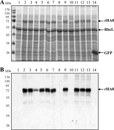
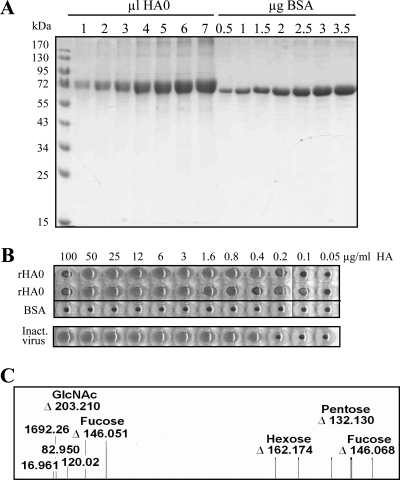
In order to optimize hemagglutinin expression levels in Nicotiana benthamiana, we have applied the magnICON (Icon Genetics, Halle, Germany) provector system (15). The expression of a full-length hemagglutinin coding sequence cloned in the 3′ module (pICH30392) (Fig. 1B) was tested in combination with different 5′ modules (Fig. 1A), providing cytosolic, chloroplastic, and apoplastic compartmentalization of rHA0. The results of the experiment are presented in Fig. 2. The Coomassie-stained gel (Fig. 2A) clearly showed that the highest expression level was obtained for apoplast-targeted rHA0, specifically for the combination with the 5′ module coding for the signal peptide of tobacco calreticulin. Expression levels were as high as 0.3 g/kg of fresh-leaf biomass. This result was confirmed by Western blot analysis (Fig. 2B). No signal was detected (Fig. 2B, lanes 1, 8, and 10) for cytosolic or chloroplast-targeted rHA0, possibly due to some of the following reasons: low expression levels, incorrect folding, and/or differences in posttranslational modifications (e.g., the absence of glycosylation for cytosolic and chloroplast-targeted rHA0). The predicted molecular mass (aglycosylated form) of rHA0 is 62.14 kDa, roughly 10 kDa less than that determined by our PAGE characterization (Fig. 2 and 3A). Taking into account that rHA0 contains eight N-glycosylation sites predicted by the NetNGlyc 1.0 program (http://www.cbs.dtu.dk/services/NetNGlyc/), the shift in molecular mass is very likely caused by a glycosylation of the recombinant hemagglutinin. Indeed, this conclusion is supported by the expression of the hemagglutinin HA1-HA2 fragment (without a transmembrane domain) with cytosolic, chloroplastic, and apoplastic compartmentalization. Weak signals of an expected molecular mass of 58 kDa (aglycosylated form) were obtained on Western blots for chloroplast-targeted and cytosolic locations, in contrast to a strong signal of ca. 70 kDa obtained for apoplast-targeted HA1-HA2 (data not shown). The estimated molecular mass of treated (deglycosylated) hemagglutinin (ca. 58 kDa, without a transmembrane domain) corresponded to the calculated one for the aglycosylated form of rHA0. For providing direct evidence of rHA0 glycosylation, MALDI-TOF MS analysis of rHA0-derived peptides was performed. The data for an arbitrary glycopeptide are shown in Fig. 3C. From the data, it followed that the fragment contained N-acetylglucosamine (GlcNAc) attached to an asparagine residue. Also, fucose is attached to GlcNAc. Very likely, α(1→3)fucose is involved, which is typical for plant glycoproteins. Indeed, this assumption was confirmed by the glycan insensitivity (in contrast to reference glycoproteins of animal origin) to cleavage with PNGase F (data not shown), as this enzyme did not release glycans that were α1,3-fucosylated at the asparagine-linked N-acetylglucosamine (1). Furthermore, a pentose was also a component of the glycosyl residue. Presumably, this pentose was xylose, which is also typical for plants. In addition, our recently reported data (2) demonstrate that apoplast-targeted recombinant proteins (immunoglobulins) produced in N. benthamiana with the help of the magnICON expression system can be glycosylated, and glycosylation is the main reason for the shift in the molecular mass (heterogeneity) of all Igs studied.
FIG. 2.
Optimization of rHA0 expression in Nicotiana benthamiana leaves. (A) Coomassie-stained SDS-PAGE gel. (B) Western blot probed with hemagglutinin-specific polyclonal antibodies. rHA0 expression levels in leaves were tested 5 days after inoculation with provector modules. Lanes 1 to 13 show 3′ module pICH30392, providing for rHA0 used in combination with different 5′ modules. The 3′ and 5′ modules were coinfiltrated with pICH14011, serving as the integrase source. Lane 1, 5′ module pICH20111, with a cytosolic location; lane 2, 5′ module pICH20155 with rice alpha-amylase SP; lane 3, 5′ module pICH20188 with Nicotiana plumbaginifolia calreticulin SP; lane 4, 5′ module pICH20199 with Phaseolus vulgaris PGIP SP; lane 5, 5′ module pICH20388 with apple pectinase SP; lane 6, 5′ module pICH20999 with barley alpha-amylase SP; lane 7, 5′ module pICH22488 with codon-optimized rice alpha-amylase SP; lane 8, 5′ module pICH20030 with chloroplast transit peptide; lane 9, 5′ module pICH22464 with N. plumbaginifolia calreticulin SP and a His6 tag; lane 10, 5′ module pICH22455 with an N-terminal His6 tag; lane 11, 5′ module pICH27757 with rice alpha-amylase SP and a His6 tag; lane 12, 5′ module pICH27784 with barley alpha-amylase SP and a His6 tag; lane 13, 5′ module pICH27770 with apple pectinase SP and a His tag; lane 14, 3′ module pICH7410 providing GFP in combination with 5′ module pICH20111. rHA0, recombinant full-length hemagglutinin from influenza virus A/NIBRG-14/2004 (H5N1); RbcL, large subunit of ribulose-1,5-bisphosphate carboxylase/oxygenase.
FIG. 3.
Analysis of purified rHA0 for estimations of concentration and purity. (A) SDS-PAGE. Aliquots of rHA0 (1, 2, 3, 4, 5, 6, and 7 μl) as well as aliquots of bovine serum albumin (0.5, 1.0, 1.5, 2.0, 2.5, 3.0, and 3.5 μg) were resolved on a 12% polyacrylamide gel and stained with Coomassie. (B) Hemagglutination assay of purified rHA0. Twofold serial dilutions of rHA0 starting with a concentration of 400 μg/ml were tested for hemagglutination activity. Twofold dilutions of inactivated NIBRG-14 virus (code 05/204; NIBSC, United Kingdom) starting with a concentration of 28 μg/ml were used as a positive control. Twofold dilutions of bovine serum albumin starting with a concentration of 400 μg/ml were used as a negative control. All sample dilutions were performed in duplicate. (C) Selected part of a processed LID-LIFT spectrum of a 3,385.570-Da glycopeptide (amino acid residues 480 to 493) from tryptic digestion of rHA0. The peptide part of this glycopeptide is 1,692.26 Da.
For animal studies, the production of rHA0 was performed by using the expression of rHA0 with the calreticulin signal peptide (pICH30392 and pICH20188). The rHA0 purification from green plant tissue required the development of a new protocol, as the adaptation of the purification method used for rHA0 recovery from animal cell cultures (25) was not possible due to the interference of chlorophyll with the ion-exchange chromatography step. Also, we preferred not to use different affinity tags such as the His tag (8) or the Strep tag (5), as the presence of irrelevant sequences in the antigen structure might be the source of undesired immune responses and might cause difficulties in vaccine approval. The new protocol used in this study included a step of enrichment in membrane-bound proteins, aqueous-micellar two-phase systems (AMTPS) for the removal of chlorophyll, and gel filtration chromatography. The yield was 20 to 30 mg of purified rHA0 per kilogram of fresh-leaf biomass; thus, further improvement in downstream efficiency still needs to be addressed. The quality of purified rHA0 was tested by comparison with the HA of inactivated virus using a hemagglutination assay (Fig. 3B) as well as a single-radial immunodiffusion assay (27; data not shown). The hemagglutination titer of plant-made rHA0 (0.1 to 0.2 μg/ml) was close to the HT value for inactivated virus (0.1 μg/ml), arguing in favor of an appropriate conformation of the plant-made recombinant protein. These data correlated with the hemagglutination titer of recombinant HA0 produced in a baculoviral system (25).
Immunogenicity and protective efficacy of plant-expressed rHA0 in chickens.
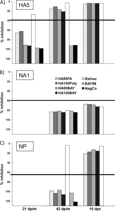
None of the chickens showed any signs of illness or adverse effects after vaccination. All prevaccination sera, as well as the sera of the control animals, were negative for AIV-specific antibodies until challenge infection (Table 2 and Fig. 4). Six weeks after the first immunization, all individuals (100%) in the HA50FA, HA100Poly, and HA50BAY groups and 78% of the HA100BAY group scored positive in the H5 ELISA used (Table 2 and Fig. 4). As expected, all animals vaccinated with the plant-expressed rHA0 showed negative results in an ELISA to detect antibodies to the NP protein, whereas chickens vaccinated with the reference vaccine (inactivated complete virions) scored positive (Table 2 and Fig. 4). Quantification of neutralizing antibodies reacting against the challenge virus was performed by a virus neutralization test (VNT). The mean 100% neutralizing dose (ND100) measured from the plant-expressed rHA0-vaccinated groups revealed relatively similar titers of about 1:32, while sera from birds vaccinated with the reference vaccine exhibited neutralizing titers of almost 1:1,024 (Table 2).
TABLE 2.
Mortality, transmission, and serology test
| Group | % survival | No. of naïve chickens infected/total no. of naïve chickensa | No. of positive chickens/total no. of chicken tested |
VNT value (log2 ND100)b |
|||||||
|---|---|---|---|---|---|---|---|---|---|---|---|
| NP ELISA |
HA5 ELISA |
NA1 ELISA |
|||||||||
| 42 days postimmunization | 10 days postchallenge | 21 days postimmunization | 42 days postimmunization | 10 days postchallenge | 42 days postimmunization | 10 days postchallenge | 42 days postimmunization | 10 days postchallenge | |||
| HA50FA | 89 | 2/2 | 0/9 | 7/8 | 5/9 | 9/9 | 8/8 | 0/9 | 1/8 | 4.8 | 10.1 |
| HA100Poly | 100 | 1/2 | 0/10 | 9/10 | 4/10 | 10/10 | 10/10 | 0/10 | 1/10 | 5.5 | 10.9 |
| HA50BAY | 90 | 2/2 | 0/10 | 9/9 | 0/10 | 10/10 | 9/9 | 0/10 | 2/9 | 5.6 | 10.7 |
| HA100BAY | 89 | 1/2 | 0/9 | 8/8 | 0/9 | 7/9 | 8/8 | 0/9 | 0/8 | 5.0 | 10.2 |
| Refvac | 100 | 0/2 | 5/5 | 5/5 | 5/5 | 5/5 | 5/5 | 0/5 | 0/5 | 9.9 | 12.1 |
| BAY98 | 0 | 2/2 | 0/5 | 0/5 | 0/5 | 0/5 | 2.2 | ||||
| negCo | 0/4 | 0/4 | 0/4 | 0/4 | 2.3 | ||||||
Two naïve chicken were integrated into each group 24 h after challenge infection.
Mean value per group (log2 ND100) against challenge virus.
FIG. 4.
DIVA serology with marker ELISAs. Sera taken from animals 3 weeks (21 days postimmunization) and 6 weeks (42 days postimmunization) after immunization and 10 days after challenge infection (10 days postchallenge) were tested with commercially available ELISAs specific for HA5 (A), NA1 (B), and NP (C), respectively. Average values for each group (“herd level”) are given as percent inhibition and are represented as a bar. Values above 40% inhibition (HA5 ELISA), 60% inhibition (N1 ELISA), or 65% inhibition (NP ELISA) scored negative. These threshold values are indicated by a horizontal line.
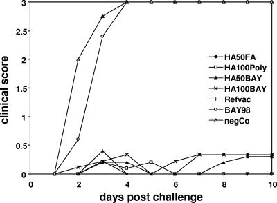
Challenge infection was performed 3 weeks after the second immunization (booster vaccination) with HPAIV A/whooper swan/Germany/R65/2006 (H5N1). A high dose (106 TCID50 per animal) of challenge virus was used, which reproducibly induced 100% mortality of naïve chickens within 4 days postinoculation. Thus, as expected, all naïve control animals succumbed to disease within this period (Fig. 5 and Table 2). Animals mock vaccinated with only the adjuvant (BAY98 group) also died within 4 days (Fig. 5 and Table 2). Clinical scoring for the rHA0-vaccinated birds revealed mean values of less than 0.5 scoring points. Nevertheless, mortality was seen in every group besides the HA100Poly and Refvac groups (Table 2). In addition, two naïve chickens placed into every group 24 h after challenge infection survived only in the Refvac group, whereas in the HA100Poly and HA100BAY groups, one out of two chickens survived (Table 2). All naïve “transmission-by-contact” controls put into the HA50FA, HA50BAY, and BAY98 groups succumbed to disease (Table 2).
FIG. 5.
Daily clinical scores after challenge infection with HPAIV H5N1. The animals were observed daily for a period of 10 days for clinical signs and scored as follows: healthy (score of 0), reduced activity (score of 0.25), slightly ill (score of 0.5), ill (score of 1), severely ill (score of 2), or dead (score of 3). A daily clinical index (CI) was calculated, which represents the mean value for all chickens per group for the given day.
Viral excretion of challenge virus by chickens immunized with plant-expressed rHA0 was strongly reduced in both oropharyngeal and cloacal swabs compared to nonimmunized or mock-vaccinated chickens (Tables 3 and 4). All control animals as well as the BAY98 group shed challenge virus until death. Analyses by real-time RT-PCR revealed mean CT values ranging from 33.1 to 19.1 for oropharyngeal swabs, representing infectious titers of about 2.3 to 6.0 log10 TCID50/ml, respectively, and 31.1 to 23.6 for cloacal swabs, corresponding to 2.8 to 4.8 log10 TCID50/ml (Tables 3 and 4). All animals immunized with plant-expressed rHA0 shed virus via oropharyngeal secretions with the lowest mean CT value of 32 (2.6 log10 TCID50/ml). However, the level of excretion detected in oropharyngeal samples from the Refvac group was still lower, with the lowest mean CT value of 34.9 (1.8 log10 TCID50/ml). Eight days after challenge infection, oropharyngeal swab samples of all but two groups (HA50BAY and HA100BAY) scored negative (Table 3). The shedding of challenge virus from cloacal swab samples was completely abolished in the HA100Poly group (as in the Refvac group) and was hardly detectable in the other vaccine groups (Table 4).
TABLE 3.
Excretion from oropharyngeal swab samples
| Day postchallenge | HA50FA |
HA100Poly |
HA50BAY |
HA100BAY |
Refvac |
BAY98 |
negCo |
|||||||
|---|---|---|---|---|---|---|---|---|---|---|---|---|---|---|
| No. of positive chickens/total no. of chickens | Avg CT (titer [log10 TCID50/ml]) | No. of positive chickens/total no. of chickens | Avg CT (titer [log10 TCID50/ml]) | No. of positive chickens/total no. of chickens | Avg CT (titer [log10 TCID50/ml]) | No. of positive chickens/total no. of chickens | Avg CT (titer [log10 TCID50/ml]) | No. of positive chickens/total no. of chickens | Avg CT (titer [log10 TCID50/ml]) | No. of positive chickens/total no. of chickens | Avg CT (titer [log10 TCID50/ml]) | No. of positive chickens/total no. of chickens | Avg CT (titer [log10 TCID50/ml]) | |
| 1 | 7/9 | 35.8 (1.6) | 9/10 | 34.1 (2.0) | 10/10 | 33.6 (2.2) | 8/9 | 34.8 (1.8) | 3/5 | 37.3 (1.2) | 5/5 | 31.6 (2.7) | 4/4 | 33.1 (2.3) |
| 2 | 9/9 | 34 (2.1) | 10/10 | 34.2 (2.0) | 10/10 | 33.9 (2.1) | 8/9 | 33.1 (2.3) | 3/5 | 36.2 (1.5) | 5/5 | 26.4 (4.1) | 4/4 | 24.8 (4.5) |
| 3 | 7/9 | 34.5 (1.9) | 9/10 | 34 (2.1) | 9/10 | 32.6 (2.4) | 9/9 | 32 (2.6) | 4/5 | 36.2 (1.5) | 3/3 | 19.5 (5.9) | 1/1 | 19.1 (6.1) |
| 4 | 8/9 | 32.9 (2.4) | 8/10 | 33.9 (2.1) | 9/10 | 32.7 (2.4) | 9/9 | 32.2 (2.5) | 5/5 | 34.9 (1.8) | ||||
| 5 | 8/9 | 33.3 (2.2) | 9/10 | 34 (2.1) | 10/10 | 32.8 (2.4) | 9/9 | 32.7 (2.4) | 4/5 | 36.4 (1.4) | ||||
| 6 | 7/9 | 35.8 (1.6) | 8/10 | 35.6 (1.6) | 10/10 | 34.2 (2.0) | 9/9 | 33.4 (2.2) | 3/5 | 37 (1.3) | ||||
| 7 | 2/8 | 39.3 (0.7) | 3/10 | 39.9 (0.5) | 6/10 | 36.8 (1.3) | 4/8 | 37.8 (1.0) | 2/5 | 38.6 (0.8) | ||||
| 8 | 0/8 | 0/10 | 3/10 | 41.1 (0.2) | 2/8 | 41 (0.2) | 0/5 | |||||||
| 9 | 0/8 | 0/10 | 4/9 | 40.5 (0.3) | 0/8 | 0/5 | ||||||||
| 10 | 0/8 | 0/10 | 0/9 | 1/8 | 42.4 | 0/5 | ||||||||
TABLE 4.
Excretion from cloacal swab samples
| Day postchallenge | HA50FA |
HA100Poly |
HA50BAY |
HA100BAY |
Refvac |
BAY98 |
negCo |
|||||||
|---|---|---|---|---|---|---|---|---|---|---|---|---|---|---|
| No. of positive chickens/total no. of chickens | Avg CT (titer [log10 TCID50/ml]) | No. of positive chickens/total no. of chickens | Avg CT (titer [log10 TCID50/ml]) | No. of positive chickens/total no. of chickens | Avg CT (titer [log10 TCID50/ml]) | No. of positive chickens/total no. of chickens | Avg CT (titer [log10 TCID50/ml]) | No. of positive chickens/total no. of chickens | Avg CT (titer [log10 TCID50/ml]) | No. of positive chickens/total no. of chickens | Avg CT (titer [log10 TCID50/ml]) | No. of positive chickens/total no. of chickens | Avg CT (titer [log10 TCID50/ml]) | |
| 1 | 0/9 | 0/10 | 0/10 | 1/9 | 42.3 | 0/5 | 0/5 | 0/4 | ||||||
| 2 | 0/9 | 0/10 | 0/10 | 0/9 | 0/5 | 5/5 | 31.1 (2.8) | 4/4 | 28.4 (3.6) | |||||
| 3 | 0/9 | 0/10 | 1/10 | 42.4 | 0/9 | 0/5 | 5/5 | 26 (4.2) | 1/1 | 23.6 (4.8) | ||||
| 4 | 0/9 | 0/10 | 0/10 | 0/9 | 0/5 | 3/3 | 25.2 (4.4) | |||||||
| 5 | 1/9 | 42.5 | 0/10 | 1/10 | 42.2 | 0/9 | 0/5 | |||||||
| 6 | 1/9 | 41.5 (0.1) | 0/10 | 2/10 | 41.5 (0.1) | 0/9 | 0/5 | |||||||
| 7 | 0/8 | 0/10 | 2/10 | 41 (0.2) | 1/8 | 42.3 | 0/5 | |||||||
| 8 | 0/8 | 0/10 | 2/10 | 41.1 (0.2) | 0/8 | 0/5 | ||||||||
| 9 | 1/8 | 41.9 | 0/10 | 1/9 | 42.5 | 0/8 | 0/5 | |||||||
| 10 | 1/8 | 42.4 | 0/10 | 1/9 | 42.2 | 0/8 | 0/5 | |||||||
In order to verify the suitability of plant-expressed recombinant rHA0 as a marker vaccine, sera of immunized and challenged chickens were investigated for the presence of influenza A virus nucleoprotein-specific antibodies and the presence of antibodies against N1 using commercially available ELISA systems. The results are summarized in Table 2 and Fig. 4. All but two individuals surviving the challenge experiment were positive by the NP ELISA, whereas only 10% scored positive by the N1 ELISA. Quantification of neutralizing antibodies against the challenge virus in sera collected 10 days after challenge infection indicated a clear boost effect, as the ND100 titers were about 1:1,024 (equal to 1:210) for the rHA0-immunized groups (Table 2).
DISCUSSION
HPAIV H5N1 is a major concern for public health, especially in regions were it is endemic, due to its cross-species transmission from animals to humans and the potentially high virulence in infected humans. Therefore, vaccination of poultry not only protects individual birds but also limits human exposure rates and, therefore, protects humans indirectly. Furthermore, benefits that result from the use of novel recombinant vaccines are the implementation of a DIVA concept (contrary to classical inactivated vaccines) and safety reasons (contrary to live vaccine viruses, which might recombine and revert to virulence). Vector vaccines based on Newcastle disease virus (NDV), fowlpox virus (FPV), or infectious laryngotracheitis virus (ILTV), which expressed rHA0, have been shown to protect chickens reliably against experimental challenge infections with homologous HPAIV isolates, and FPV- as well as NDV-derived vaccines have already been used in practice (3, 4, 10, 17, 18, 22, 23, 24). Additionally, rHA0 expressed in a baculovirus system could protect chickens against homologous challenge infection, indicating the capability of purified rHA0 to induce a protective immune response (6, 14). However, none of the recombinant platforms currently available compare with plant-based transient expression in terms of flexibility, speed, and cost. We show for the first time that plant-produced recombinant rHA0 of HPAIV H5N1, administered with potent adjuvants, is highly immunogenic and can fully protect chicken against lethal challenge infection with heterologous HPAIV H5N1 of 96% homology to rHA0. However, only the use of a sophisticated expression strategy with apoplast-targeted rHA0 with an optimized 5′ module allowed the production of a correctly folded and processed full-length protein.
Only very few of the chickens immunized with the novel plant-expressed rHA0 developed minor clinical signs after infection with HPAIV A/whooper swan/Germany/R65/2006 (H5N1), and real-time RT-PCR as well as serological tests indicated only marginally increased challenge virus replication compared to animals immunized with the inactivated full-virus reference vaccine. Therefore, the level of protection of the most effective rHA0-adjuvant combination is similar to or even better than those of available vector or subunit vaccines (10). RNA replication of HPAIV challenge viruses was strongly inhibited in immunized animals and, interestingly, restricted to the respiratory tract. Like most other recombinant vaccines, plant-derived rHA0 also allowed an easy serological differentiation of vaccinated from AIV-infected animals, since antibodies against influenza virus NP were not detectable after immunization but appeared in most animals after HPAIV challenge infection. Interestingly, replication was obviously reduced in the rHA0-vaccinated chickens to an extent such that antibodies against the second glycoprotein N1 could not be detected consistently after challenge infection, and also, the missing seroconversion against NP in two of the vaccinated animals indicates a very high level of protection. Whether these results could also be achieved by immunizing animals only once or at most further increased by using different adjuvants will be a subject of future studies.
In conclusion, a novel rHA0 protein from HPAIV H5N1, rHA0/NIBRG-14, was engineered and produced in N. benthamiana plants. The newly developed rHA0 protein induced marked immune responses with neutralizing antibodies in chickens and protective immunity against lethal challenge infection, demonstrating the potential of plant-produced rHA0-based influenza virus vaccines fulfilling the DIVA concept at the herd level. The results are also a promising model for the use of plant-derived antigens for the immunization of humans against influenza A viruses such as the pandemic H1N1/2009 strain.
Acknowledgments
We thank Mareen Grawe and Thorsten Arnold for excellent technical assistance. We also thank Anne-Katrin Paschke and Claudia Stein for densitometric analysis of purified rHA0 samples.
The study was funded by Bayer HealthCare, Leverkusen, Germany.
Footnotes
Published ahead of print on 1 September 2010.
REFERENCES
- 1.Altmann, F., S. Schweiszer, and C. Weber. 1995. Kinetic comparison of peptide:N-glycosidases F and A reveals several differences in substrate specificity. Glycoconj. J. 12:84-93. [DOI] [PubMed] [Google Scholar]
- 2.Bendandi, M., S. Marillonnet, R. Kandzia, F. Thieme, A. Nickstadt, S. Herz, R. Frode, S. Inoges, A. Lopez-Diaz de Cerio, E. Soria, H. Villanueva, G. Vancanneyt, A. McCormick, D. Tuse, J. Lenz, J. E. Butler-Ransohoff, V. Klimyuk, and Y. Gleba. 21 May 2010, posting date. Rapid, high-yield production in plants of individualized idiotype vaccines for non-Hodgkin's lymphoma. Ann. Oncol. doi: 10.1093/annonc/mdq256. [DOI] [PubMed]
- 3.Capua, I., and D. J. Alexander. 2004. Avian influenza: recent developments. Avian Pathol. 33:393-404. [DOI] [PubMed] [Google Scholar]
- 4.Capua, I., and D. J. Alexander. 2007. Animal and human health implications of avian influenza infections. Biosci. Rep. 27:359-372. [DOI] [PubMed] [Google Scholar]
- 5.Cornelissen, L. A., R. P. de Vries, E. A. de Boer-Luijtze, A. Rigter, P. J. Rottier, and C. A. de Haan. 2010. A single immunization with soluble recombinant trimeric hemagglutinin protects chickens against highly pathogenic avian influenza virus H5N1. PLoS One 5:e10645. [DOI] [PMC free article] [PubMed] [Google Scholar]
- 6.Crawford, J., B. Wilkinson, A. Vosnesensky, G. Smith, M. Garcia, H. Stone, and M. L. Perdue. 1999. Baculovirus derived hemagglutinin vaccines protect against lethal influenza infections by avian H5 and H7 subtypes. Vaccine 17:2265-2274. [DOI] [PubMed] [Google Scholar]
- 7.D'Aoust, M. A., P. O. Lavoie, M. M. Couture, S. Trépanier, J. M. Guay, M. Dargis, S. Mongrand, N. Landry, B. J. Ward, and L. P. Vézina. 2008. Influenza virus-like particles produced by transient expression in Nicotiana benthamiana induce a protective immune response against a lethal viral challenge in mice. Plant Biotechnol. J. 6:930-940. [DOI] [PubMed] [Google Scholar]
- 8.Dorokhov, Y. L., P. A. Ivanov, V. K. Novokov, A. A. Agranovsky, S. Y. Morozov, V. A. Efimov, R. Casper, and J. G. Atabekov. 1994. Complete nucleotide sequence and genome organization of a tobamovirus infecting cruciferae plants. FEBS Lett. 350:5-8. [DOI] [PubMed] [Google Scholar]
- 9.Fricke, B. 1993. Phase separation of nonionic detergents by salt addition and its application to membrane proteins. Anal. Biochem. 212:154-159. [DOI] [PubMed] [Google Scholar]
- 10.Fuchs, W., A. Römer-Oberdörfer, J. Veits, and T. C. Mettenleiter. 2009. Novel avian influenza virus vaccines. Rev. Sci. Tech. 28:319-332. [DOI] [PubMed] [Google Scholar]
- 11.Giritch, A., S. Marillonnet, C. Engler, G. van Eldik, J. Botterman, V. Klimyuk, and Y. Gleba. 2006. Rapid high-yield expression of full-size IgG antibodies in plants coinfected with noncompeting viral vectors. Proc. Natl. Acad. Sci. U. S. A. 103:14701-14706. [DOI] [PMC free article] [PubMed] [Google Scholar]
- 12.Hoffmann, B., T. Harder, E. Starick, K. Depner, O. Werner, and M. Beer. 2007. Rapid and highly sensitive pathotyping of avian influenza A H5N1 virus by using real-time reverse transcription-PCR. J. Clin. Microbiol. 45:600-603. [DOI] [PMC free article] [PubMed] [Google Scholar]
- 13.Laemmli, U. K. 1970. Cleavage of structural proteins during the assembly of the head of bacteriophage T4. Nature 227:680-685. [DOI] [PubMed] [Google Scholar]
- 14.Lin, Y. J., M. C. Deng, S. H. Wu, Y. L. Chen, H. C. Cheng, C. Y. Chang, M. S. Lee, M. S. Chien, and C. C. Huang. 2008. Baculovirus-derived hemagglutinin vaccine protects chickens from lethal homologous virus H5N1 challenge. J. Vet. Med. Sci. 70:1147-1152. [DOI] [PubMed] [Google Scholar]
- 15.Marillonnet, S., A. Giritch, M. Gils, R. Kandzia, V. Klimyuk, and Y. Gleba. 2004. In planta engineering of viral RNA replicons: efficient assembly by recombination of DNA modules delivered by Agrobacterium. Proc. Natl. Acad. Sci. U. S. A. 101:6852-6857. [DOI] [PMC free article] [PubMed] [Google Scholar]
- 16.Marillonnet, S., C. Thoeringer, R. Kandzia, V. Klimyuk, and Y. Gleba. 2005. Systemic Agrobacterium tumefaciens-mediated transfection of viral replicons for efficient transient expression in plants. Nat. Biotechnol. 23:718-723. [DOI] [PubMed] [Google Scholar]
- 17.Nicolson, C., D. Major, J. M. Wood, and J. S. Robertson. 2005. Generation of influenza vaccine viruses on Vero cells by reverse genetics: an H5N1 candidate vaccine strain produced under a quality system. Vaccine 23:2943-2952. [DOI] [PubMed] [Google Scholar]
- 18.Pavlova, S. P., J. Veits, G. M. Keil, T. C. Mettenleiter, and W. Fuchs. 2009. Protection of chickens against H5N1 highly pathogenic avian influenza virus infection by live vaccination with infectious laryngotracheitis virus recombinants expressing H5 hemagglutinin and N1 neuraminidase. Vaccine 27:773-785. [DOI] [PubMed] [Google Scholar]
- 19.Shoji, Y., H. Bi, K. Musiychuk, A. Rhee, A. Horsey, G. Roy, B. Green, M. Shamloul, C. E. Farrance, B. Taggart, N. Mytle, N. Ugulava, S. Rabindran, V. Mett, J. A. Chichester, and V. Yusibov. 2009. Plant-derived hemagglutinin protects ferrets against challenge infection with the A/Indonesia/05/05 strain of avian influenza. Vaccine 27:1087-1092. [DOI] [PubMed] [Google Scholar]
- 20.Shoji, Y., C. E. Farrance, H. Bi, M. Shamloul, B. Green, S. Manceva, A. Rhee, N. Ugulava, G. Roy, K. Musiychuk, J. A. Chichester, V. Mett, and V. Yusibov. 2009. Immunogenicity of hemagglutinin from A/Bar-Headed Goose/Qinghai/1A/05 and A/Anhui/1/05 strains of H5N1 influenza viruses produced in Nicotiana benthamiana plants. Vaccine 27:3467-3470. [DOI] [PubMed] [Google Scholar]
- 21.Swayne, D. E., and D. L. Suarez. 2000. Highly pathogenic avian influenza. Rev. Sci. Tech. 19:463-482. [DOI] [PubMed] [Google Scholar]
- 22.Swayne, D. E. 2009. Avian influenza vaccines and therapies for poultry. Comp. Immunol. Microbiol. Infect. Dis. 32:351-363. [DOI] [PubMed] [Google Scholar]
- 23.Taylor, J., R. Weinberg, Y. Kawaoka, R. G. Webster, and E. Paoletti. 1988. Protective immunity against avian influenza induced by a fowlpox virus recombinant. Vaccine 6:504-508. [DOI] [PubMed] [Google Scholar]
- 24.Veits, J., D. Wiesner, W. Fuchs, B. Hoffmann, H. Granzow, E. Starick, E. Mundt, H. Schirrmeier, T. Mebatsion, T. C. Mettenleiter, and A. Römer-Oberdörfer. 2006. Newcastle disease virus expressing H5 hemagglutinin gene protects chickens against Newcastle disease and avian influenza. Proc. Natl. Acad. Sci. U. S. A. 103:8197-8202. [DOI] [PMC free article] [PubMed] [Google Scholar]
- 25.Wang, K., K. M. Holtz, K. Anderson, R. Chubet, W. Mahmoud, and M. M. Cox. 2006. Expression and purification of an influenza hemagglutinin—one step closer to a recombinant protein-based influenza vaccine. Vaccine 24:2176-2185. [DOI] [PMC free article] [PubMed] [Google Scholar]
- 26.Weber, S., T. Harder, E. Starick, M. Beer, O. Werner, B. Hoffmann, T. C. Mettenleiter, and E. Mundt. 2007. Molecular analysis of highly pathogenic avian influenza virus of subtype H5N1 isolated from wild birds and mammals in northern Germany. J. Gen. Virol. 88:554-558. [DOI] [PubMed] [Google Scholar]
- 27.Wood, J. M., G. C. Schild, R. W. Newman, and V. Seagroatt. 1977. An improved single-radial-immunodiffusion technique for the assay of influenza haemagglutinin antigen: application for potency determinations of inactivated whole virus and subunit vaccines. J. Biol. Stand. 5:237-247. [DOI] [PubMed] [Google Scholar]
- 28.World Organisation for Animal Health. 2009. Terrestial animal health code. World Organisation for Animal Health, Paris, France.
- 29.Wu, H., N. K. Singh, R. D. Locy, K. Scissum-Gunn, and J. J. Giambrone. 2004. Immunization of chickens with VP2 protein of infectious bursal disease virus expressed in Arabidopsis thaliana. Avian Dis. 48:663-668. [DOI] [PubMed] [Google Scholar]
- 30.Wu, J., L. Yu, L. Li, J. Hu, J. Zhou, and X. Zhou. 2007. Oral immunization with transgenic rice seeds expressing VP2 protein of infectious bursal disease virus induces protective immune responses in chickens. Plant Biotechnol. J. 5:570-578. [DOI] [PubMed] [Google Scholar]
- 31.Wu, H., K. Scissum-Gunn, N. K. Singh, and J. J. Giambrone. 2009. Toward the development of a plant-based vaccine against reovirus. Avian Dis. 53:376-381. [DOI] [PubMed] [Google Scholar]


