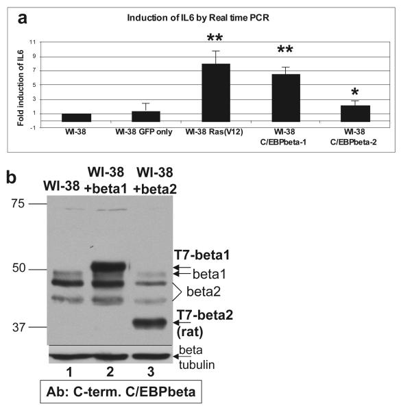
Figure 5.
C/EBPbeta1 induces IL6 expression in WI-38 human fibroblasts a. WI-38 cells were infected with LZRS-GFP, pBABE Ras(V12)-puromycin, LZRS-T7-C/EBPbeta1-IRES-eGFP, or LZRS-T7-C/EBPbeta2-IRES-eGFP. Six days post-infection RNA was prepared. Total RNA was isolated using the RNeasy Mini kit and RNase-Free DNase kit (Qiagen). cDNA was synthesized with the high capacity cDNA reverse transcription kit according to the manufacturer’s instructions (Applied Biosystems). Taqman real-time PCR was performed to determine the relative levels of targets, using GAPDH as the internal control and an IL6 primer (Hs00985639_m1, Applied Biosystems). Results are presented as fold induction of IL6 as compared to uninfected WI-38. This assay was repeated three times with standard deviation represented by the error bars. p values were calculated using the student t test. ** p < 0.02, * p < 0.03. b. Immunoblot analysis of T7-C/EBPbeta1 versus T7-C/EBPbeta2 protein levels in infected WI-38 cells. Cell lysates from WI-38 cells infected with LZRS-T7-C/EBPbeta1-IRES-eGFP or LZRS-T7-C/EBPbeta2-IRES-eGFP were analyzed via 10% SDS-PAGE. Lane 1 is uninfected WI-38 cells, lane 2 is WI-38-T7-C/EBPbeta1 cells, and lane 3 WI-38-T7-C/EBPbeta2 cells. Equal amounts of total protein were loaded in each lane. Immunoblot analysis was performed with an anti-C/EBPbeta C-terminal antibody (Santa Cruz C-19) at 1:5000. Arrows indicate the particular isoforms of C/EBPbeta. T7-beta indicates the exogenously expressed protein. C/EBPbeta2 is smaller than the endogenous because the T7-tagged rat protein is being expressed, which is smaller than human. Immunoblot analysis for beta-tubulin was performed as a loading control (Sigma T7816). (beta = C/EBPbeta)

