
Figure 3.
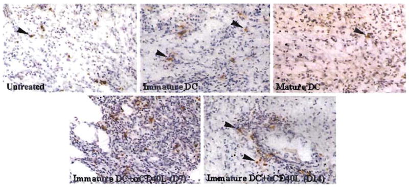
Detection of TUNEL+ apoptotic cells within B10 cardiac allografts of untreated C3H mice or mice pretreated (day –7) with either immature donor-derived DC±anti-CD40L mAb, or mature donor DC. Results obtained on day 7 posttransplant, with the exception of bottom right (day 14). The comparatively high incidence of apoptotic cells seen in the graft-infiltrating cell (GIC) population of mice pretreated with immature donor DC+anti-CD40L mAb correlated with significant elevations (2-fold) in DNA fragmentation detected by spectrofluorometric assay in freshly isolated GIC compared with untreated controls 7 days post transplant (see Table 1) (magnification × 100). Counterstained with hematoxylin.