Abstract
Bacillus anthracis, the etiological agent of anthrax, has a dormant stage in its life cycle known as the endospore. When conditions become favorable, spores germinate and transform into vegetative bacteria. In inhalational anthrax, the most fatal manifestation of the disease, spores enter the organism through the respiratory tract and germinate in phagosomes of alveolar macrophages. Germinated cells can then produce toxins and establish infection. Thus, germination is a crucial step for the initiation of pathogenesis. B. anthracis spore germination is activated by a wide variety of amino acids and purine nucleosides. Inosine and l-alanine are the two most potent nutrient germinants in vitro. Recent studies have shown that germination can be hindered by isomers or structural analogues of germinants. 6-Thioguanosine (6-TG), a guanosine analogue, is able to inhibit germination and prevent B. anthracis toxin-mediated necrosis in murine macrophages. In this study, we screened 46 different nucleoside analogues as activators or inhibitors of B. anthracis spore germination in vitro. These compounds were also tested for their ability to protect the macrophage cell line J774a.1 from B. anthracis cytotoxicity. Structure-activity relationship analysis of activators and inhibitors clarified the binding mechanisms of nucleosides to B. anthracis spores. In contrast, no structure-activity relationships were apparent for compounds that protected macrophages from B. anthracis-mediated killing. However, multiple inhibitors additively protected macrophages from B. anthracis.
Anthrax is as an often-fatal, acute infectious disease caused by the rod-shaped, Gram-positive, endospore-forming bacterium Bacillus anthracis (17). The highly resistant Bacillus anthracis spores remain dormant in soil, occasionally entering mammals through skin abrasions, ingestion, or inhalation (27). The severity of B. anthracis pathogenicity is dependent on the uptake route. In inhalational anthrax, the most deadly form of the disease, spores enter the host organism through the respiratory tract (27). Once inside the lungs, spores are phagocytosed by alveolar macrophages and germinate inside the phagosome (15, 16). The germinated spores become metabolically active and secrete anthrax toxins, the bacterium's primary virulence factors (3, 4, 6). Vegetative bacilli rapidly proliferate within the lymphatic system and disseminate throughout the bloodstream (18). The continuous secretion of toxins leads to fatal septicemia.
Although spore germination is a critical step in the establishment of anthrax infection (18), very little is known about the signaling pathways involved in spore germination (28, 32). The first step in the germination process is most commonly the binding of metabolites by germination (Ger) receptors (8, 23, 38). These receptors are membrane proteins mostly encoded by tricistronic operons. Up to seven Ger receptors have been characterized in B. anthracis (13). Combinations of Ger receptors may be involved in different interacting pathways for germination (13, 30). Usually a purine and an amino acid are required for the efficient germination in vitro of B. anthracis spores (2, 23, 37). Once germination is activated, a series of degradative events break up spore-specific structures and proteins (24, 29, 34). Germination is followed by a period of outgrowth, during which actively dividing cells are regenerated (19, 20, 22).
It has been observed that Bacillus and Clostridium spore germination can be blocked by alcohols (11, 36), ion channel blockers (26), protease inhibitors (9), sulfhydryl reagents (14), and other miscellaneous compounds (10). Most of these studies targeted specific germination pathways in different organisms and are not directly comparable. A more recent study tested the activities of subsets of the different types of compounds against Bacillus subtilis and Bacillus megaterium germination (10). Research from many groups, including ours, has shown that nucleoside and amino acid analogues act as competitive inhibitors of B. anthracis spore germination in vitro (2, 21, 25). Of these inhibitors, d-alanine (d-Ala) and d-histidine (amino acid analogues) and 6-thioguanosine (6-TG; a nucleoside analogue) were shown to also protect macrophages from B. anthracis-mediated necrosis (2, 21, 25).
In this study, we screened 46 different nucleoside analogues for their abilities to inhibit B. anthracis spore germination in vitro and in macrophage cultures. Structure-activity relationship analysis allowed identification of epitopes necessary for nucleoside recognition by B. anthracis spores. However, we found no correlation between in vitro germination inhibition and the ability of nucleosides to protect macrophages from B. anthracis cytotoxicity. We also showed that a nucleoside analogue (6-TG) and an amino acid analogue (d-Ala) combined to increase macrophage protection from B. anthracis cytotoxicity.
MATERIALS AND METHODS
Cell lines, reagents, and equipment.
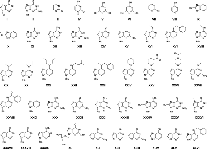
Murine macrophage J774A.1 cells were a generous gift from Jürgen Brojatsch (Albert Einstein College of Medicine, NY). B. anthracis Sterne 34F2 strain was a generous gift from Arturo Casadevall (Albert Einstein College of Medicine, NY). Immunicillin H (IH; compound XXXVIII) was a generous gift from Vern Schramm (Albert Einstein College of Medicine, NY). Nucleoside analogues of 6-benzylthioinosine (6-BTI; compound XVII), 6-N-diethyladenosine (6-DEA; XX), 6-N-dipropyladenosine (6-DPA; XXI), 6-N-benzylmethyladenosine (6-BMA; XXIII), 6-N-piperidineadenosine (6-PPA; XXIV), 6-N-((3-ethylacetyl)cyclohexyl) adenosine (6-ECA; XXV), and ribofuranosyl-((9H-purin-6-yl)pipierazin-1-yl)ethanone (6-PPER; XXVI) were synthesized by Kyungae Lee from NSRB. Other compounds were purchased from Sigma-Aldrich Corporation (St. Louis, MO) or Berry & Associates Inc. (Brea, CA) (Fig. 1). In vitro spore germination and macrophage viability were monitored in a Tecan Infinite M200 multimode microplate reader.
FIG. 1.
Compounds tested as B. anthracis spore germination inhibitors in vitro and in cell culture (with the compound number shown in roman numerals in parentheses): INO (I), 6-TG (II), 2-mercaptopyrimidine (III), 2-thiouracil (2-TU; IV), trithiocyanuric acid (TTCA; V), 2,4-diamino-6-mercaptopyrimidine (DAMPy; VI), 2-mercaptopyridine (VII), 4-mercaptopyridine (VIII), 2-mercaptobenzimidazole (2-MBI; IX), 2-methylmercaptobenzimidazole (2-MMBI; X), 6-TI (XI), ADE (XII), GUA (XIII), 6-CPR (XIV), 2-APR (XV), 6-MMPR (XVI), 6-BTI (XVII), 6-methylaminopurine riboside (6-MAPR; XVIII), 6-N-dimethyladenosine (6-DMA; XIX), 6-DEA (XX), 6-DPA (XXI), 6-DMAA (XXII), 6-BMA (XXIII), 6-PPA (XXIV), 6-ECA (XXV), 6-PPER (XXVI), 6-OMG (XXVII), 6-NBA (XXVIII), NEB (XXIX), 2-AADE (XXX), 6-CG (XXXI), ISO (XXXII), XAN (XXXIII), 8-AADE (XXXIV), 8-HG (XXXV), 7-MI (XXXVI), APR (XXXVII), IH (XXXVIII), TU (XXXIX), GCV (XL), 6-Tg (XLI), 6-Mp (XLII), 6-Cg (XLIII), 6-cyanopurine (6-CYp; XLIV), allopurinol (Ap, XLV), 6-BAp (XLVI).
Synthesis of 6-BTI (compound XXVII), 6-DEA (XX), 6-DPA (XXI), 6-BMA (XXIII), 6-PPA (XXIV), 6-ECA (XXV), and 6-PPER (XXVI).
N-6- and S-6-subtituted purine ribosides were synthesized by substituting 6-chloropurine riboside with various amines or thiols in ethanol or dimethylformamide (DMF), as previously reported (35). In brief, a mixture of 6-chloropurine riboside (1 eq), the corresponding amine or thiol (3 eq), and triethylamine (72 μl; 0.51 mmol) in ethanol or DMF was heated at 60 to 90°C for 18 h and cooled to room temperature. After evaporation of the solvent, the residue was dissolved in methanol (1 ml). Upon standing overnight, crystals of the desired product were formed, and these were collected by filtration (50 to 88% yield). Compounds were characterized by 1H nuclear magnetic resonance and mass spectrometry.
B. anthracis spore preparation.
B. anthracis cells were plated in nutrient agar (EMD Chemicals Inc.) and incubated at 37°C to yield single cell clones. Individual B. anthracis colonies were grown in nutrient broth and replated to obtain bacterial lawns. Plates were incubated for 5 days at 37°C. The resulting bacterial lawns were collected by flooding with ice-cold deionized water. Spores were pelleted by centrifugation and resuspended in fresh deionized water. After two washing steps, spores were separated from vegetative and partially sporulated cells by centrifugation through a 20%-to-50% HistoDenz gradient (1). Spores were resuspended in water and washed three times before storage at 4°C. Spores in all preparations were more than 95% pure as determined by microscopic observation of Schaeffer-Fulton-stained aliquots. Spore viability was assessed by heat treatment followed by serial dilution plating in nutrient agar. Spore viability was retested every 15 to 20 days.
Activation of B. anthracis spore germination.
B. anthracis spore germination was monitored spectrophotometrically whereby the loss in light diffraction following addition of a germinant was reflected by a decreased optical density at 580 nm (OD580). All germination experiments were carried out in a Tecan Infinite M200 multimode microplate reader in UV-visible mode with its monochromator set at 580 nm. The final volume of each reaction mixture was 0.2 ml. Experiments were carried out in triplicate on at least two different days with two different spore preparations. Standard deviations of germination rates were calculated from these six independent assays. Spores were heat activated at 70°C for 30 min before resuspension in germination buffer (50 mM Tris-HCl [pH 7.5], 10 mM NaCl) to an OD580 of 1. The spore suspension was monitored for autogermination based on the OD580 after 1 h. No autogermination was detected in any sample used. Lack of autogermination was confirmed by phase-contrast microscopy on selected samples. Spore suspensions were individually supplemented with 250 μM concentrations of compounds I to XLVI and 40 μM l-alanine to test for compounds that activate B. anthracis spore germination. Germination was monitored by following the decrease in OD580 for 60 min. Optical densities were normalized by dividing each data point by the absorbance at the time of analogue addition and were reported as the relative OD580. Germination rates were calculated from the initial linear region of the germination curves (2). Germination rates were set to 100% for spores treated with 250 μM inosine and 40 μM l-alanine. These inosine and l-alanine concentrations represent the lowest concentrations that induce the maximal germination rate. Percent germination for other nucleosides was calculated as the fraction of germination rate compared to these conditions. Germination was confirmed in selected samples by Schaeffer-Fulton staining.
Inhibition of B. anthracis spore germination.
To test for antagonists of B. anthracis spore germination, spore aliquots were individually supplemented with various concentrations of compounds II to XLVI and incubated for 15 min at room temperature. Spore-analogue mixtures were then treated with 250 μM inosine and 40 μM l-alanine to induce germination. As above, optical densities were normalized by dividing each data point by the absorbance at the time of inosine addition and were reported as the relative OD580. Germination rates were calculated from the initial linear region of the germination curves, as described above. Germination inhibition was assessed as the reduction of germination rate with increased analogue concentration. Inhibited germination rates were plotted against inhibitor concentrations to calculate 50% inhibitory concentration (IC50) values using SigmaPlot v.11 software.
Macrophage viability assay.
Macrophage J774A.1 cells were cultured using Dulbecco modified Eagle's medium (DMEM) without l-glutamine (Cellgro). The medium was supplemented with 10% fetal bovine serum (FBS; HyClone), 2 mM Glutamax (Invitrogen Corp.), and 100 U of penicillin-streptomycin. The cells were incubated at 37°C and 5% CO2 until cultures reached approximately 80% confluence. Cells were subcultured three times before use in cell killing assays. Near-confluent macrophages were washed twice with ice-cold phosphate-buffered saline (PBS) and then detached by treating for 15 min with PBS supplemented with 5 mM EDTA. Detached cells were resuspended in DMEM without FBS or antibiotics and seeded at 8 × 10⁵ cells/ml on 96-well transparent plates. After the cells had adhered, compounds I to XLVI were individually added at different final concentrations. The multiwell plate was incubated for 30 min at 37°C. Subsequently, B. anthracis spores, suspended in Hanks balanced saline solution (HBSS; HyClone, Logan, UT), were added to macrophages at a multiplicity of infection of 5. To control for compound toxicity, some cells were treated with analogues but not infected with B. anthracis spores. Infected macrophages were incubated at 37°C and 5% CO2 for 75 min. To remove nonphagocytosed spores, all wells were washed twice with 200 μl HBSS and then filled with 100 μl of fresh medium containing identical analogue concentrations. At 3 h postinfection, cells were treated with 20 μM propidium iodide (PI). Cell necrosis was assessed by increases in PI fluorescence. Fluorescence was measured every hour for up to 7 h postinfection by following PI fluorescence at 617 nm. Relative fluorescence was calculated as the quotient of the difference of each data point and the fluorescence value at the time of PI addition, divided by the fluorescence value at the time of PI addition. Relative fluorescence intensities were plotted against inhibitor concentrations to calculate IC50 values by using SigmaPlot v.11 software.
Testing for compounds that enhance macrophage killing.
Macrophages were individually treated with potential enhancers to a 100 μM final concentration. B. anthracis spores were then added to each sample. PI fluorescence was determined 5 h postinfection. Macrophage killing enhancement was calculated by dividing the PI fluorescence intensity observed from infected macrophages treated with specific compounds by the PI fluorescence intensity obtained from infected untreated macrophages. GraphPad online calculators were used to run Student's t test and to obtain P values.
Treatment with combinations of 6-thioguanosine and d-alanine.
Macrophages were treated with 1 mM d-Ala, 50 μM d-Ala, 0.1 mM 6-TG, 0.1 μM 6-TG, or 50 μM d-Ala-0.1 μM 6-TG. B. anthracis spores were then added to each sample. Macrophage killing was determined 5 h postinfection based on the increase of PI fluorescence intensity, as described above. GraphPad online calculators were used to run Student's t test and to obtain P values.
RESULTS AND DISCUSSION
Effects of 6-TG.
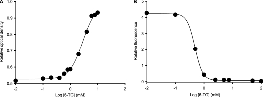
Our previous studies showed that purine analogues can inhibit inosine (INO; compound I)-l-alanine-triggered Bacillus anthracis spore germination in vitro (2). Furthermore, one compound, 6-TG (compound II), was also able to inhibit intracellular spore germination and thus protect murine RAW264.7 macrophages from B. anthracis cytotoxicity (2). As previously shown, 6-TG is a modest inhibitor of spore germination in vitro, with an IC50 of 1 mM (Fig. 2A). This inhibition was not due to the reducing properties of the thiol group, as compounds III to X, with free sulfhydryls, were unable to inhibit spore germination. The inabilities of compounds III to X to affect B. anthracis spore germination also suggest that the purine ring is necessary for recognition by the germination machinery. 6-TG was also able to protect J774a.1 macrophages from B. anthracis cytotoxicity, with an IC50 of 3.5 μM (Fig. 2B). Thus, 6-TG is approximately 300-fold more effective at preventing spore germination intracellularly than in vitro. The reason for the discrepancies in IC50 values is not clear. In vitro spore germination assays require high spore concentrations to obtain reliable optical density changes. As a result, millimolar concentrations of inosine and l-alanine are needed to activate germination. In contrast, B. anthracis spores will encounter low inosine concentrations in the macrophage intracellular milieu (7). Low intracellular inosine concentrations might be the reason for the higher efficiency of 6-TG at blocking germination of B. anthracis spores in macrophages. It is conceivable that inosine is the principal germinant inside cells and that 6-TG prevents germination by competing with endogenous inosine.
FIG. 2.
(A) B. anthracis spores were treated with different concentrations of 6-TG, and germination was monitored by decreases in the optical density at 580 nm. The IC50 for 6-TG (1 mM) was determined based on these data. (B) Macrophages were treated with B. anthracis spores and different concentrations of 6-TG. Macrophage killing was monitored based on increases of PI fluorescence intensity.
B. anthracis spore germination in vitro.
Of the 46 compounds screened as agonists and antagonists of B. anthracis spore germination, 5 were able to act as cogerminants with l-alanine, and 18 were able to inhibit germination of B. anthracis spores treated with inosine and l-alanine. None of the pyrimidine analogues (compounds III to VIII) was able to activate B. anthracis spore germination in vitro.
Position 6 of the purine ring seems to have a significant role in the interaction of germinants with the B. anthracis spore germination machinery. INO, 6-thioinosine (6-TI; compound XI), adenosine (ADE; compound XII), guanosine (GUA; compound XIII), and 6-chloropurine riboside (6-CPR; compound XIV) are germinants for B. anthracis spores. All five compounds have 6-exocyclic groups with unsubstituted electronegative atoms directly bound to C-6 of the purine ring.
Electronegative groups are also present in every inhibitor of B. anthracis spore germination in vitro, with the exception of 2-amino riboside (2-APR; compound XV) (Table 1). However, nucleoside inhibitors normally possess aliphatic or aromatic chains linked to an electronegative atom. For example, 6-TI (compound XI), with a nonsubstituted 6-thiol (-SH), was a weak germinant in vitro. In contrast, 6-mercaptopurine riboside (6-MMPR; compound XVI) and 6-BTI (XVII) are B. anthracis spore germination inhibitors, with IC50s of 318 μM and 50 μM, respectively. A similar correlation was observed across other 6-substituted germination inhibitors (Table 1). A characteristic pattern of C-6-X-R (where X is oxygen, sulfur, or nitrogen and R is a hydrophobic group) is observed for inhibitors of B. anthracis spore germination. Hence, while nonsubstituted compounds act as germinants, addition of a hydrophobic side chain to the 6-exocyclic heteroatom is sufficient to convert a germinant structure into an inhibitor (Fig. 1, compounds XVI to XXVIII). However, there was no correlation between side chain physical properties and strength of inhibition. As an example, 2-APR (compound XV), 6-MMPR (XVI), 6-DEA (XX), 6-(γ,γ-dimethylallyl amino)purine riboside (DMAA; XXII), and 6-O-methylguanosine (6-OMG; XXVII) showed similar IC50 values (Table 1). This suggests that binding of inhibitors in B. anthracis spores is mostly driven by nonspecific hydrophobic interactions. A similar pattern has been previously observed for the inhibition of Bacillus cereus spore germination in vitro (12).
TABLE 1.
Antagonists of B. anthracis spore germination in vitroa
| Antagonist | Acronym | IC50 (mM) |
|---|---|---|
| 6-Thioguanosine | 6-TG | 1.00 |
| 6-Chloroguanosine | 6-CG | 0.89 |
| 6-Benzylthioinosine | 6-BTI | 0.05 |
| 6-Methylmercaptopurine riboside | 6-MMPR | 0.32 |
| 6-O-Methylguanosine | 6-OMG | 0.35 |
| 2-Aminoadenosine | 2-AADE | 0.70 |
| Xanthosine | XAN | 7.19 |
| 6-Methylaminopurine riboside | 6-MAPR | 0.19 |
| 2-Aminopurine riboside | 2-APR | 0.38 |
| Ribofuranosyl-((9H-purin-6-yl) piperazin-1-yl)ethanone | 6-PPER | 0.05 |
| 6-N-Benzylmethyladenosine | 6-BMA | 0.12 |
| 6-N-Benzylaminopurine | 6-BAP | 1.55 |
| 6-(γ,γ-Dimethylallyl amino)purine riboside | 6-DMAA | 0.36 |
| 6-N-Benzoyladenosine | 6-NBA | 1.14 |
| 6-N-Piperidineadenosine | 6-PPA | 1.09 |
| 6-N-Diethyladenosine | 6-DEA | 0.31 |
| 6-N-Dipropyladenosine | 6-DPA | 0.76 |
| 6-N-((3-Ethylacetyl)cyclohexyl) adenosine | 6-ECA | 0.70 |
| d-Alanine | d-Ala | 0.02 |
Spore suspensions were individually supplemented with various concentration of compounds II to XLVI and then treated with 250 μM inosine and 40 μM l-alanine. Compounds that did not inhibit germination are not shown in this table.
In vitro assays showed that the presence of a 2-amino exocyclic group in the purine ring also contributes to spore germination inhibition. This is exemplified by 6-TI (compound XI), a 6-TG (compound II) analogue that lacks the 2-amino group and does not inhibit, but activates, spore germination in vitro. Similarly, nebularine (NEB; compound XXIX), a nonsubstituted purine riboside, does not affect B. anthracis spore germination, while 2-APR (compound XV), with an exocyclic amino group at position 2 of the purine ring, inhibits spore germination with an IC50 of 370 μM (Table 1). Furthermore, ADE (XII), with an amino group at the 6-position, is a good germinant in vitro. Meanwhile, 2-aminoadenosine (2-AADE; XXX), which has amino groups at both the 2- and 6-positions, is a germination inhibitor, with an IC50 of 700 μM. Similarly, 6-chloroguanosine (6-CG; XXXI) is also an inhibitor, with an IC50 of 893 μM. In contrast, isoguanosine (ISO; XXXII), a guanosine positional isomer, and xanthosine (XAN; XXXIII), with carbonyl groups at the 2-positions, are inactive in spore germination assays.
8-AADE (compound XXIV) is a positional isomer of 2-AADE (XXX) and has similar physical and chemical properties. However, 8-AADE did not activate or inhibit spore germination under any conditions tested. Similarly, 8-hydroxyguanosine (8-HG; XXXV) had no effect on B. anthracis spore germination. The inabilities of 8-AADE and 8-HG to influence B. anthracis spore germination could be due to steric hindrance at the 8-position.
The inosine derivative 7-methylinosine (7-MI; compound XXXVI) showed no effect on B. anthracis spore germination. Similarly, inosine analogues allopurinol riboside (APR; XXVII) and IH (XXXVIII) do not affect spore germination. Furthermore, the adenosine analogue tubericidin (TUB; XXXIX) is inactive in B. anthracis spore germination assays. Hence, the presence of an unsubstituted N-7 heterocyclic nitrogen in the purine ring also affects the germination properties of a compound.
Similar to the purine ring, the ribosyl group seems to be required for germination inhibition of B. anthracis spores. Ganciclovir (GCV; compound XL), a purine riboside analogue that lacks the 2′-position, fails to either trigger or hinder B. anthracis spore germination. Similarly, purine bases lacking the ribosyl group (compounds XLI to XLVI), with the exception of 6-benzylaminopurine (BAp; XLVI), had no effect on germination in vitro. The aromatic side chain at the C-6 position may be responsible for the modest inhibition shown by BAp against the germination of B. anthracis spores in vitro. Therefore, an entire purine nucleoside frame is required for optimal germination inhibition in vitro.
Intracellular B. anthracis germination in J774a.1 macrophages.
Murine J774a.1 macrophages can readily phagocytose endospores of B. anthracis. After infection, spore germination occurs inside the phagolysosome (18). Three hours postinfection, necrosis starts to be apparent, and approximately 20% cell death occurs. Cell death then increases rapidly, reaching almost 100% at 5 h postinfection (Fig. 3A). The timing of cell death is variable, but most cells have entered necrosis at 5 h postinfection. This variability is probably due to nonuniformity in spore intake by macrophages, which leads to heterogeneous cell killing (33).
FIG. 3.
Kinetics of macrophage killing by B. anthracis spores. (A) Macrophages were treated with medium (○) or B. anthracis spores (•). Macrophage necrosis was monitored over time by the increase of PI fluorescence intensity. Error bars represent standard deviations of six independent measurements. (B) Macrophages were observed under a microscope in white light (upper panels) or fluorescence (lower panels) modes. Macrophages showed limited cell death 3 h postinfection with B. anthracis spores (a and b). In contrast, macrophages showed extensive necrosis at 7 h postinfection with B. anthracis spores (c and d). Macrophage-engulfed spores (e.g., black circle in panel c) and vegetative B. anthracis cells (e.g., black arrow in panel c) were also seen under these conditions. Macrophages treated with 6-TG were protected from B. anthracis cytotoxicity even at 7 h postinfection with B. anthracis spores (e and f). No vegetative B. anthracis cells were observed.
Due to the heterogeneity of necrosis onset, compounds were tested for their ability to reduce B. anthracis-mediated killing at 5 h postinfection. Of the 46 compounds screened, only 8 were able to protect macrophages from B. anthracis cytotoxicity (Table 2). No compound showed toxic effects in the absence of B. anthracis spores. The protective compounds were either purine nucleoside or purine base derivatives. The purine nucleoside derivatives 6-TG (II), 6-TI (XI), 6-BTI (XVII), 6-CG (XXXI), and APR (XXXVII) protected macrophages, with IC50 values ranging from 3.5 μM (6-TG; II) to near 1 mM (6-BTI; XVII) (Table 2). 6-TG (II) and 6-thioguanine (6-Tg; XLI) were the most efficient analogues and protected macrophages from necrosis even 6 h after infection. The remaining nucleoside analogues started to lose effectiveness at 6 h postinfection. Thus, these compounds did not completely inhibit intracellular spore germination but were able to delay the onset of the cytotoxic effect.
TABLE 2.
Protection of macrophages from B. anthracis-mediated killinga
| Antagonist | Acronym | IC50 (mM) |
|---|---|---|
| 6-Thioguanosine | 6-TG | 0.004 |
| 6-Chloroguanosine | 6-CG | 0.19 |
| 6-Thioinosine | 6-TI | 0.04 |
| Allopurinol riboside | APR | 0.65 |
| 6-Benzylthioinosine | 6-BTI | 1.04 |
| 6-Thioguanine | 6-Tg | 0.002 |
| 6-Chloroguanine | 6-Cg | 0.20 |
| 6-Mercaptopurine | 6-Mp | 0.40 |
| d-Alanine | d-Ala | 0.02 |
Macrophage cultures were individually supplemented with various concentrations of compounds II to XLVI and then infected with B. anthracis spores. Compounds that did not protect macrophages from anthrax cytotoxicity are not shown in this table.
Three purine analogues, 6-Tg (XLI), 6-mercaptopurine (6-Mp; XLII), and 6-chloroguanine (6-Cg; XLIII), were also able to prevent cell necrosis. The protective properties of these compounds are intriguing, since in vitro assays showed no effect on B. anthracis spore germination. It is worth noting that these compounds are the corresponding bases for the protective nucleosides 6-TG (II), 6-TI (XI), and 6-CG (XXXI), respectively. Furthermore, the IC50s for nucleosides and free bases were similar in macrophage protection assays (Table 2). Thus, both nucleosides and their free bases are able to protect macrophages from B. anthracis cytotoxicity, even though they behave differently in spore germination assays.
Similar to in vitro germination assays, the presence of the 2-amino side group of the base favors the action of the compounds in macrophage protection assay. Analogues lacking this group showed decreased macrophage protective effects compared to their substituted counterparts. For example, 6-TG (II) had an IC50 value of 3.5 μM, while the unsubstituted analog, 6-TI (XI), had an IC50 value of 35 μM, a 10-fold decrease in protection ability. Similarly, 6-CG (XXXI) had an IC50 value of 189 μM, while 6-CPR (XIV) was unable to prevent cell killing.
Comparing microscopy observations with viability assay results showed that macrophages in which B. anthracis progressed from spores to vegetative cells also showed up as PI-positive necrotic cells. On the other hand, in cell cultures treated with an efficient inhibitor, spores did not progress to vegetative bacteria and the integrity of mammalian cells was maintained (Fig. 3B). Thus, endospores per se do not show toxin-related killing toward macrophages. This suggests that macrophage necrosis was either stopped or slowed when B. anthracis spores did not germinate efficiently upon treatment with germination inhibitors.
Compounds that enhance macrophage killing.
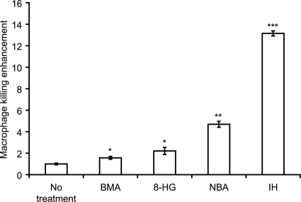
Cell viability screening allowed the identification of compounds that cooperated with B. anthracis spores to increase the rate of macrophage killing (Fig. 4). The compounds that enhanced cell killing more efficiently were 6-BMA (XXIII), 6-benzoyladenosine (6-NBA; XXVIII), 8-hydroxyguanosine (8-HG; XXXV), and IH (XXXVIII). This cell killing enhancement was not due to compound toxicity, since these compounds do not induce cell necrosis in the absence of B. anthracis spores (data not shown). Surprisingly, some of the compounds that enhanced cell killing were inhibitors in vitro. Currently, the mechanism by which these compounds are able to accelerate macrophage killing in the presence of B. anthracis spores is not known.
FIG. 4.
Compounds that enhance B. anthracis cytotoxicity. BMA, 8-HG, NBA, and IH were added to macrophages to a 100 μM final concentration. B. anthracis spores were then added to each sample. Macrophage necrosis, based on increased PI fluorescence intensity, was monitored at 5 h postinfection. Macrophage killing enhancement was calculated by normalizing the PI fluorescence intensity determined for each compound treatment by the PI fluorescence intensity for untreated macrophages. Error bars represent standard deviations of six independent measurements. *, P < 0.05; **, P < 0.01; ***, P < 0.001 (all for comparisons to untreated samples).
Effect of d-alanine and nucleoside analogue combinations.
d-Ala is a natural antigerminant for B. anthracis spores (21). This molecule is biosynthesized by an alanine racemase present in the spore and can modulate germination pathways that require l-alanine as a germinant (25). Two research groups have already shown that d-Ala protects macrophages from B. anthracis toxin-mediated necrosis by inhibiting spore germination (21, 25). We corroborated the antigerminant ability of d-Ala both in vitro (IC50, 16.0 μM) and in the macrophage killing protection assay (IC50, 22.9 μM).
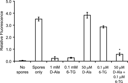
Since both inosine and l-alanine are strong germinants of B. anthracis spores, a combination of a nucleoside inhibitor (6-TG; II) and an amino acid inhibitor (d-Ala) could increase effectiveness by simultaneously blocking multiple Ger receptors. Indeed, combinations of 6-TG and d-Ala were able to protect macrophages from B. anthracis-mediated cytotoxicity, even at suboptimal concentrations of both inhibitors (Fig. 5). Similar results were obtained with d-Ala and 6-CG (XXXI) (data no shown).
FIG. 5.
Macrophage protection by 6-TG and d-Ala combinations. Macrophages were treated with B. anthracis spores, and killing was monitored at 5 h postinfection based on increased PI fluorescence intensity. Addition of 1 mM d-Ala or 0.1 mM 6-TG significantly reduced macrophage killing. In contrast, treatment with 50 μM d-Ala or 0.1 μM 6-TG did not prevent cell killing. Simultaneous treatment with 50 μM d-Ala and 0.1 μM 6-TG protected macrophages from B. anthracis cytotoxicity. Error bars represent standard deviations of six independent measurements. *, significantly decreased killing in the presence of both 50 μM d-Ala and 0.1 μM 6-TG compared to samples treated with 50 μM d-Ala alone (P < 0.005) or samples treated with 0.1 μM 6-TG alone (P < 0.0001).
Conclusions.
Spore germination is the first necessary step in the establishment of B. anthracis infection (15). Hence, inhibition of B. anthracis spore germination could serve as a prophylactic approach to prevent anthrax in exposed personnel (5). Antigermination therapy will delay infection onset and hence allow antibiotic and/or vaccine treatments to be more effective. We had previously tested a limited number of purine nucleosides as inhibitors of B. anthracis spore germination (2). In this study, we tested a larger collection of purine nucleosides, purine bases, and pyrimidine bases for their abilities to prevent spore germination in vitro and protect macrophages from B. anthracis-mediated killing. All compounds (but one) that inhibited spore germination in vitro were purine nucleoside derivatives, showing that the sugar moiety is important for inhibitor recognition. Furthermore, hydrophobic-substituted electronegative exocyclic groups at the 6-position and an amine at the 2-position of the purine ring were necessary functional groups for efficient germination inhibition in vitro.
Molecular interactions between the spore germination machinery and the nucleoside inhibitors require specific contacts. Hydrogen bonding and/or ionic interactions are probably involved in the recognition of the amino group at the 2-position of purine nucleoside inhibitors. Similarly, hydrogen bonds could also serve to recognize essential sugar hydroxyls and the exocyclic heteroatom at position 6. Hydrophobic and π-π interactions would increase binding affinities for nucleosides with alkyl and aromatic side chains.
The efficiency of spore germination in vitro did not correlate with the capacity to protect macrophages from intracellular spore germination and cytotoxicity. Some compounds were able to inhibit spore germination in vitro but did not protect macrophages against spore-mediated killing (e.g., 6-MMPR [compound XVI] and 6-OMG [XXVII]). Conversely, other compounds were unable to curtail spore germination in vitro but were able to prevent macrophage necrosis (e.g., 6-TI [XI] and APR [XXXVII]). Yet another compound subset was composed of efficient in vitro inhibitors that enhanced macrophage killing by B. anthracis spores (e.g., 6-BMA [XXIII] and 6-NBA [XXVIII]).
The lack of correlation between in vitro and intracellular B. anthracis spore germination assay results was not due to drug toxicity. These two assays measured spore germination under different conditions. In vitro germination assays are only dependent on the ability of compounds to cross the spore coat and interact with their respective Ger receptor. In contrast, intracellular germination assays are complicated by the additional requirement for compounds to cross both the mammalian cytoplasmic and phagosomal membranes. Furthermore, potential germination inhibitors may have to contend with cellular catabolism, active drug efflux pumps, and other mammalian cellular processes.
It was noticeable that some purine bases that did not hinder germination in vitro were effective antigerminants in macrophages. These bases (6-Tg [XLI], 6-Mp [XLII], and 6-Cg [XLIII]) prevented cell killing with efficiencies similar to those of their nucleoside derivatives (Table 2). Previous research has shown that 6-Tg (XLI), 6-Mp (XLII), and probably 6-Cg (XLIII) are transformed into 6-TG (II), 6-TI (XI), and 6-CG (XXXI) by the purine salvage pathway (7, 31). We postulate that this transformation allows the bases to be converted to nucleosides and to block spore germination inside the phagolysosome.
Similarly, two purine nucleosides, 6-TI (XI) and APR (XXXVII), did not affect germination in vitro but prevented macrophage necrosis. Thus, there must be a structural change in both 6-TI and APR that interferes with the germination of the B. anthracis endospores inside cells. We postulate that the nucleotide metabolism pathways allow these compounds to be converted to active inhibitors in the macrophage cytoplasm (7, 31).
In summary, analyses of B. anthracis spore germination activation and of inhibition in vitro allow mapping of important contacts necessary for spore binding. However, in vitro spore germination assays do not provide a good guide for the behavior of antigerminants inside cells. The multiplicity and variety of germinants and germination pathways in vivo make it difficult to find a single “ideal” molecule to block B. anthracis spore germination. Even though nucleosides show complex intracellular behavior, combining active nucleoside and amino acid germination inhibitors showed an additive effect in macrophage protection. Hence, simultaneous blocking of multiple germination pathways protects macrophages from B. anthracis cytotoxicity, even at suboptimal concentrations of the individual inhibitors.
Acknowledgments
This work was supported by the RING-TRUE III 0447416 award from NSF-EPSCoR and by NIH grant no. P20 RR-016464 from the INBRE Program of the National Center for Research Resources. Support for E.A.-S. was provided by the Ellison Medical Foundation Young Scholar Award in Global Infectious Diseases and by Public Health Service grant 1R01A1GM053212 from the National Institutes of Health. Support to K.L. was provided by the National Screening Laboratory for the Regional Centers of Excellence in Biodefense and Emerging Infectious Diseases (NIAID U54 AI057159).
We thank Su Chiang for helpful discussions.
Footnotes
Published ahead of print on 4 October 2010.
REFERENCES
- 1.Abel-Santos, E., and T. Dodatko. 2007. Differential nucleoside recognition during Bacillus cereus 569 (ATCC 10876) spore germination. New J. Chem. 31:748-755. [Google Scholar]
- 2.Akoachere, M., R. C. Squires, A. M. Nour, L. Angelov, J. Brojatsch, and E. Abel-Santos. 2007. Identification of an in vivo inhibitor of Bacillus anthracis Sterne spore germination. J. Biol. Chem. 282:12112-12118. [DOI] [PubMed] [Google Scholar]
- 3.Alileche, A., E. R. Serfass, S. M. Muehlbauer, S. A. Porcelli, and J. Brojatsch. 2005. Anthrax lethal toxin-mediated killing of human and murine dendritic cells impairs the adaptive immune response. PLoS Pathog. 1:e19. [DOI] [PMC free article] [PubMed] [Google Scholar]
- 4.Alileche, A., R. C. Squires, S. M. Muehlbauer, M. P. Lisanti, and J. Brojatsch. 2006. Mitochondrial impairment is a critical event in anthrax lethal toxin-induced cytolysis of murine macrophages. Cell Cycle 5:100-106. [DOI] [PubMed] [Google Scholar]
- 5.Alvarez, Z., and E. Abel-Santos. 2007. Potential use of inhibitors of bacteria spore germination in the prophylactic treatment of anthrax and Clostridium difficile-associated disease. Expert Rev. Anti Infect. Ther. 5:783-792. [DOI] [PubMed] [Google Scholar]
- 6.Banks, D. J., M. Barnajian, F. J. Maldonado-Arocho, A. M. Sanchez, and K. A. Bradley. 2005. Anthrax toxin receptor 2 mediates Bacillus anthracis killing of macrophages following spore challenge. Cell. Microbiol. 7:1173-1185. [DOI] [PubMed] [Google Scholar]
- 7.Barankiewicz, J., and A. Cohen. 1985. Purine nucleotide metabolism in resident and activated rat macrophages in vitro. Eur. J. Immunol. 15:627-631. [DOI] [PubMed] [Google Scholar]
- 8.Barua, S., M. McKevitt, K. DeGiusti, E. E. Hamm, J. Larabee, S. Shakir, K. Bryant, T. M. Koehler, S. R. Blanke, D. Dyer, A. Gillaspy, and J. D. Ballard. 2009. The mechanism of Bacillus anthracis intracellular germination requires multiple and highly diverse genetic loci. Infect. Immun. 77:23-31. [DOI] [PMC free article] [PubMed] [Google Scholar]
- 9.Boschwitz, H., Y. Milner, A. Keynan, H. O. Halvorson, and W. Troll. 1983. Effect of inhibitors of trypsin-like proteolytic enzymes Bacillus cereus T spore germination. J. Bacteriol. 153:700-708. [DOI] [PMC free article] [PubMed] [Google Scholar]
- 10.Cortezzo, D. E., B. Setlow, and P. Setlow. 2004. Analysis of the action of compounds that inhibit the germination of spores of Bacillus species. J. Appl. Microbiol. 96:725-741. [DOI] [PubMed] [Google Scholar]
- 11.Craven, S. E., and L. C. Blankenship. 1985. Activation and injury of Clostridium perfringens spores by alcohols. Appl. Environ. Microbiol. 50:249-256. [DOI] [PMC free article] [PubMed] [Google Scholar]
- 12.Dodatko, T., M. Akoachere, N. Jimenez, Z. Alvarez, and E. Abel-Santos. 2010. Dissecting interactions between nucleosides and germination receptors in Bacillus cereus 569 spores. Microbiology 156:1244-1255. [DOI] [PMC free article] [PubMed] [Google Scholar]
- 13.Fisher, N., and P. Hanna. 2005. Characterization of Bacillus anthracis germinant receptors in vitro. J. Bacteriol. 187:8055-8062. [DOI] [PMC free article] [PubMed] [Google Scholar]
- 14.Foster, S. J., and K. Johnstone. 1986. The use of inhibitors to identify early events during Bacillus megaterium KM spore germination. Biochem. J. 237:865-870. [DOI] [PMC free article] [PubMed] [Google Scholar]
- 15.Guidi-Rontani, C., M. Levy, H. Ohayon, and M. Mock. 2001. Fate of germinated Bacillus anthracis spores in primary murine macrophages. Mol. Microbiol. 42:931-938. [DOI] [PubMed] [Google Scholar]
- 16.Guidi-Rontani, C., M. Weber-Levy, E. Labruyere, and M. Mock. 1999. Germination of Bacillus anthracis spores within alveolar macrophages. Mol. Microbiol. 31:9-17. [DOI] [PubMed] [Google Scholar]
- 17.Gursky, E., T. V. Inglesby, and T. O'Toole. 2003. Anthrax 2001: observations on the medical and public health response. Biosecur. Bioterror. 1:97-110. [DOI] [PubMed] [Google Scholar]
- 18.Hanna, P. C., and J. A. Ireland. 1999. Understanding Bacillus anthracis pathogenesis. Trends Microbiol. 7:180-182. [DOI] [PubMed] [Google Scholar]
- 19.Harry, E. J. 2001. Coordinating DNA replication with cell division: lessons from outgrowing spores. Biochimie 83:75-81. [DOI] [PubMed] [Google Scholar]
- 20.Horsburgh, M. J., P. D. Thackray, and A. Moir. 2001. Transcriptional responses during outgrowth of Bacillus subtilis endospores. Microbiology 147:2933-2941. [DOI] [PubMed] [Google Scholar]
- 21.Hu, H., J. Emerson, and A. I. Aronson. 2007. Factors involved in the germination and inactivation of Bacillus anthracis spores in murine primary macrophages. FEMS Microbiol. Lett. 272:245-250. [DOI] [PubMed] [Google Scholar]
- 22.Huang, C. M., C. A. Elmets, D. C. Tang, F. Li, and N. Yusuf. 2004. Proteomics reveals that proteins expressed during the early stage of Bacillus anthracis infection are potential targets for the development of vaccines and drugs. Genom. Proteom. Bioinform. 2:143-151. [DOI] [PMC free article] [PubMed] [Google Scholar]
- 23.Ireland, J. A., and P. C. Hanna. 2002. Amino acid- and purine ribonucleoside-induced germination of Bacillus anthracis ΔSterne endospores: gerS mediates responses to aromatic ring structures. J. Bacteriol. 184:1296-1303. [DOI] [PMC free article] [PubMed] [Google Scholar]
- 24.Makino, S., and R. Moriyama. 2002. Hydrolysis of cortex peptidoglycan during bacterial spore germination. Med. Sci. Monit. 8:RA119-RA127. [PubMed] [Google Scholar]
- 25.McKevitt, M. T., K. M. Bryant, S. M. Shakir, J. L. Larabee, S. R. Blanke, J. Lovchik, C. R. Lyons, and J. D. Ballard. 2007. Effects of endogenous d-alanine synthesis and autoinhibition of Bacillus anthracis germination on in vitro and in vivo infections. Infect. Immun. 75:5726-5734. [DOI] [PMC free article] [PubMed] [Google Scholar]
- 26.Mitchell, C., J. F. Skomurski, and J. C. Vary. 1986. Effect of ion channel blockers on germination of Bacillus megaterium spores. FEMS Microbiol. Lett. 34:211-214. [Google Scholar]
- 27.Mock, M., and A. Fouet. 2001. Anthrax. Annu. Rev. Microbiol. 55:647-671. [DOI] [PubMed] [Google Scholar]
- 28.Moir, A. 2006. How do spores germinate? J. Appl. Microbiol. 101:526-530. [DOI] [PubMed] [Google Scholar]
- 29.Popham, D. L. 2002. Specialized peptidoglycan of the bacterial endospore: the inner wall of the lockbox. Cell. Mol. Life Sci. 59:426-433. [DOI] [PMC free article] [PubMed] [Google Scholar]
- 30.Ross, C., and E. Abel-Santos. 2010. The Ger receptor family in sporulating bacteria. Curr. Issues Mol. Biol. 12:147-158. [PMC free article] [PubMed] [Google Scholar]
- 31.Scazzocchio, C. 1994. The purine degradation pathway, genetics, biochemistry and regulation. Prog. Ind. Microbiol. 29:221-257. [PubMed] [Google Scholar]
- 32.Setlow, P. 2003. Spore germination. Curr. Opin. Microbiol. 6:550-556. [DOI] [PubMed] [Google Scholar]
- 33.Stojkovic, B., E. M. Torres, A. M. Prouty, H. K. Patel, L. Zhuang, T. M. Koehler, J. D. Ballard, and S. R. Blanke. 2008. High-throughput, single-cell analysis of macrophage interactions with fluorescently labeled Bacillus anthracis spores. Appl. Environ. Microbiol. 74:5201-5210. [DOI] [PMC free article] [PubMed] [Google Scholar]
- 34.Takamatsu, H., and K. Watabe. 2002. Assembly and genetics of spore protective structures. Cell. Mol. Life Sci. 59:434-444. [DOI] [PMC free article] [PubMed] [Google Scholar]
- 35.Trávnícek, Z., J. Mikulík, M. Cajan, R. Zboril, and I. Popa. 2008. Novel iron complexes bearing N6-substituted adenosine derivatives: synthesis, magnetic, 57Fe Mössbauer, DFT, and in vitro cytotoxicity studies. Bioorg. Med. Chem. 16:8719-8728. [DOI] [PubMed] [Google Scholar]
- 36.Trujillo, R., and N. Laible. 1970. Reversible inhibition of spore germination by alcohols. Appl. Microbiol. 20:620-623. [DOI] [PMC free article] [PubMed] [Google Scholar]
- 37.Weiner, M. A., T. D. Read, and P. C. Hanna. 2003. Identification and characterization of the gerH operon of Bacillus anthracis endospores: a differential role for purine nucleosides in germination. J. Bacteriol. 185:1462-1464. [DOI] [PMC free article] [PubMed] [Google Scholar]
- 38.Wolgamott, G. D., and N. N. Durham. 1971. Initiation of spore germination in Bacillus cereus: a proposed allosteric receptor. Can. J. Microbiol. 17:1043-1048. [DOI] [PubMed] [Google Scholar]


