Fig. 5.
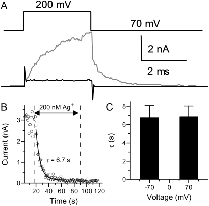
The modification of BK channels by Ag+ at positive potential. A, macroscopic currents evoked by 200-mV test pulses in 0 μM Ca2+ solution before (gray) and after (black) perfusion in 200 nM Ag+. The holding potential was 70 mV. B, time course of Ag+ modification of BK channels at 70 mV. The Ag+ concentration was 200 nM. The modification started at the 17th second and ended at 90th second. C, the histogram of mean modification time constants by 200 nM Ag+ at −70 (6.7 ± 1.3 s, n = 6) or 70 mV (6.8 ± 1.2 s, n = 4).

