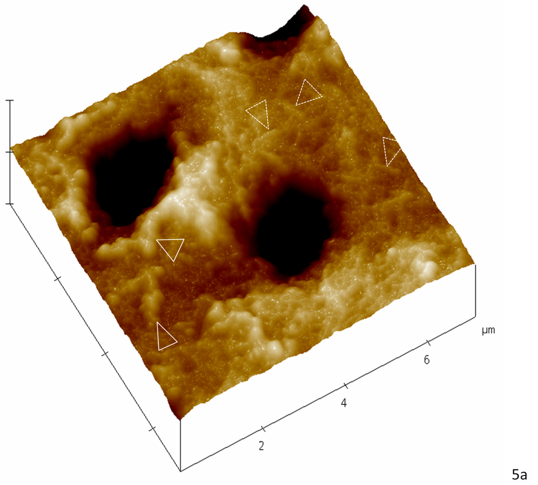
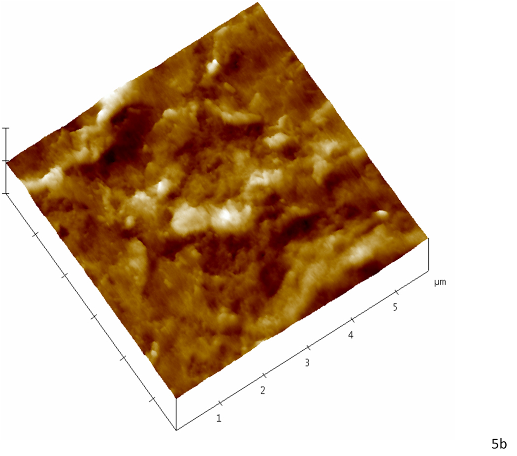
Figure 5.
Higher magnification AFM scans in the contact mode of demineralized dentin after remineralization for five consecutive days. (a) Specimen treated with the continuous approach; white arrowheads show zones suggestive of higher mineral content attached to the collagen network while dotted arrowheads show zones suggestive collagen with less mineral attached. (b) Specimen treated with the static approach; the precipitate completely covers the intertubular dentin hiding any structure that resembled the collagen network seen in ‘a’.


