

Necroptosis is a form of programmed necrotic cell death mediated by the activity of receptor-interacting protein 1 (RIP1) kinase (1). This type of cell death is caspase-independent and marked by early membrane permeabilization and does not exhibit any other hallmarks of apoptotic cell death such as phosphatidylserine externalization or DNA strand breaks (1). Thus, the markers available to characterize apoptotic cell death cannot be used to analyze necroptosis. Only one marker of necrosis has been identified, and that is the release of the chromatin protein high mobility group B1 (HMGB1) (2). However, we have found the release of HMGB1 to occur at a late stage of death. In this study we have found that cyclophilin A (CypA), a cytosolic peptidyl-prolyl cis-trans isomerase, is released early in necroptosis. We propose that the release of CypA may be used as a biomarker for necroptosis and other cell death processes when the integrity of the plasma membrane is compromised.
Since necroptotic cells exhibit early permeabilization of the plasma membrane (1), we hypothesized that the release of intracellular protein(s) may be used as a biomarker for necroptosis. Treatment of L929 cells with zVAD.fmk induces necroptosis, which can be inhibited by treatment with the RIP1 kinase inhibitor, necrostatin-1 (Nec-1) (1). We observed the appearance of a strong band around 17 kD by SDS-PAGE in the supernatant of zVAD-treated L929 cells at an early stage of cell death (Supplemental Figure 1, top panel). Mass spectrometry analysis identified this band as cyclophilin A (CypA), a soluble cytosolic protein that catalyzes the cis-trans isomerization of proline bonds. The release of CypA was further confirmed by western blot (Supplemental Figure 1, bottom panel). In contrast to the necrosis marker HMGB1, the timing of CypA release precedes or occurs around the same time as that of HMGB1 (Figure 1A). Nec-1 blocks the release of CypA into the supernatant (Figure 1B).
Figure 1.
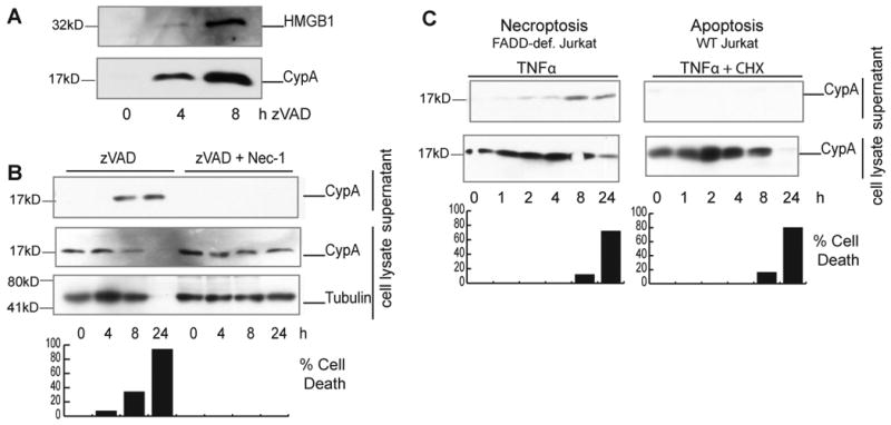
Cyclophilin A is released from necroptotic cells as a result of cytoplasmic membrane permeabilization. A, L929 cells were treated with 100μM zVAD.fmk for indicated hrs. The supernatants were collected, concentrated, and analyzed by SDS-PAGE followed by western blotting using anti-CypA and anti-HMGB1. B, L929 cells were treated with 100μM zVAD.fmk in the presence or absence of 30μM Nec-1. The cell lysate and supernatant were analyzed by western blotting using anti-CypA. Anti-Tubulin was used as a control. Cell viability was determined by ATP assay and indicated as a bar graph below. C, FADD-deficient Jurkat cells (left panel) or wt Jurkat cells (right panel) were treated with 10ng/ml hTNFα to induce necroptosis, or 10ng/ml hTNFα + 0.5μg/ml CHX to induce apoptosis, respectively. The cell lysate and supernatant were analyzed by western blotting using anti-CypA. The percentages of cell death were determined by ATP assay and indicated below.
To determine if the release of CypA from the cells was specific to necroptosis, we tested the supernatant of apoptotic cells. We were unable to detect CypA released from Jurkat cells treated with TNFα and CHX to induce apoptosis, even though intracellular CypA appears to be degraded at 24h when cell death was completed (Figure 1C). However, FADD-deficient Jurkat cells, which undergo necroptosis in response to TNFα, did release CypA (Figure 1C). We also tested different inducers of apoptosis for the ability to release CypA. Treatment of doxorubicin and staurosporine caused a small amount of CypA release at later stages of cell death, hours after caspase-3 activation (Supplemental Figures 2-3). Thus, CypA may also be released after permeabilization of the plasma membrane in late phase of apoptotic cell death. However, since apoptotic cells are efficiently engulfed in vivo, the release of CypA by apoptotic cells may not occur in vivo.
To examine if CypA could be released by permeabilization of the cells, we treated cells with the detergent digitonin, which led to cell membrane permeabilization. We found that digitonin permeabilized cells also release CypA (Supplemental Figure 4). This may explain why CypA is released during necroptosis, which exhibit early permeabilization of the plasma membrane (1).
Elevated serum CypA levels have been found in inflammatory diseases such as rheumatoid arthritis and CypA is secreted in vivo by progressive solid tumors (3, 4). CypA release from necrotic cells may play a role in the effects of necrosis on surrounding tissue. Necrosis is typically associated with inflammation. Necroptosis occurs in models of neuronal ischemia and traumatic brain injury (1, 5). Treatment with Nec-1 significantly reduces cell death and inhibits the recruitment of neutrophils to the injured area (5). Extracellular CypA has been shown to induce a rapid inflammatory response upon injection in vivo and to recruit neutrophils. This recruitment is dependent on the CypA receptor, CD147 (EMMPRIN) (3). Signaling of CypA through CD147 is proposed to contribute to the migration of neutrophils into the joints in rheumatoid arthritis and possibly in the destruction of bone and cartilage in the disease (6).
We propose that CypA may be used as a biomarker for necroptosis. The release of CypA, in combination with Nec-1, a specific inhibitor of necroptosis, may be used as an assay for the activation of necroptosis, both in vitro and in vivo.
Supplementary Material
Supplemental Figure 1. Identification of CypA release from necroptotic L929 cells. L929 cells were treated with 100μM zVAD.fmk for indicated time and the supernatant collected, concentrated and subjected to SDS-PAGE. The gel was silver stained (upper panel) and mass spectrometry analysis identified the indicated band as CypA. The bottom panel is a western blot using anti-CypA to confirm the identity of this band as CypA.
Supplemental Figure 2. CypA is not released from apoptotic cells. NIH 3T3 cells treated with 50μg/ml doxorubicin (Dox). The presence of CypA in supernatant and cell lysate was determined by western blotting using anti-CypA. Anti-Tubulin as used as a control. The percentages of cell death were determined by ATP assay and indicated below.
Supplemental Figure 3. CypA is not released from apoptotic cells. NIH 3T3 cells treated with 10μg/ml staurosporine (STS). Apoptosis induced by STS was confirmed by caspase-3 cleavage. The presence of CypA in supernatant and cell lysate was determined by western blotting using anti-CypA. Anti-Tubulin as used as a control. The percentages of cell death were determined by ATP assay and indicated below.
Supplemental Figure 4. Permeabilization of cytoplasmic membrane is sufficient to release CypA. L929 cells were trypsinized and suspended in PBS. Digitonin was added to the suspension and the cells incubated at room temperature for 5 minutes. Permeabilization was measured by Trypan blue staining of a small aliquot of the cell suspension. The cells were spun down out of solution and lysed and the supernatants concentrated and each component was analyzed by western blotting using anti-CypA.
Acknowledgments
This work was supported in part by a NIH Director's Award and a R37 Merit Award (to JY) and a NIH F31Predoctoral Fellowship (to DC). Mass spectrometry analysis was conducted in the Taplin Mass Spectrometry Facility.
Footnotes
The authors declare no conflict of interest.
References
- 1.Degterev A, Huang Z, Boyce M, Li Y, Jagtap P, Mizushima N, et al. Chemical inhibitor of nonapoptotic cell death with therapeutic potential for ischemic brain injury. Nat Chem Biol. 2005 Jul;1(2):112–9. doi: 10.1038/nchembio711. [DOI] [PubMed] [Google Scholar]
- 2.Bianchi ME, Manfredi A. Chromatin and cell death. Biochimica et biophysica acta. 2004 Mar 15;1677(1-3):181–6. doi: 10.1016/j.bbaexp.2003.10.017. [DOI] [PubMed] [Google Scholar]
- 3.Arora K, Gwinn WM, Bower MA, Watson A, Okwumabua I, MacDonald HR, et al. Extracellular cyclophilins contribute to the regulation of inflammatory responses. J Immunol. 2005 Jul 1;175(1):517–22. doi: 10.4049/jimmunol.175.1.517. [DOI] [PMC free article] [PubMed] [Google Scholar]
- 4.Huang CM, Ananthaswamy HN, Barnes S, Ma Y, Kawai M, Elmets CA. Mass spectrometric proteomics profiles of in vivo tumor secretomes: capillary ultrafiltration sampling of regressive tumor masses. Proteomics. 2006 Nov;6(22):6107–16. doi: 10.1002/pmic.200600287. [DOI] [PubMed] [Google Scholar]
- 5.You Z, Savitz SI, Yang J, Degterev A, Yuan J, Cuny GD, et al. Necrostatin-1 reduces histopathology and improves functional outcome after controlled cortical impact in mice. J Cereb Blood Flow Metab. 2008 Sep;28(9):1564–73. doi: 10.1038/jcbfm.2008.44. [DOI] [PMC free article] [PubMed] [Google Scholar]
- 6.Wang CH, Dai JY, Wang L, Jia JF, Zheng ZH, Ding J, et al. Expression of CD147 (EMMPRIN) on neutrophils in rheumatoid arthritis enhances chemotaxis, matrix metalloproteinase production and invasiveness of synoviocytes. J Cell Mol Med. 2010 May 3; doi: 10.1111/j.1582-4934.2010.01084.x. [DOI] [PMC free article] [PubMed] [Google Scholar]
Associated Data
This section collects any data citations, data availability statements, or supplementary materials included in this article.
Supplementary Materials
Supplemental Figure 1. Identification of CypA release from necroptotic L929 cells. L929 cells were treated with 100μM zVAD.fmk for indicated time and the supernatant collected, concentrated and subjected to SDS-PAGE. The gel was silver stained (upper panel) and mass spectrometry analysis identified the indicated band as CypA. The bottom panel is a western blot using anti-CypA to confirm the identity of this band as CypA.
Supplemental Figure 2. CypA is not released from apoptotic cells. NIH 3T3 cells treated with 50μg/ml doxorubicin (Dox). The presence of CypA in supernatant and cell lysate was determined by western blotting using anti-CypA. Anti-Tubulin as used as a control. The percentages of cell death were determined by ATP assay and indicated below.
Supplemental Figure 3. CypA is not released from apoptotic cells. NIH 3T3 cells treated with 10μg/ml staurosporine (STS). Apoptosis induced by STS was confirmed by caspase-3 cleavage. The presence of CypA in supernatant and cell lysate was determined by western blotting using anti-CypA. Anti-Tubulin as used as a control. The percentages of cell death were determined by ATP assay and indicated below.
Supplemental Figure 4. Permeabilization of cytoplasmic membrane is sufficient to release CypA. L929 cells were trypsinized and suspended in PBS. Digitonin was added to the suspension and the cells incubated at room temperature for 5 minutes. Permeabilization was measured by Trypan blue staining of a small aliquot of the cell suspension. The cells were spun down out of solution and lysed and the supernatants concentrated and each component was analyzed by western blotting using anti-CypA.