

Abstract
Purpose
Physicians should be aware of the physiological variations of the knee angle in the local population to avoid unnecessary intervention in normal children. The normal development of the knee angle in children has been studied in various ethnic groups. However, there is a scarcity of such literature for Indian children.
Methods
Using clinical methods, the tibiofemoral angles (TFAs) were measured in 215 healthy Indian children ranging from 2 to 15 years of age. A record of the intermalleolar distance (IMD) and intercondylar distance (ICD) was also kept of all of the subjects.
Results
We found that physiological varus rarely persists beyond 2 years of age in Indian children. A progressive increase in knee valgus occurs after 2 years of age, with peak knee valgus averaging almost 8° at around 6 years of age. Thereafter, the valgus at the knee decreases and, after the age of 10 years, stabilizes to around 4–5° in most of the children. Indian girls show, overall, more valgus alignment of the knees as compared to boys.
Conclusions
The overall pattern of development might be slightly different in Indian children, especially in Indian girls, with early reversal of physiological varus (<2 years of age) and a late peak of maximal valgus at the knee (6 years of age). Varus after 3 years seems atypical for Indian children. We provide an elaborate set of data for the mean TFA of different age groups and believe that this data could be of potential benefit to the physicians while evaluating lower limb alignment in Indian children aged 2–15 years.
Keywords: Indian, Tibiofemoral angle, Intermalleolar distance, Intercondylar distance, Knee angle
Introduction
Development of the knee angle from bowlegs (varus) in the infant to knock knees (valgus) in early childhood as a part of normal and physiological development is well known [1–5]. This physiological variation in knee angle (tibiofemoral angle [TFA]) often causes apprehension amongst the parents [1, 2, 6–8]. Several workers have studied knee angle variation in various parts of the world and many of them have tried to set standards for certain ethnic/social groups [3, 4, 9–11]. Despite such a varied literature on the subject, misunderstandings regarding the physiological ranges of knee angles persist and often lead to unnecessary therapeutic interventions (such as bracing) on the part of physicians. This problem of improper understanding of the normal development of the knee angle in children might be more common in countries like India because of a scarcity of literature defining normal physiological ranges of knee angles in Indian children.
The TFA has been described as the angle defined by the mechanical axis of the femur intersecting the mechanical axis of the tibia [12]. Radiologic, photographic, and clinical techniques have been used to assess the normal limits of the TFA [1, 3–5, 13]. However, very few studies [4, 10] have reported the normal limits of the TFA and measured the intercondylar distance (ICD) or intermalleolar distance (IMD) in normal children from the beginning of walking age to the end of the adolescent period. Such studies are especially lacking for the Indian population. The present study, thus, aimed at determining the mean values and normal ranges for the TFA, ICD, and IMD in Indian children. We used clinical techniques for measurements of the TFA, ICD, and IMD as we believe that such methods are safe from the radiation exposure point of view, inexpensive, and easily reproducible. With our study, we attempted to describe normal development of the knee angle in Indian children and tried to relate our findings with previous studies so as to document differences that might exist between various social/ethnic groups.
Materials and methods
A total of 430 lower limbs from 215 Indian children (136 boys and 79 girls) were included in this study. The ages of the children ranged from 2 to 15 years. The chronological age of the subject was rounded off to the nearest integer. For example, children from the age of 2 years and 7 months to the age of 3 years and 6 months were included in the 3 years age group. During the data collection, an effort was made to calculate the statewise distribution of the children.
Children were mostly selected from the normal relatives of patients admitted to the hospital. Also included were the children admitted to the pediatric ward for ailments not related to the musculoskeletal system. Children with skeletal or extraskeletal disorders (that might have affected the alignment of the lower extremity) and orthopedic disorders such as developmental dislocations of the hip, various skeletal dysplasias, cerebral palsy, neuromuscular disorders, foot deformities, and metabolic diseases influencing the musculoskeletal system were excluded from the present study. A high index of suspicion was kept in mind for rickets and radiographs were done to rule out rickets in suspected patients. A metabolic workup was also done in such patients. Patients with suspected rickets were excluded from the study. We also excluded children with known personal or family history of musculoskeletal disorders.
Both of the limbs of each subject were evaluated sequentially by the three examiners and the patients were re-examined in cases with discrepancy in the results. The clinical TFA was measured using a goniometer, while the ICD and IMD were measured using a measuring tape. The lower limbs were carefully positioned during the assessment (Fig. 1). The children were made to stand ensuring full extension and neutral rotation at the hips and knees, with the knees or ankles touching each other closely. First, the anterior superior iliac spines, the center of the patella, and the midpoint of the ankle joint were marked with a pen (Fig. 1a). The femoral and tibial axes were then palpated and marked by skin dots. While the examiner measured the TFA, an assistant stabilized the position of the subject (Fig. 2a). A valgus TFA was considered as positive, while a varus angle was given a negative value. The bony ICD in bow-legged children or the IMD in children with knock knees was then measured with a measuring tape (Fig. 2b). In assessing the IMD, the positioning of the knees was done in such a way as to press the bony points (the medial condyles) of both the limbs against each other. This helped us overcome erroneous measurement in obese patients with fatty thighs. We also kept a record of the body mass and heights of the patients, so as investigate their correlation with the TFA and the ICD and IMD. We used a millimeter scale, but all of the distances were rounded off to the nearest 0.5 cm. Body mass was measured in grams and rounded off to the nearest 0.5 kg.
Fig. 1.
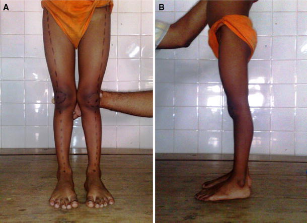
Marking of the femoral tibial axis and patella in the standing position (a) and careful positioning with full extension of the knee (b)
Fig. 2.
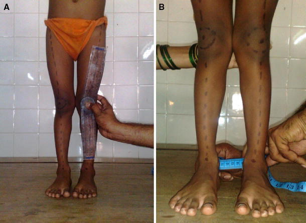
Measurement of the tibiofemoral angle(TFA) (a) and measurement of the intermalleolar distance (IMD) with the femoral condyle closely approximated (b)
No child below walking age was included in our study. Because all measurements were taken in the standing position with hips and knees in full extension and the ankles or knees of both sides touching each other, such measurements are not possible in children less than 1 year of age who cannot stand on their own. We also believe that alignment problems in most instances are picked up after the child starts walking, and this usually occurs after the age of 1.5–2 years. Thus, our study population included children greater than 1.5 years of age and we rounded off their age to the nearest integer.
Statistical analysis was conducted using SPSS version 16.0 for Windows. A paired t-test was used to assess differences between the right and left TFAs of the subjects. Student’s t-test was used to assess variables for differences between different age groups of boys and girls. Correlations between the TFA, IMD, and ICD with age, weight, and standing height measurements were studied by performing Pearson’s correlation test. A P-value of less than 0.05 was considered to be statistically significant.
Results
The age and sex distribution of the subjects, along with the mean TFA ± standard deviation (SD) at various ages and 95% confidence intervals (CIs) are highlighted in Table 1.
Table 1.
Age and sex distribution with the mean tibiofemoral angle (TFA ± standard deviation [SD]) and 95% confidence intervals (CIs)
| Age in years | Number of limbs | Males | Females | Mean TFA (±SD) | 95% CI of TFA |
|---|---|---|---|---|---|
| 2 | 34 | 24 | 10 | 1.0 ± 3.2 | (−0.12, 2.12) |
| 3 | 42 | 26 | 16 | 2.3 ± 3.1 | (1.36, 3.13) |
| 4 | 30 | 16 | 14 | 4.3 ± 1.4 | (3.77, 4.82) |
| 5 | 38 | 20 | 18 | 6.7 ± 2.1 | (6.00, 7.41) |
| 6 | 30 | 18 | 12 | 7.9 ± 2.4 | (7.01, 8.84) |
| 7 | 26 | 14 | 12 | 6.1 ± 1.7 | (5.37, 6.78) |
| 8 | 36 | 20 | 16 | 6.2 ± 2.2 | (5.41, 6.91) |
| 9 | 26 | 18 | 8 | 5.5 ± 1.9 | (4.73, 6.26) |
| 10 | 44 | 28 | 16 | 5.4 ± 2.1 | (4.75, 6.06) |
| 11 | 22 | 12 | 10 | 4.9 ± 2.4 | (3.85, 5.96) |
| 12 | 30 | 24 | 6 | 4.8 ± 2.0 | (4.06, 5.53) |
| 13 | 26 | 16 | 10 | 4.8 ± 1.6 | (4.19, 5.49) |
| 14 | 22 | 16 | 6 | 4.7 ± 1.3 | (4.11, 5.25) |
| 15 | 24 | 20 | 4 | 4.6 ± 2.0 | (3.79, 5.45) |
| Total | 430 | 272 | 158 |
Table 2 shows the mean TFA in male and female children of different age groups, with Figure 3 showing the graphical illustration. Among boys, the maximum mean TFA (7.5°) was seen in the age group of 5 years, while among girls, the maximum mean TFA (9.0°) was seen in the age group of 6 years. Thus, in Indian girls, the peak value of valgus at the knees reaches a peak at a somewhat later age than boys.
Table 2.
Mean TFA ± SD distribution amongst male and female children at different ages
| Age in years | Mean TFA of males (±SD) | Mean TFA of females (±SD) | P-value |
|---|---|---|---|
| 2 | 1.0 ± 3.1 | 1.0 ± 3.6 | 1.00 |
| 3 | 1.6 ± 3.6 | 3.4 ± 1.5 | 0.033* |
| 4 | 3.9 ± 1.4 | 4.8 ± 1.3 | 0.077 |
| 5 | 7.5 ± 2.2 | 5.9 ± 1.8 | 0.022* |
| 6 | 7.2 ± 2.6 | 9.0 ± 1.9 | 0.038* |
| 7 | 5.8 ± 1.5 | 6.4 ± 2.0 | 0.379 |
| 8 | 5.7 ± 2.5 | 6.7 ± 1.8 | 0.198 |
| 9 | 5.3 ± 2.2 | 5.9 ± 1.0 | 0.395 |
| 10 | 5.3 ± 2.3 | 5.6 ± 1.9 | 0.601 |
| 11 | 4.58 ± 2.35 | 5.30 ± 2.49 | 0.500 |
| 12 | 4.70 ± 2.15 | 5.16 ± 0.98 | 0.451 |
| 13 | 4.68 ± 1.95 | 5.10 ± 0.87 | 0.471 |
| 14 | 4.56 ± 1.45 | 5.00 ± 0.63 | 0.340 |
| 15 | 4.55 ± 2.13 | 5.00 ± 0.81 | 0.487 |
| Total | 4.61 ± 2.96 | 5.39 ± 2.45 | 0.003* |
* Significant
Fig. 3.

Comparative chart showing development of the knee angle in males and females, with valgus being charted as negative values, while varus is shown as positive knee angles (TFA)
We found statistically significant differences between the mean TFA of boys and girls in the age groups of 3, 5, and 6 years (Table 2). The mean TFA was significantly higher in Indian girls of age groups 3 and 6 years, while Indian boys of age group 5 years showed a significantly higher mean TFA. After the age of 6 years, there were no statistically significant differences between the mean TFA of Indian boys and girls.
The maximum valgus of 11° was recorded in a perfectly normal girl of age 6 years. The average mean TFA was never found to be negative (varus knee) in Indian children over 2 years of age. At the age of 2 years, the knee angle was found to be almost the same in boys and girls, with mean value 1.0°. No statistically significant differences were seen in the TFA of the left and right knees of the subjects as seen on paired t-tests.
On analysis of the ICD and IMD, we found a fair degree of correlation of these with the age of the subjects. The correlation coefficient of age with ICD (r = −0.223, P = 0.001) and IMD (r = −0.192, P = 0.005) was found to be statistically significant. Both the ICD and IMD progressively decrease with age, with the ICD decreasing at a faster rate with age.
A fair degree of correlation was also obtained between the TFA and the ICD and IMD, being negative for ICD and positive for IMD. The correlation coefficient of the mean TFA with the ICD was r = −0.247 (P = 0.000) and with the IMD, it was r = 0.162 (P = 0.018). As expected, the ICD decreases with the increase in TFA, while the IMD increases with the increase in TFA.
Height at a specific age group was not found to have any statistically significant correlation with the mean TFA or the ICD or IMD. Neither was the body mass of a child at any age group found to be statistically related to the mean TFA or the IMD. However, the body mass of the subjects was found to have a negative correlation with the ICD in the subjects (r = −0.153, P = 0.025).
Discussion
Many children present to physicians because of their parents’ apprehension about the knee angle of their children. Knowing the normal range of the knee angle is of utmost importance to avoid unnecessary radiation exposure and therapeutic interventions (orthotics or bracing) on the part of the physician. A reassurance on the part of the treating physician also helps alleviate the tension of the parents worried about the “bending of the knees” of their children. The knee angles at different age groups differ and ethnic variations are expected to be present. The physician should, therefore, be aware of these variations in the local population in order to make appropriate treatment decisions. There are studies available regarding normal development of the knee angle in whites, Chinese, Nigerians, Koreans, and Turkish children [3, 4, 9–11]. These studies have been summarized in Table 3. However, such a study defining the normal range of the knee angle in healthy Indian children is lacking in the literature.
Table 3.
Previous studies highlighting the variations of knee angles in healthy children of different ethnic groups
| Title | Authors (year) | Number of children | Age composition | Results | Conclusion |
|---|---|---|---|---|---|
| Angular and rotational profile of the lower limb in 2,630 Chinese children [4] | Cheng et al. (1991) | 2,630 | Newborn to 12 years of age | Most newborns had bowlegs, with a mean ICD of 3 cm, ranging from 0 to 5.5 cm. At 1 year of age, the ICD decreased to a mean of 0 cm, followed by a knock-knee phase with the maximum IMD of 2.8 cm at age 3 years, followed by gradual reversion back to the plateau of 0 cm at age 8 years | The largest study carried out to date in the Chinese population. It recorded bowlegs (varus) at birth, genu rectus at 1 year, maximum valgus at 3 years, and a rapid decrease in the mean IMD after 3 years of age in Chinese children, reaching 0 cm at age 8 years, with a normal range of ±3 cm |
| Knee angles and rickets in Nigerian children [11] | Oginni et al. (2004) | 2,036 | Newborn to 12 years of age | The majority of the knees were bowed (varus) in the first 6 months. At 21–23 months, the distribution of angles became strongly bimodal: about half being varus and half being valgus (knock-kneed). After this, the knee angle was found to be valgus in most of the children | Change from varus to valgus in individual infants might be sudden (a few weeks), although this changeover of the whole population appeared smooth and gradual. Varus knee alignment was uncommon after 2 years of age in Nigerian children and large knee angles between 2 and 5 years of age suggested rickets |
| The development of the tibiofemoral angle in children [5] | Salenius and Vankka (1975) | 1,279 | Newborn to 16 years of age | Pronounced varus position was recorded before the age of 1 year, which changed in valgus between 18 months and 3 years of age. After that, the valgus corrected spontaneously to about 5–6°. No gender variation was seen | There is a wide range of normal development of the knee angle which is physiological, so an operative procedure to correct the angle in normal children is seldom indicated |
| Normal development of the tibiofemoral angle in children: a clinical study of 590 normal subjects from 3 to 17 years of age [10] | Arazi et al. (2001) | 590 Turkish children | 3–17 years of age | Children aged between 3 and 17 years were found to exhibit up to 11° physiologic valgus. The maximal mean valgus angle was 9.6° at 7 years of age for boys and 9.8° at 6 years of age for girls | A measurable varus angle or a valgus angle higher than 11° only should be considered as abnormal. These higher normal values should be considered as racial differences between Turkish children and those of other races |
| Development of tibiofemoral angle in Korean children [9] | Yoo et al. (2008) | 818 limbs (433 boys and 385 girls) | Younger than 16 years old | Genu varum was found before 1 year of age, progressing to neutral at 1.5 years of age. This was followed by increasing genu valgum, with a maximum of 7.8° at 4 years, followed by a gradual decrease to approximately 5–6° of genu valgum of the adult level at 7–8 years of age | Overall patterns of the chronological changes in the knee angle were similar to those described previously in western or Asian children, but the knee angle development was delayed |
| Normal limits of knee angle in white children—genu varum and genu valgum [3] | Heath and Staheli (1993) | 196 | 6 months to 11 years | Maximum bow-leg was noted at an age of 6 months, which progressed towards neutral knee angles by the age of 18 months. The greatest mean knock knee of 8° was found at an average age 4 years, followed by a gradual decrease to a mean of <6° at 11 years of age | The presence of varus during the ages 2–12 years is considered as abnormal. Normal children also display ICDs <3 cm and IMDs <8 cm |
| Development of the clinical tibiofemoral angle in normal adolescents. A study of 427 normal subjects from 10 to 16 years of age [13] | Cahuzac (1995) | 427 | 10–16 years | Girls were found to have a constant valgus and displayed an IMD <8 cm or an ICD <4 cm. Contrary to this, boys had a varus evolution (4.4°) during the last 2 years of growth and displayed an IMD <4 cm or an ICD <5 cm | The development of the TFA follows different paths in boys and girls between 10 and 16 years of age |
Several methods [1, 3–5, 13–16] have been used to measure the knee angles in children. Radiographic methods [5, 14–16], although used most commonly, are time-consuming and have ethnical issues related to unnecessary radiation exposure in healthy children. Further, a malrotation of the limb, if not taken care of, might lead to significant errors in the measurement of the TFA. Clinical methods [3, 4, 10, 13] of measuring the knee angles are acceptable and reproducible, with the advantage of being cheap and radiation-free. We used fixed bony points to calculate the TFA and the ICD and IMD. Palpation of such fixed bony landmarks is practical and easy. Also, the measurements are less likely to be erroneous, even in obese children and in children with marked femoral bowing which might preclude the exact location of the femur.
The development of the TFA can be divided into three phases: phase I, during which knee alignment changes from an infantile physiologic varus to maximum valgus; phase II, when valgus knee alignment decreases in amount; and phase III, during which knee alignment remains stationary, i.e., the adult pattern of genu valgus. However, the age ranges at which these phases come in children has been found to be different in children with different ethnic groups [3, 4, 9–11].
In our present study, we found that none of the subjects had a varus knee alignment in the more than 3 years age groups. Also, even for the children in the 2 years age group, the mean TFA was positive, indicating a mean valgus alignment in Indian children, even at the age of 2 years. This was contrary to the findings of the largest study carried to date by Cheng et al. [4] in the Chinese population, which showed a mean varus TFA at the age of 2 years. After 3 years, the authors noted a rapid decrease in the mean IMD in the Chinese children, reaching 0 cm at the age of 8 years, with a normal range of ±3 cm. Conversely, we observed the highest mean IMD of 4.5 cm in children aged 5 years, with a minimum mean of 1 cm at the age of 9 years.
We also found that the maximum mean valgus was seen at the age of 6 years in Indian children. Thereafter, the valgus alignment decreased in Indian children and the mean TFA stabilized to lie between 4 and 5° in most of the children after the age of 10 years. Indian girls overall showed more valgus at the knees than boys (P = 0.003) and the results were statistically significant, especially at the age of 6 years (P = 0.038), when peak valgus was achieved in Indian children. The maximum TFA of 11° was seen in a 6-year-old perfectly healthy girl. We also found a boy with a valgus knee angle of 10.5° at the age of 5 years. Our study results, thus, strongly compare to those of Salenius and Vankka [5], who carried out one of the largest and the first study regarding the development of the TFA in healthy Caucasian children. As concluded by Salenius and Vankka, we agree that there is a wide variation in the normal development of the knee angle, which might be physiological. Thus, an operative procedure to correct the angle in normal children is seldom indicated.
Our results were somewhat similar to those of Oginni et al. [11], who measured the knee angles of 2,036 normal Nigerian children up to an age of 12 years. In their study, they found the majority of the knees to be bowed (varus) in the first 6 months. At 21–23 months of age, the distribution of angles became strongly bimodal: about half being varus and half being valgus (knock-kneed). After this, the knee angle was found to be valgus in most of the children. They concluded that the change from varus to valgus in individual infants might be sudden (a few weeks), although the changeover of the whole population appeared smooth and gradual. The authors also concluded that varus knee alignment was uncommon after 2 years in Nigerian children and large knee angles between 2 and 5 years suggested rickets. However, in their study, the children became maximally and uniformly knock-kneed (−7.1° ± 1.4°) between 3 and 3.5 years, with little change thereafter. This is contrary to our findings in Indian children, in whom the maximum valgus has been found to occur at an age of 5–6 years.
Our results were also comparable to those in Turkish children, as shown in a study by Arazi et al. [10]. Just like us, they noted a significantly higher degree of valgus angle than that in previous reports. The maximal mean valgus angle was 9.6° at 7 years of age for boys and 9.8° at 6 years of age for girls. These differences were considered to be racial differences between Turkish children and those of other races. Turkish children aged between 3 and 17 years were found to exhibit up to 11° physiologic valgus. The authors concluded that a measurable varus angle or a valgus higher than 11° during this period should be considered as abnormal.
Various other studies have outlined different patterns of knee development for different ethnic groups. In a study conducted by Heath and Staheli [3] in white children, using clinical measurements of the TFA and the ICD and IMD, children were maximally bow-legged at the age of 6 months and progressed toward approximately neutral knee angles (0.0°) by the age of 18 months. They found the greatest mean knock knee of 8° at the age of 4 years, which was followed by a gradual decrease to a mean of <6° at 11 years of age. Normal children aged 2–11 years had knock knee up to 12° and IMD up to 8 cm. Just like our results, the existence of bowlegs after the age of 2 years was found to be abnormal.
Yoo et al. [9] carried out a study on normal healthy Korean children, with full-length anteroposterior view standing radiographs. They found that the overall patterns of the chronological changes in the knee angle were similar to those described previously in western or Asian children, but the knee angle development was delayed, i.e., genu varum before 1 year, neutral at 1.5 years, increasing genu valgum with a maximum value of 7.8° at 4 years, followed by a gradual decrease to approximately 5–6° of genu valgum of the adult level at 7–8 years of age.
Although the use of different techniques to estimate the knee angles might be responsible for variations in observations in different studies, it is more likely that these variations are possibly due to the ethnical and racial differences that might exist in different population groups. Thus, we attempted to describe the normal development of the knee angle in Indian children which could be used as a practical and accurate screening reference, thereby, influencing decisions regarding the necessity for further clinical and/or radiologic assessment in Indian children.
From our present study, we found that physiological varus rarely persists beyond 2 years of age in Indian children. We believe that a varus alignment of the knee in Indian children after the age of 3 years might be atypical and needs detailed evaluation. The knee angle at the age of 2 years averages around 1° of valgus and is almost the same for both Indian girls and boys. Thereafter, a progressive increase in knee valgus occurs, with peak knee valgus averaging almost 8°, reached at around 6 years of age. After that, the valgus at the knee decreases and stabilizes to around 4–5° in most of the children after the age of 10 years. Indian girls show, overall, more valgus alignment of the knees as compared to boys. We believe that a valgus angle of up to 11° might be normal in Indian children in the age group 5–6 years and needs only observation. We have also provided 95% CIs of the mean TFA of Indian children in various age groups (Table 1), and these values could be used as a reference for Indian children in future studies.
We did not find the height or weight to have any significant correlation with the mean TFA in Indian children in any age group. However, we found the weight of children at a specific age to have a negative correlation with the ICD of the subject. However, this correlation was found to be weak (r = −0.153) and although statistically significant, it might not be of any clinical significance. The only possible explanation to this might be the relatively thick thighs of the heavier children, which is expected to subjectively decrease the ICD. However, considering the fact that fixed body landmarks on the body should not change even in obese children, this result is hard to explain.
There were many potential weaknesses of our study, the first being its cross-sectional nature. A cohort study would have been more meaningful and reliable, but that would have required repeated examinations and would have added expense to the study. Moreover, the compliance of the normal subjects for a regular continuous follow up for so many years is doubtful. Also, our study placed emphasis on the representation of the knee angle in different age groups rather than on sequential changes in a specific cohort group.
A second potential weakness of our study was the wide range of data and the large standard deviations encountered in our results, which hinder the establishment of a ‘cut-off’ value for different age groups. Although general trends in knee angle changes were detected by the study, the determination as to whether a specific limb is malaligned or not is difficult. Nevertheless, we believe that our study provides a fair assessment of the development of the knee angle and further evaluation could be carried out in children with markedly varying knee angles.
Despite the fact that the study was carried out in a single tertiary hospital in Mumbai, India, the study population was found to be representative of the entire Indian population. This is because Mumbai (the capital of Maharashtra) is a cosmopolitan city that homes people from all over the country. Statewise distribution revealed a surprise finding of less than 25% of the children belonging to natives of the state of Maharashtra. The northern states of Bihar, Uttar Pradesh, Punjab, and Rajasthan contributed to almost 40% of the subjects. This might be due to a high influx of immigrants from these states into the metropolitan city of Mumbai. This finding of ours, in turn, helps us generalize our results to the entire Indian population. We agree that only a multi-institutional study from all parts of the country involving a large number of children can exactly predict the normal development of the knee angle in healthy children of India. However, at the same time, we believe that such a large-scale study might not be possible and our study, despite its potential limitations, reasonably represents the highly diverse Indian population.
Another limitation of our study could be its dependence on clinical measurements whose reliability and reproducibility deserve attention. However, during our study, we ensured that the measurements were taken by three separate examiners who were blinded with respect to each other’s measurements. We always re-evaluated the children in which significant discrepancies were found in the measurements by different examiners, and if these discrepancies persisted, the children were excluded from our study. Although exact reproducibility of the data might be doubted, we believe that the general trends of the development of the TFA and the ICD and IMD have been well represented in our study.
To summarize, we provide the data of the normal development of the knee angle in a small group of healthy Indian children using clinical measurements. Our study shows that the overall pattern of development might be slightly different in Indian children, especially in Indian girls, with early reversal of physiological varus (<2 years of age) and a late peak of the maximal valgus at the knee (6 years of age). We also provide an elaborate data of the mean TFA for different age groups. We believe that this data, though not totally representative of the general trend in Indian children, could still be considered as a guide while evaluating lower limb alignment in Indian children from 2 to 15 years of age.
References
- 1.Engel GM, Staheli LT. The natural history of torsion and other factors influencing gait in childhood. A study of the angle of gait, tibial torsion, knee angle, hip rotation, and development of the arch in normal children. Clin Orthop Relat Res. 1974;99:12–17. doi: 10.1097/00003086-197403000-00002. [DOI] [PubMed] [Google Scholar]
- 2.Hachiya M. A roentgenographical study on chronological changes in genu varum and valgum in children (author’s transl) Nippon Seikeigeka Gakkai Zasshi. 1981;55:31–43. [PubMed] [Google Scholar]
- 3.Heath CH, Staheli LT. Normal limits of knee angle in white children—genu varum and genu valgum. J Pediatr Orthop. 1993;13(2):259–262. [PubMed] [Google Scholar]
- 4.Cheng JC, Chan PS, Chiang SC, Hui PW. Angular and rotational profile of the lower limb in 2,630 Chinese children. J Pediatr Orthop. 1991;11(2):154–161. doi: 10.1097/01241398-199103000-00003. [DOI] [PubMed] [Google Scholar]
- 5.Salenius P, Vankka E. The development of the tibiofemoral angle in children. J Bone Joint Surg Am. 1975;57(2):259–261. [PubMed] [Google Scholar]
- 6.McDade W. Bow legs and knock knees. Pediatr Clin North Am. 1977;24(4):825–839. doi: 10.1016/s0031-3955(16)33501-5. [DOI] [PubMed] [Google Scholar]
- 7.Morley AJ. Knock-knee in children. Br Med J. 1957;2(5051):976–979. doi: 10.1136/bmj.2.5051.976. [DOI] [PMC free article] [PubMed] [Google Scholar]
- 8.Sherman M. Physiologic bowing of the legs. South Med J. 1960;53:830–836. doi: 10.1097/00007611-196007000-00002. [DOI] [PubMed] [Google Scholar]
- 9.Yoo JH, Choi IH, Cho TJ, Chung CY, Yoo WJ. Development of tibiofemoral angle in Korean children. J Korean Med Sci. 2008;23(4):714–717. doi: 10.3346/jkms.2008.23.4.714. [DOI] [PMC free article] [PubMed] [Google Scholar]
- 10.Arazi M, Oğün TC, Memik R. Normal development of the tibiofemoral angle in children: a clinical study of 590 normal subjects from 3 to 17 years of age. J Pediatr Orthop. 2001;21(2):264–267. [PubMed] [Google Scholar]
- 11.Oginni LM, Badru OS, Sharp CA, Davie MW, Worsfold M. Knee angles and rickets in Nigerian children. J Pediatr Orthop. 2004;24(4):403–407. doi: 10.1097/01241398-200407000-00011. [DOI] [PubMed] [Google Scholar]
- 12.Mesa PA, Yamhure FH. Percutaneous hemi-epiphysiodesis using transphyseal cannulated screws for genu valgum in adolescents. J Child Orthop. 2009;3(5):397–403. doi: 10.1007/s11832-009-0203-8. [DOI] [PMC free article] [PubMed] [Google Scholar]
- 13.Cahuzac JP, Vardon D, Sales de Gauzy J. Development of the clinical tibiofemoral angle in normal adolescents. A study of 427 normal subjects from 10 to 16 years of age. J Bone Joint Surg Br. 1995;77(5):729–732. [PubMed] [Google Scholar]
- 14.Sabharwal S, Zhao C, Edgar M. Lower limb alignment in children: reference values based on a full-length standing radiograph. J Pediatr Orthop. 2008;28(7):740–746. doi: 10.1097/BPO.0b013e318186eb79. [DOI] [PubMed] [Google Scholar]
- 15.Levine AM, Drennan JC. Physiological bowing and tibia vara. The metaphyseal-diaphyseal angle in the measurement of bowleg deformities. J Bone Joint Surg Am. 1982;64(8):1158–1163. [PubMed] [Google Scholar]
- 16.Sharrard WJW. Knock knees and bow legs. Br Med J. 1976;1(6013):826–827. doi: 10.1136/bmj.1.6013.826. [DOI] [PMC free article] [PubMed] [Google Scholar]