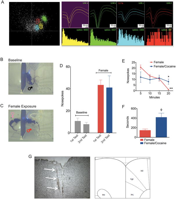
Figure 1.
Single unit recordings in mPFC of awake behaving male rats presented with a sexually receptive female. A) Representative single units isolated from the medial prefrontal cortex. B) Nosepoke responses before and C) following presentation of a sexually receptive female. D) Total amounts of nosepokes made during two baseline sessions and two female presentation sessions. E) Nosepokes over time during the presentation of a receptive female rats before and after cocaine administration (*significant difference between female and female/cocaine; **significant difference between 20 and 5 minutes). F) Latency in seconds (± standard error) to initiate nosepoke behavior after the introduction of a receptive female on the other side of the dividing wall (φ significant difference between female and female/cocaine). G) Cresyl violet stained section of the mPFC showing an example electrode lesion and its corresponding anatomical location according the Paxinos and Watson atlas of the rat brain.

