Abstract
Objective
This study describes a method for inducing osteopenia using bilateral ovariectomy (OVX), which causes significant changes in bone mineral density (BMD) in rats.
Methods
Twenty-five 10-week-old female Sprague Dawley rats were used. Five rats were euthanized after two weeks, and BMD was measured in their femora. The other 20 rats were assigned to one of two groups : a sham group (n = 10), which underwent a sham operation, and an OVX group (n = 10), which underwent bilateral OVX at 12 weeks of age. After six weeks, five rats from each group were euthanized, and BMD was measured in their femora. The same procedures were performed in the remaining rats form each group eight weeks later.
Results
The femur BMD was significantly lower in the six-week OVX group than in the six-week sham group, and in the eight-week OVX group than in the eight-week sham group.
Conclusion
Bilateral OVX is a safe method for creating an osteopenic rat model. The significant decrease in BMD appears six weeks after bilateral OVX.
Keywords: Animal model, Osteoporosis, Rat, Ovariectomy
INTRODUCTION
The incidence of osteoporosis, which affects bone strength and increases the risk of fracture, is higher in postmenopausal women and elderly men8). Spinal fusion is the most common procedure used in various bone graft surgeries and the gold standard treatment for degenerative spine disease associated with severe back pain2,12). The number of spinal fusion has increased annually as population ages. Elderly patients with bone fragility caused by osteoporosis are treated with anti-resorptive drugs such as bisphosphonate and selective estrogen receptor modulators after spinal arthrodesis3,9,12,15,16). It is important to understand the effects of the anti-resorptive drugs on patients with osteoporosis. Several articles have been written on the use of antiresorptive treatment in osteoporotic animal models4,7,12). Before assessing the effects of anti-resorptive drugs in an osteoporotic animal model, it is important to identify whether the animal model produce an osteoporotic state. The procedure for creating an animal model of osteoporosis and the duration of osteoporosis bave not been described in detail in any articles. Researchers wishing to use such procedures for their own study bone mineral density (BMD) need such details. In this study, we focused on a method for inducing osteopenia in a rat model using bilateral ovariectomy (OVX) and identified the time when BMD decreased significantly in this model.
MATERIALS AND METHODS
Animals
Twenty-five female Sprague Dawley rats, 10 weeks of age (Orientbio, Gyeonggi, Korea) were housed, two per ventilated cage, in a specific pathogen-free room with a 12-hour light-dark cycle. The rats were allowed free access to tap water and commercially standard rodent food.
Experimental design
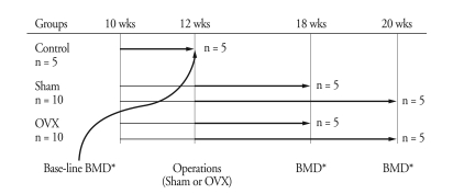
After two weeks, five 12-week-old rats (control group) were euthanized, their femora were removed, and BMD was measured in the femora. The other 20 rats were assigned to one of two groups. The sham group (n = 10), underwent a sham operation at 12 weeks of age, and the OVX group (n = 10), underwent bilateral OVX, to induce osteopenia, at 12 weeks of age. Six weeks after the OVX or sham operation, five rats from each of the two groups were euthanized, and the femora were removed and BMD was measured. Eight weeks after the OVX or sham operation, the remaining rats in each group were euthanized and BMD of their femora was measured (Fig. 1). All rats were euthanized using CO2 inhalation.
Fig. 1.
Experimental groups and time schedule. Twenty-five 10-week-old female rats are housed initially. Two weeks later, five rats are sacrificed obtaining femora to determine base-line bone mineral density (BMD). Remained rats are randomly divided into two groups and each group undergoes sham operation and ovariectomy (OVX) respectively. Six and 8 weeks later, five rats from the each two groups are euthanized to get femora that are used in measuring BMD after OVX. *The time of euthanasia
OVX procedure
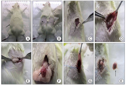
Anesthesia was induced with 5% isoflurane and maintained with 2.5% isoflurane; Oxygen was supplied through a coaxial nose cone during anesthesia. We performed bilateral OVX using a double dorso-lateral approach6). The anesthetized rat was laid prone on the operating table and fixed using sticking plaster. The bulged area on the back was shaved bilaterally (Fig. 2A). The ovaries were found on both sides of the abdomen, a little below the kidney; we chose as the skin incision site a position just medial to the most bulging part of the back6). This site is less obvious in very young or thin rats, which may lack the bulge. To make the incision, a thumb was placed at the uppermost proximal area of the thigh. The medial portion of the base of the distal phalanx was the incision site (Fig. 2B). A 1.5 cm skin incision was made to expose the dorsolateral abdominal muscles such as the external oblique muscle (Fig. 2C). Entrance to the peritoneal cavity was gained by dissecting the muscle, which revealed the adipose tissue surrounding the ovary (Fig. 2D). The adipose tissue was pulled away until the ovary and uterine tube were identified (Fig. 2E, F). The periovarian fat with the ovary was pulled away from the incision site gently to prevent detachment of a small piece of ovary, which may fall into the abdominal cavity where it may be reimplanted and carry on its normal function11). After identifying the ovary and uterine horn, ligation was performed at the distal uterine horn to remove the ovarian tissue completely in one action (Fig. 2G, H)6). The horn was returned to the abdominal cavity and the muscle and skin were sutured.
Fig. 2.
Procedures of ovariectomy in rat. Anesthetized rat is laid prone on operating table. Thick black arrow : shaving site (A). Skin incision point is located just medial portion of the most bulged area (★) or a thumb used to find the incision point (dotted arrow) (B). External oblique muscle is exposed after skin incision. Thick black arrow : External oblique muscle (C). After the muscle dissection, peritoneal space and adipose tissue surrounding ovary are exposed. Dotted arrow : adipose tissue surrounding ovary (D). The surrounding fat must be gently pulled to avoid detachment of small pieces of ovary (E). This shows ovary (thick black arrow) and uterine horn (dotted arrow) surrounded by fat (F). Ligation must undergo at distal uterine horn in order to get rid of total ovary at a time (G). Ovary surrounded by fat is removed totally (thick black arrow) (H).
Femur BMD measurement
Rats were sacrificed at 12, 18, and 20 weeks, and both femora from each rat were harvested. BMD was measured in the femora using a dual-energy X-ray absorptiometer (DEXA; Lunar PIXImus, Madison, WI, USA) and Lunar PIXImus 2 2.0 software.
Statistical analysis
Statistical analysis was performed using SPSS software (version 12.0, Chicago, IL, USA). The Mann-Whitney test was used to compare values between two groups (sham and OVX), and the Wilcoxon signed-rank test was used to compare mean values of BMD within the same group. Probability values < 0.05 were considered significant.
RESULTS
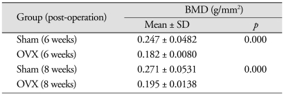
There was no procedure-related death in this study. Baseline BMD measured in the 12-week-old rats (control group) was 0.184 ± 0.0098 g/mm2. The mean femur BMD was significantly lower six weeks after OVX in the OVX rats than in the corresponding sham rats (0.184 ± 0.0098 g/mm2 vs. 0.247 ± 0.0482 g/mm2, p = 0.000). The mean femur BMD was also significantly lower eight weeks after OVX in the OVX rats than in the corresponding sham rats (0.195 ± 0.0138 g/mm2 vs. 0.271 ± 0.0531 g/mm2, p = 0.000) (Table 1).
Table 1.
The changes of BMD of Sham and OVX groups
BMD : bone mineral density, OVX : ovariectomy, SD : standard deviation
DISCUSSION
Many elderly women experience postmenopausal osteoporosis, and understanding the progression of osteoporosis is helpful for achieving a good outcome after spinal fusion in patients with osteoporosis. Animal models of osteoporosis are crucial for anticipating a treatment's efficacy and safety as measured by bone quality. OVX is the most frequently used model for studying the events associated with postmenopausal osteopenia10). The rat skeleton is more sensitive to the loss of ovarian hormones, and an OVX rat bone loss model is suitable for researching issues that are relevant to postmenopausal bone loss1,5,10). However, the exact procedures for creating an OVX rat model have not been clearly published in detail, thus we describe our procedures in this article.
Ovariectomy can be performed in some different methods. The surgical ways depend on ways of approach and incision. Approach has done through dorsal and ventral (abdominal) sides and the ways of skin incision compromise single midline dorsal, double dorso-lateral and single transverse lateral incisions6,11,14). We performed ovariectomy of rat using double dorso-lateral skin incisions. Because the double dorso-lateral skin incisions need not to suture muscle and have shorter length of skin incision than that of single midline dorsal skin incision, we chose the double dorso-lateral skin incisions6,14). This article reports the stepwise description of OVX using the double dorso-lateral skin incisions.
When using an OVX rat model, researchers may be uncertain about the appropriate age and when BMD decreases significantly after OVX. The skeletal response to OVX is more responsive in the growing than in the aged female rat13). Because the endocrine system and genital gland in rats mature at the age of three months and the muscles and skeleton are well formed at this time, an OVX model should use rats older than three months5,8). Therefore, we selected 12-week-old rats as the experimental model in this study. Some reports have shown a significant decrease in bone mass four weeks after OVX in the rat and marked decreases in BMD eight or 10 weeks after OVX1,8). Although we observed a significant decrease in BMD six weeks after OVX, the typical osteoporotic profile is seen in bone marrow eight weeks after OVX8). Therefore, a better rat model would be to measure BMD and other bone-related variables eight weeks after OVX when evaluating antiresorptive drugs or procedures. We did not evaluate the change in cellularity in the bone marrow after OVX, and bone marrow pathology should be evaluated in future studies.
CONCLUSION
Using bilateral OVX, the osteopenic rat model can be achieved easily, which we described in detail. A significant decrease in femur BMD occurs within six weeks after bilateral OVX in the rat.
Acknowledgements
All procedures in this study were approved by Institutional Animal Care and Use Committee of Clinical Research Institute, Seoul National University Hospital.
References
- 1.Bauss F, Dempster DW. Effects of ibandronate on bone quality : preclinical studies. Bone. 2007;40:265–273. doi: 10.1016/j.bone.2006.08.002. [DOI] [PubMed] [Google Scholar]
- 2.Boden SD. Overview of the biology of lumbar spine fusion and principles for selecting a bone graft substitute. Spine (Phila Pa 1976) 2002;27:S26–S31. doi: 10.1097/00007632-200208151-00007. [DOI] [PubMed] [Google Scholar]
- 3.Bridwell KH, Sedgewick TA, O'Brien MF, Lenke LG, Baldus C. The role of fusion and instrumentation in the treatment of degenerative spondylolisthesis with spinal stenosis. J Spinal Disord. 1993;6:461–472. doi: 10.1097/00002517-199306060-00001. [DOI] [PubMed] [Google Scholar]
- 4.Huang RC, Khan SN, Sandhu HS, Metzl JA, Cammisa FP, Jr, Zheng F, et al. Alendronate inhibits spine fusion in a rat model. Spine (Phila Pa 1976) 2005;30:2516–2522. doi: 10.1097/01.brs.0000186470.28070.7b. [DOI] [PubMed] [Google Scholar]
- 5.Kalu DN. The ovariectomized rat model of postmenopausal bone loss. Bone Miner. 1991;15:175–191. doi: 10.1016/0169-6009(91)90124-i. [DOI] [PubMed] [Google Scholar]
- 6.Lasota A, Danowska-Klonowska D. Experimental osteoporosis-different methods of ovariectomy in female white rats. Rocz Akad Med Bialymst. 2004;49(Suppl 1):129–131. [PubMed] [Google Scholar]
- 7.Lehman RA, Jr, Kuklo TR, Freedman BA, Cowart JR, Mense MG, Riew KD. The effect of alendronate sodium on spinal fusion : a rabbit model. Spine J. 2004;4:36–43. doi: 10.1016/s1529-9430(03)00427-3. [DOI] [PubMed] [Google Scholar]
- 8.Lei Z, Xiaoying Z, Xingguo L. Ovariectomy-associated changes in bone mineral density and bone marrow haematopoiesis in rats. Int J Exp Pathol. 2009;90:512–519. doi: 10.1111/j.1365-2613.2009.00661.x. [DOI] [PMC free article] [PubMed] [Google Scholar]
- 9.McGuire RA, Amundson GM. The use of primary internal fixation in spondylolisthesis. Spine (Phila Pa 1976) 1993;18:1662–1672. doi: 10.1097/00007632-199309000-00015. [DOI] [PubMed] [Google Scholar]
- 10.Miller SC, Bowman BM, Jee WS. Available animal models of osteopenia--small and large. Bone. 1995;17:117S–123S. doi: 10.1016/8756-3282(95)00284-k. [DOI] [PubMed] [Google Scholar]
- 11.Parhizkar S, Ibrahim R, Latiff LA. Incision choice in laparatomy : a comparison of two incision techniques in ovariectomy of rats. World Apple Sci J. 2008;4:537–540. [Google Scholar]
- 12.Takahata M, Ito M, Abe Y, Abumi K, Minami A. The effect of anti-resorptive therapies on bone graft healing in an ovariectomized rat spinal arthrodesis model. Bone. 2008;43:1057–1066. doi: 10.1016/j.bone.2008.08.124. [DOI] [PubMed] [Google Scholar]
- 13.Thompson DD, Simmons HA, Pirie CM, Ke HZ. FDA Guidelines and animal models for osteoporosis. Bone. 1995;17:125S–133S. doi: 10.1016/8756-3282(95)00285-l. [DOI] [PubMed] [Google Scholar]
- 14.Waynforth HB. Experimental and surgical technique in the rat. New York: Academic press; 1980. pp. 161–163. [Google Scholar]
- 15.West JL, 3rd, Bradford DS, Ogilvie JW. Results of spinal arthrodesis with pedicle screw-plate fixation. J Bone Joint Surg Am. 1991;73:1179–1184. [PubMed] [Google Scholar]
- 16.Zdeblick TA. A prospective, randomized study of lumbar fusion. Preliminary results. Spine (Phila Pa 1976) 1993;18:983–991. doi: 10.1097/00007632-199306150-00006. [DOI] [PubMed] [Google Scholar]


