Cough-induced abdominal intercostal hernias are extremely rare, with only seven cases described in the literature. We report the case of a 55-year-old man with a painless right-sided abdominal lump, who had previously fractured a rib due to severe coughing. A chest X-ray was inconclusive, but ultrasound and computed tomography scans showed herniation of the liver between the seventh and eighth ribs on the right.
Case report
A 55-year-old man presented with a five-week history of a painless enlarging lump in the right upper quadrant. He recalled being told he had a fractured rib several years earlier after a particularly severe coughing episode related to chronic obstructive pulmonary disease (COPD).
Physical examination revealed a soft reducible mass on the lateral aspect of the right chest between the seventh and eighth ribs. The mass measured 10 cm × 8 cm. A cough impulse was felt, as well as a foreshortened rib anteriorly. No bowel sounds were audible over the mass.
The man had an eight-year history of COPD, and had continued to smoke 30 cigarettes a day. This required long-term inhaled corticosteroid therapy. He had a number of co-morbidities including obesity, hypertension, kypho-scoliosis and longstanding back pain. He was registered disabled, with dyspnoea and back pain limiting his exercise tolerance to 20 yards on the flat.
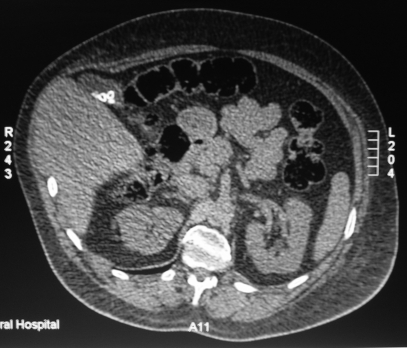
Chest radiographs were inconclusive, showing an old fracture of the eighth rib and right lower chest wall deformity. An ultrasound scan found a swelling with deviation of the antero-lateral abdominal wall in keeping with a hernia. Computed tomography revealed a defect in the right lower chest wall between the seventh and eighth ribs with the liver herniating through it (Figure 1). There was also a defect in the 11th rib posteriorly.
Figure 1.
CT scan showing herniation of the liver through the seventh intercostal space
As multiple co-morbidities put him at high risk for surgical intervention, it was decided not to operate but to follow him up clinically with advice to report any symptoms promptly.
Discussion
Most intercostal hernias involve penetrating or blunt abdominal injury, and so cough-induced abdominal intercostal hernia is a rare occurrence. To date, there are only seven cases described in the literature.
All of these occurred in patients over the age of 50 years, only two of them in women. Four of the cases were on the right, and three on the left. The hernial contents in these patients were lung in two, liver in two, small bowel in two, and stomach in one case. All had predisposing factors such as pneumonia, COPD, asthma and steroid therapy.
The first case was reported by Testlin and Ledon in 1970. The patient, who 25 years earlier had been involved in a car accident while a prisoner of war in Poland, developed pneumonia associated with a severe cough with pain and bruising in the lower right ribcage. The enlargement appeared over the same location a few days later, and was found to be caused by herniation of the inferior lobe of the right lung. The hernia was repaired and the breach sutured up.1
There are two previous cases involving herniation of the liver. The first was recorded in 1993, in a 75-year-old woman with asthma. The woman also reported a recent injury to the right side of her chest wall. Chest radiograph and computed tomogram confirmed both lung and liver herniation between the ninth and tenth ribs, with associated rib fractures. The hernia was reduced and a Gore-Tex® (polytetrafluoroethylene) mesh sewn onto the extrathoracic wall of the defect.2
One case occurred in a 51-year-old smoker who had experienced a severe coughing spell two months earlier, and DEXA scan later revealed low bone mineral density. In this patient, magnetic resonance imaging (MRI) was used to show protrusion of the liver through the chest wall. Initial surgery using endogenous tissue reinforcement failed, requiring the patient to undergo a second procedure four months later, this time with insertion of a Marlex mesh.3
Similar instances have taken place in patients with COPD, sarcoidosis and severe asthma exacerbation. Following incarceration, two of these patients were treated with and two without mesh repair.4–7
Development of an intercostal hernia may occur acutely or over a number of years. Chronic severe coughing can tear the intercostal muscles, and even fracture underlying ribs. Positive intrathoracic pressure which occurs during expiration, coughing, vomiting and defecation, then forces the contents out through weakened areas of the chest wall.
The chest wall is anatomically weaker from the chostochondral junction to the sternum anteriorly due to lack of external intercostal muscles, and from the costal angle to the vertebrae posteriorly due to lack of internal intercostal muscles.8 Areas which have previously suffered trauma are particularly susceptible, something we noted in a number of the cases we reviewed. As in any intercostal hernia the hernial contents are at risk of subsequent incarceration and strangulation, but there is little evidence as to the frequency of this complication.9
Intercostal hernias are suggested by the patient's history and examination, the usual finding being a reducible bulge on the thoracoabdominal wall. Both ultrasound and CT have been used successfully to show the hernia. Ultrasound will not determine the exact contents of the hernia, and so CT may be preferable when contemplating surgery. Chest X-ray is unlikely to provide enough information on the hernia itself, but may show evidence of previous injury. The unusual site for such a lump as well as the absence of any history of trauma may cause diagnostic difficulty, but careful examination and appropriate use of imaging can overcome this. Incarceration is the key indication for surgical intervention. One approach to treatment has been to reduce the hernia and suture the defect, with rib approximation if appropriate. However, this has been associated with recurrence, and reinforcement with a prosthetic mesh may be preferred.
DECLARATIONS
Competing interests
None declared
Funding
None
Ethical approval
Written informed consent to publication has been obtained from the patient or next of kin
Guarantor
AC
Contributorship
ATC reviewed the evidence, drafted and revised the paper; EM performed the literature search and revised the paper
Acknowledgements
None
Reviewer
Stefan Limmer
References
- 1.Testelin GM, Ledon F, Giordano A A propos d'un cas de hernie intercostale abdominale. Mem Acad Chir (Paris) 1970;96:569–70 [PubMed] [Google Scholar]
- 2.Fiane AE, Nordstrand K Intercostal pulmonary hernia after blunt thoracic injury: two case reports. Eur J Surg 1993;159:379–81 [PubMed] [Google Scholar]
- 3.Losanoff JE, Richman BW, Jones JW Recurrent intercostal herniation of the liver. Ann Thorac Surg 2004;77:699–701 [DOI] [PubMed] [Google Scholar]
- 4.Croce EJ, Mehta VA Intercostal pleuroperitoneal hernia. Thorac Cardiovasc Surg 1979;77:856–7 [PubMed] [Google Scholar]
- 5.Cole FH, Miller MP, Jones CV Transdiaphragmatic intercostal hernia. Ann Thorac Surg 1986;41:565–6 [DOI] [PubMed] [Google Scholar]
- 6.Rogers FB, Leavitt BJ, Jensen PE Traumatic transdiaphragmatic intercostal hernia secondary to coughing: case report and review of the literature. J Trauma 1996;41:902–3 [DOI] [PubMed] [Google Scholar]
- 7.Kallay N, Crim L, Dunagan DP, Kavanagh PV, Meredith W, Haponik EF Massive left diaphragmatic separation and rupture due to coughing during an asthma exacerbation. South Med J 2000;93:729–31 [PubMed] [Google Scholar]
- 8.Unlu E, Temizoz O, Cagli B Acquired spontaneous intercostal abdominal hernia: Case report and a comprehensive review of the world literature. Australasian Radiology 2007;51:163–7 [DOI] [PubMed] [Google Scholar]
- 9.Nielsen JS, Jurik AG Spontaneous Intercostal hernia with subsegmental incarceration. European J Cardiothorac Surg 1989;3:562–4 [DOI] [PubMed] [Google Scholar]


