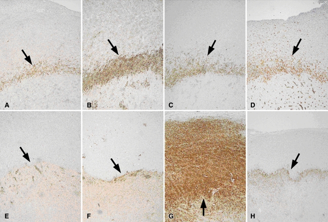
Figure 2:
Apoptosis following combined RF ablation and liposomal chemotherapy. A–H, Photomicrographs show rim staining of cleaved caspase-3 (arrows) surrounding zone of coagulation. (Original magnification, ×4.) A–D, Sections from tumors excised at 4 hours. E–H, Sections from tumors excised at 24 hours. A, E, RF ablation alone. B, F, RF ablation–doxorubicin. C, G, Paclitaxel–RF ablation. D, H, Paclitaxel–RF ablation–doxorubicin. At 4 hours, RF ablation–doxorubicin demonstrated the strongest staining for cleaved caspase-3 expression. At 24 hours, paclitaxel–RF ablation demonstrated the greatest overall staining, seen extending to tumor margin on G.

