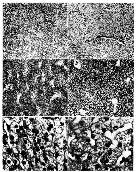
Fig. 3.
Photomicrographs of sections from a liver which had been subjected to partial portacaval transposition. The part of the liver which received vena caval blood (panels on left) shows shrinkage of the liver lobules, depletion of centrilobular glycogen, and atrophy of hepatocytes when compared with the part which received portal blood (panels on right). (Upper, reticulin stain. ×30. Middle, periodic acid–Schiff. ×40. Lower, hematoxylin and eosin. ×250.)

