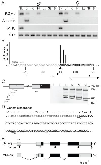
Figure 1. Establishing mouse RGMc gene structure.
A. RGMc mRNA is expressed in striated muscle and in the liver. Results of RT-PCR experiments for RGMc, albumin, skeletal muscle myosin heavy chain polypeptide 3 (MHC), and S17 mRNAs using RNA from tissues (Sk, skeletal muscle (gastrocnemius); Li, liver; K, kidney; Ht, heart; Lu, lung; St, stomach; Br, brain) of adult male (left) and female (right) mice. B. Mapping the 5′ end of the mouse RGMc gene by 5′ RACE using mouse skeletal muscle RNA. The number of clones is graphed on the y-axis above the corresponding location of the 5′ residue on the x-axis. The putative transcription start site is denoted as +1 (arrowhead), with exon 1 in upper case letters. A potential TATA box is labeled, and primers II - IV used in (C) are indicated below the sequence. C. Mapping the 5′ end of the mouse RGMc gene by RT-PCR with cDNA from mouse skeletal muscle RNA and overlapping PCR primers located in different parts of RGMc exon 1, as seen on the gene map to the left (see Supplemental Table 1 for DNA sequences of primers). Exons 1 and 2 are depicted as boxes, with the 5′ UTR in gray and the protein coding region in black, and introns and flanking DNA as horizontal lines. Results are seen to the right, and molecular weight markers are indicated (see Supplemental Fig. 1A for results with heart and liver RNA). In addition to mapping the 5′ end of exon 1, the results also show that alternative RNA splicing occurs between exons 1 and 2. D. DNA sequence of the junction between intron 1 and exon 2 of the mouse RGMc gene. Exon 2 is in upper case letters; the locations of alternative RNA splicing are noted by chevrons, with the –AG splice-acceptor residues underlined. E. Organization of the mouse RGMc gene and mRNAs. The gene contains 4 exons (boxes) and three introns (thin lines). The transcription start site is denoted as a bent arrow, and the polyadenylation site as a vertical arrow. The three RGMc mRNAs are diagramed below, and result from use of alternative splice acceptor sites at the 5′ end of exon 2.

