Abstract
The Matthiass posture test is a clinical test to detect posture changes in children and adolescents. Aim of this study was to objectify this test using a dynamic rasterstereographic measuring device. We examined 31 healthy athletes during a modified Matthiass test with a dynamic rasterstereographic measuring system. Hereby the trunk inclination, kyphosis and lordosis angle were measured. The trunk inclination decreased by about 50% of the basic value just by raising the arms. Additional weight loads of only 5% body weight (bw) resulted in significant changes of the posture (lordosis and kyphosis angle) during this test. With this rasterstereographic measuring device it seems to be possible to determine spinal posture changes under dynamic conditions. The results suggest that additional weights of 5% bw during the Matthias-test are enough to create significant deviations in posture parameters, even in healthy subjects.
Keywords: Spine, Posture, Rasterstereography, Dynamic examination, Matthiass test
Introduction
The analysis of the human back shape, especially in children and adolescents, is very demanding because of the complex interaction between anatomical, muscular and psychological factors. To prevent spine deformities and posture asymmetries an early and objective diagnosis of posture changes is essential [21]. Until today the gold standard in the detection of pathological changes of the spine shape, e.g. like in scoliosis, are the clinical examination along with radiological techniques such as X-ray, computer tomography and MRI [5, 8, 9, 15]. A considerable disadvantage of these procedures is the radiation which leads, particularly in young patients, to a very critical deployment for follow-up examinations [1, 14, 19]. Simple but inaccurate and poor repeatable methods were developed for follow-up examinations of the spine like the kyphometer or the curve ruler. Further, more advanced methods for the detection of the spinal shape like, e.g. the video system VICON® or the ultrasound-based Zebris-System®, are based upon the placement of marker on the skin over bony landmarks of the patient [20, 24]. But all these systems tend to lead to measuring errors due to a possible shift of the skin over the underlying bony structures. A precise, radiation-free and inexpensive method for detecting the spinal posture, especially for follow-up examinations, is the so-called rasterstereography [2–4, 6, 8, 22, 23]. The surface analysis occurs from the projection of light-line-patterns on the back of the patient. Anatomical landmarks can be detected automatically by assigning concave and convex areas to curved light patterns. By means of certain anatomical fixed points the system is able to calculate a three-dimensional model of the human spine. From this model, clinical relevant parameters such as e.g. the trunk inclination, the kyphosis or lordosis angle of the spine can be determined [2–4, 6, 10, 11, 22, 23]. In addition to the static analysis of the spinal shape for evaluating asymmetries and imbalances, the evaluation of spinal posture changes over a defined time period seems to be important [5]. A clinical test which makes posture changes visible and which has proved itself as a valid tool in posture diagnosis is the Matthiass posture test [7, 16, 18]. Up to now the subjective evaluation of the posture by the examiner seems to be problematic [13]. Therefore, the aim of this present study was to combine a modified Matthiass test with a dynamic rasterstereographic examination to possibly objectify this posture test.
Methods
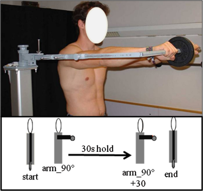
Thirty-one youth athletes (6 female, 25 male) with an average age of 14.3 years (SD 1.6 years) participated in this examination. The mean weight of the test subjects was 56.68 kg (SD 12.6 kg) and the mean body height was 166.48 cm (SD 12.9 cm). The parents of all adolescents gave informed consent and the local ethics board of the Eberhard Karls University of Tubingen (Germany) approved the study protocol. Before participating in the study every participant was examined orthopedically and was found with normal posture. In our study we modified the posture test according to Matthiass–Groeneveld [7]. To provide a standardized arm position during the test we attached different weights on a lever arm. The lever arm was then adapted to the individual arm length and placed in the centre of rotation of each probands shoulder joint (Fig. 1). During the modified Matthiass test the participants lifted weights that were related to the body weight (bw) of +0, +5, +7.5 and +10% bw, into different test positions. The measurement started from a still position (start) by raising the arms about 90° into anteversion. Then the athletes were instructed to keep their arms with the specific extra weight for 30 s in this position (arm_90°). 30 s later an acoustic signal from a timekeeper signalled the end of the holding period (arm_90° + 30). Thereafter the probands were instructed to lower their arms controlled and slowly. When the starting point was reached again the examination was stopped (end). For further analysis, all recorded images were saved on a personal computer. All examinations were performed with the rasterstereographic measuring system formetric 4D®, an advancement of the static rasterstereographic system formetric 3D® (Diers International GmbH, Schlangenbad, Germany). The device used in this study was a prototype for dynamic rasterstereographic measurements. It was equipped with a digital video camera, which was able to measure with a maximum recording frequency of 15 Hz. For the analysis of the recordings we used a computer software (Draco, Diers International GmbH, Schlangenbad, Germany) that was able to calculate certain parameters from each frame of the dynamic measurement. Based on the automatic detection of the dorsal process of the seventh vertebral body (VP) and of the right and left lumbar dimple (DR and DL), the software was able to calculate the trunk length, the trunk inclination as well as the kyphosis and lordosis angle automatically. The trunk length accorded the distance between the VP and the middle of the two lumbar dimples (DM) and the trunk inclination was set as the angle in ° between the vertical and the connection line between VP and DM. 0° trunk inclination means that the VP is positioned exactly vertical over the DM. The kyphosis angle is calculated by the tangent produced from the coordinates of the VP and the reversal point from kyphosis to lordosis (ITL). The lordosis angle is determined by the tangent produced from ITL and DM [5]. In pre-examinations, we determined the accuracy of the measuring system under dynamic conditions with values of 3.29 mm (mean value) for the trunk length and 1.04 mm (mean value) for the dimple distance.
Fig. 1.
Male test person during the Matthiass test using the lever arm with an extra weight. All extra weights were related to the body weight of the test persons. The schematic drawing shows the different phases of the Matthiass test that were analysed with the rasterstereographic measuring device
Using an ANOVA test we calculated the mean values and standard deviations using the statistical programme SPSS® (Version 14 SPSS, Chicago, IL, USA). The significance level was set at P < 0.05. Statistical differences were calculated by the dependant t test.
Results
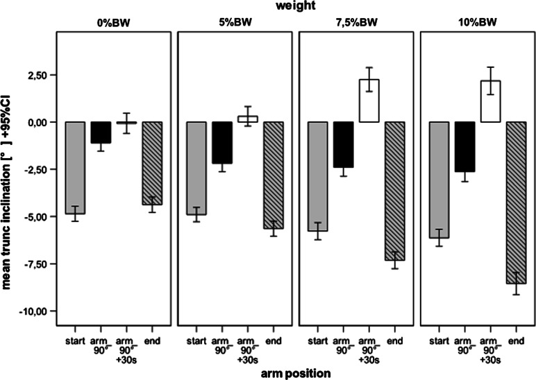
The trunk inclination for 0% bw in the starting position of the modified Matthiass test was −4.7° (SD 3.4°) and for 10% bw −5.8° (SD 3.9°) (Fig. 2). These values were reduced by 50% elevating both arms into an anteversion of 90°. During the subsequent 30 s arm holding phase, a straightening of the spine resulted for all different weight loads, shown by the positive values in Fig. 2 (Arm_90 + 30 s). It could also be shown that the trunk inclination at the end of the 90 s arm holding phase increases. The increase of the trunk inclination correlated with the lifted weights. For 0% bw we detected an inclination of 1.3° (SD 2.1°), respectively, 4.7° (SD 3.6) for 10% bw. These differences are significant between 0 and 7.5% bw but not between 0 and 5% bw. At the end of the modified Matthiass test the trunk tilts forward, exceeding the values of the starting position (Fig. 2).
Fig. 2.
Mean values and 95% confidence intervals of the parameter trunk inclination during the different phases of the Matthiass test. For all measured weights the back was straightened up during the 30 s of the arm holding phase. The changes of the posture are significant between 0 and 7.5% bw as well as for 10% bw
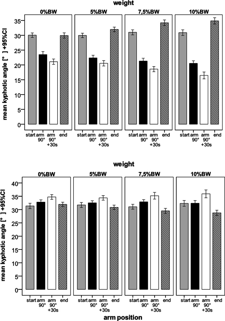
The detected kyphosis angle at the beginning of the Matthiass test ranged from 30° (0 and 5% bw) to 31° (7.5 and 10% bw). A significant difference could not be found. Elevating the arms led to a significant reduction of the kyphosis angle. The greatest changes occurred when higher weights were lifted (2° at 0 and 5% bw, 4° at 7.5 and 10% bw). During the arm holding phase, the kyphosis angle decreased significantly for all four weight loads.
The lordosis angle at the beginning of the modified Matthiass test was measured between 32° and 33°. Elevating the arms led to an increase of the lordosis angle only at 5% bw, while the other weight loads did not change the measured lordosis angle (Fig. 3). During the 30-s arm holding phase the lordosis angle increased significantly in all measurements. At the end of the modified Matthiass test the lordosis angle decreased for all weights, except for 0% bw.
Fig. 3.
Mean values and 95% confidence intervals of the parameters kyphosis angle (upper illustration) and lordosis angle (lower illustration) during the different phases of the Matthiass test. A significant reduction of the kyphosis angle and a significant increase of the lordosis angle can be found in all measured weight loads during the 30 s arm holding phase
In summary the results showed that an additional weight of 5% bw changes the posture (kyphosis, lordosis and trunk inclination) significantly, even in healthy people. Additionally, the results confirmed that the modified Matthiass test can be objectified by dynamic rasterstereography.
Discussion
An objective evaluation of posture deficiencies is sometimes only conditionally possible. The Matthiass-posture-test is indeed an easy way to measure the posture, but it is strongly influenced by the examiner. A quantification of clinically relevant posture parameters is desirable, but it is very difficult to apply in daily use because practicable test procedures are often time consuming and expensive. Rasterstereography offers an alternative way of detecting spinal posture parameters because it is side effect free, non-invasive and objective. Several studies have proved the high to very high reliability and validity of this method [2, 3, 6, 8, 22, 23]. A disadvantage is that thus far it was not possible to record sequentially, so that the measurements had to take place during the phases of standing still. During longer periods of analysis or during phases of highly ambitious motoric coordination it is necessary to count on a natural fluctuation of the bodies’ centre of gravity, which cannot be detected by a single frame analysis [5]. Using a prototype which was developed during this study, it was for the first time possible to measure the human posture rasterstereographically and dynamically over a period of 30 s with a recording frequency of 15 Hz. The results of this study prove that the system is able to measure with high accuracy, even under dynamic conditions.
During the Matthiass test a straightening of the dorsum results by elevating the arms to a position of 90° anteversion. This can be seen by a reduced trunk inclination as well as by a reduction of the kyphosis angle. These results are in contrast to the results found by Junghans and Groeneveld [7, 12]. They noticed an increase of the kyphosis angle of posture-healthy children under these conditions. A possible explanation might be that the arm lever used in this study could have forced the adolescents into a trunk stabilizing position. A compensating motion with layback and increase of the lordosis can be avoided, which could have led to only slight changes in the lordosis angle. From a biomechanical perspective lifting the arms leads to a relocation of the centre of gravity to ventral [17]. This relocation can be compensated by a posture-healthy proband by creating an antidromic moment of torque in the ankle joint, which keeps the bodies centre of gravity over the feet. It can be achieved by a strong stretching of the intrinsic back muscles, which also counteracts an increase of the lordosis. Our results with weight loads show that additional weights of 5% bw are enough to create significant differences during an arm holding phase. It can be concluded that posture deficiencies can already be detected in the period right after elevating the arms by using additional weights. The arm holding phase of 30 s leads to further changes in the parameters for all different weight loads, caused by an expected fatigue effect. Considering the fact that right after the elevation of the arms changes of the posture occur, it should be discussed if it would be possible to detect posture weaknesses by letting patients raise an additional weight and then detect the percentage of change compared to the posture test without weights. The results of this study are based on measurements with healthy, active adolescents. It must be concluded that posture weak patients would show more considerable changes of the posture, but this will have to be evaluated in further studies.
Conclusions
The rasterstereographic system used in this study allows an objectification of the modified Matthiass test under dynamic conditions. Additional weight loads of only 5% bw result in significant changes of the posture during this test, even in healthy people. The use of the modified Matthiass test in combination with the dynamic rasterstereographic system could lead to a fast, objective and safe detection of posture weaknesses.
Acknowledgments
This study was supported by operating grants from EU Craft Project 4D Body Scan Project No 1999-71038.
References
- 1.Chodick G, Ronckers CM, Shalev V, Ron E. Excess lifetime cancer mortality risk attributable to radiation exposure from computed tomography examinations in children. Isr Med Assoc J. 2007;9:584–587. [PubMed] [Google Scholar]
- 2.Drerup B, Hierholzer E. Evaluation of frontal radiographs of scoliotic spines—part II. Relations between lateral deviation, lateral tilt and axial rotation of vertebrae. J Biomech. 1992;25:1443–1450. doi: 10.1016/0021-9290(92)90057-8. [DOI] [PubMed] [Google Scholar]
- 3.Drerup B, Hierholzer E. Evaluation of frontal radiographs of scoliotic spines—part I. Measurement of position and orientation of vertebrae and assessment of clinical shape parameters. J Biomech. 1992;25:1357–1362. doi: 10.1016/0021-9290(92)90291-8. [DOI] [PubMed] [Google Scholar]
- 4.Drerup B, Hierholzer E. Assessment of scoliotic deformity from back shape asymmetry using an improved mathematical model. Clin Biomech. 1996;11:376–383. doi: 10.1016/0268-0033(96)00025-3. [DOI] [PubMed] [Google Scholar]
- 5.Drerup B, Ellger B, Meyer zu Bentrup FM, Hierholzer E. Functional rasterstereographic images: a new method for biomechanical analysis of skeletal geometry. Orthopade. 2001;30:242–250. doi: 10.1007/s001320050603. [DOI] [PubMed] [Google Scholar]
- 6.Frobin W, Hierholzer E. Analysis of human back shape using surface curvatures. J Biomech. 1982;15:379–390. doi: 10.1016/0021-9290(82)90059-8. [DOI] [PubMed] [Google Scholar]
- 7.Groeneveld HB. Metric detection and definition of back shape and posture of the human. Stuttgart: Hippokrates Verlag; 1976. [Google Scholar]
- 8.Hackenberg L, Liljenqvist U, Hierholzer E, Halm H. Scanning stereographic surface measurement in idiopathic scoliosis after VDS (ventral derotation spondylodesis) Z Orthop Ihre Grenzgeb. 2000;138:353–359. doi: 10.1055/s-2000-10162. [DOI] [PubMed] [Google Scholar]
- 9.Hierholzer E, Hackenberg L. Three-dimensional shape analysis of the scoliotic spine using MR tomography and rasterstereography. Stud Health Technol Inform. 2002;91:184–189. [PubMed] [Google Scholar]
- 10.Huysmans T, Haex B, Van Audekercke R, Vander Sloten J, Van der Perre G. Three-dimensional mathematical reconstruction of the spinal shape, based on active contours. J Biomech. 2004;37:1793–1798. doi: 10.1016/j.jbiomech.2004.01.020. [DOI] [PubMed] [Google Scholar]
- 11.Huysmans T, Van Audekercke R, Vander Sloten J, Bruyninckx H, Van der Perre G. A three-dimensional active shape model for the detection of anatomical landmarks on the back surface. Proc Inst Mech Eng H. 2005;219:129–142. doi: 10.1243/095441105X9309. [DOI] [PubMed] [Google Scholar]
- 12.Junghans H. The spine under the influences of daily life, of leisure time, of sports. Stuttgart: Hippokrates; 1986. [Google Scholar]
- 13.Klee A. Predictive value of Matthiass’ arm-raising test. Z Orthop Ihre Grenzgeb. 1995;133:207–213. doi: 10.1055/s-2008-1039439. [DOI] [PubMed] [Google Scholar]
- 14.Levy AR, Goldberg MS, Hanley JA, Mayo NE, Poitras B. Projecting the lifetime risk of cancer from exposure to diagnostic ionizing radiation for adolescent idiopathic scoliosis. Health Phys. 1994;66:621–633. doi: 10.1097/00004032-199406000-00002. [DOI] [PubMed] [Google Scholar]
- 15.Liljenqvist U, Halm H, Hierholzer E, Drerup B, Weiland M. 3-dimensional surface measurement of spinal deformities with video rasterstereography. Z Orthop Ihre Grenzgeb. 1998;136:57–64. doi: 10.1055/s-2008-1044652. [DOI] [PubMed] [Google Scholar]
- 16.Mahlknecht JF. The prevalence of postural disorders in children and adolescents: a cross sectional study. Z Orthop Unfall. 2007;145:338–342. doi: 10.1055/s-2007-965256. [DOI] [PubMed] [Google Scholar]
- 17.Matthiass H. Measuring methods of the spine in the diagnostic of spine diseases. Stuttgart, Jena, New York: Gustav Fischer; 1961. [Google Scholar]
- 18.Matthiass H. Maturation, growth and disturbances of growth of the posture and the musculoskeletal system of adolescents. Basel: Karger; 1966. [Google Scholar]
- 19.Morin Doody M, Lonstein JE, Stovall M, Hacker DG, Luckyanov N, Land CE. Breast cancer mortality after diagnostic radiography: findings from the U.S. Scoliosis Cohort Study. Spine (Phila Pa 1976) 2000;25:2052–2063. doi: 10.1097/00007632-200008150-00009. [DOI] [PubMed] [Google Scholar]
- 20.Quinlan JF, Mullett H, Stapleton R, FitzPatrick D, McCormack D. The use of the Zebris motion analysis system for measuring cervical spine movements in vivo. Proc Inst Mech Eng H. 2006;220:889–896. doi: 10.1243/09544119JEIM53. [DOI] [PubMed] [Google Scholar]
- 21.Salminen JJ, Erkintalo M, Laine M, Pentti J. Low back pain in the young. A prospective three-year follow-up study of subjects with and without low back pain. Spine. 1995;20:2101–2107. doi: 10.1097/00007632-199510000-00006. [DOI] [PubMed] [Google Scholar]
- 22.Schulte TL, Liljenqvist U, Hierholzer E, Bullmann V, Halm HF, Lauber S, Hackenberg L. Spontaneous correction and derotation of secondary curves after selective anterior fusion of idiopathic scoliosis. Spine. 2006;31:315–321. doi: 10.1097/01.brs.0000197409.03396.24. [DOI] [PubMed] [Google Scholar]
- 23.Schulte TL, Hierholzer E, Boerke A, Lerner T, Liljenqvist U, Bullmann V, Hackenberg L. Raster stereography versus radiography in the long-term follow-up of idiopathic scoliosis. J Spinal Disord Tech. 2008;21:23–28. doi: 10.1097/BSD.0b013e318057529b. [DOI] [PubMed] [Google Scholar]
- 24.Windolf M, Gotzen N, Morlock M. Systematic accuracy and precision analysis of video motion capturing systems–exemplified on the Vicon-460 system. J Biomech. 2008;41:2776–2780. doi: 10.1016/j.jbiomech.2008.06.024. [DOI] [PubMed] [Google Scholar]


