Abstract
A technique of bile diversion by tube choledochoureterostomy has been devised for the purpose of studying the role of bile in the intestinal absorption of drugs, This method was used in six dogs, No technical difficulties or major complications developed, as are inevitable with alternative methods, including external fistula.
Keywords: Bile, bile duct, pharmacokinetics, dogs
There have been many attempts to exclude the bile from the intestine to study the role of bile on the absorption of food or drugs. The usual method is creation of an external fistula. However, this method has several major disadvantages. The most important of them is the difficulty in carrying the animals for a long period without complications, such as development of infection and incomplete drainage.1–3
Another approach is anastomosis of the biliary tract to the urinary tract, allowing excretion of the bile into the urine. One method used to achieve this is choledochoureterostomy, and another is cholecystonephrostomy.
Choledochoureterostomy was first described in 1913.4 In this study, a right nephrectomy was performed, followed by anastomosis of the right ureter to the common bile duct directly. Half of the animals developed a major complication, such as rupture or stricture of the anastomosis.
Cholecystonephrostomy was first described in 1924.5 In this procedure, an anastomosis is performed directly between the gallbladder and the right renal pelvis. This method appears to have fewer complications but has not achieved widespread use because the operation is technically difficult.6
Our method of tube choledochoureterostomy is easy to perform and provides uncomplicated long-term bile diversion without any technical complications.
Materials and Methods
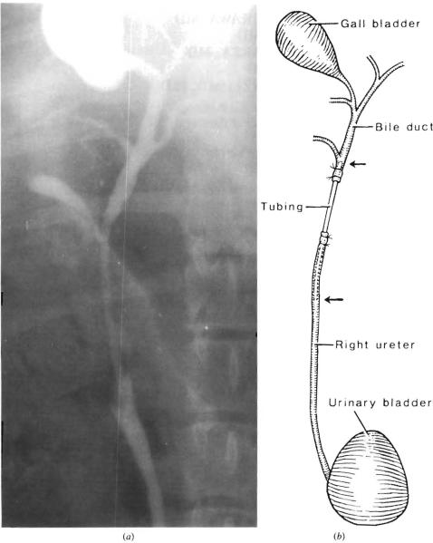
Six female beagle dogs (9.5–12.3 kg) were used. The animals were allowed water but not food beginning the evening prior to the operation. General anesthesia was induced with intravenous sodium pentobarbiturate sodium (25 mg/kg) and maintained with oxygen, nitrous oxide, and halothane. A midline abdominal incision was made. The right kidney was freed from its bed, the pedicle was exposed, and the right renal artery, vein, and ureter were dissected free from surrounding tissue. The renal artery and vein were doubly ligated and divided. The ureter was divided at the level of ureteropelvic junction and the right kidney was removed. The bile duct was dissected and ligated at the level of the upper edge of the duodenum. A choledochostomy was made right above the ligation. One end of a 5- to 7-cm piece of polyurethane tubing (2.0 mm outside diam, 1.5 mm inside diam) was inserted through the choledochostomy toward the liver. This end of tubing was placed below the lowest hepatic branch and secured with 2-0 silk ligatures. The other end was inserted into the ureter 3 em distally and secured with 2-0 silk ligatures. The midline incision was then closed (Fig 1a and b), Postoperatively, Cephamandol 1 g im was given for 3 days, with the first dose given upon closing the wound. Serum GOT, GPT, total bilirubin, alkaline phosphatase, creatinine, and BUN were measured once a week. The urine mixed with bile was collected daily. The volume, total bilirubin, and cholesterol were measured. The animals were killed 33 days after the operation. Autopsy was performed, and cultures of the content in the bile duct and bladder were obtained.
Figure 1.
(a) Retrograde ureterogram showing continuity of the right ureter to the biliary system. There was no leak and obstruction at the anastomosis. (b) Schematic diagram of (a). Arrows indicate the upper and lower ends of the tube.
Results
Immediately after the operation, bile was present in the urine. The animals soon recovered from effects of operation and began eating on postoperative day (POD) 1. The stools became clay-colored and free of bile pigment. There were no surgical complications and no deaths. Postoperatively, two dogs showed a minimal increase in serum GPT and four dogs showed a minimal to mild elevation in serum alkaline phosphatase, which fell to normal after POD 7. Total bilirubin and cholesterol were present in the urine immediately after the procedure and achieved stable levels by POD 5 (Fig 2). Autopsy done on POD 33 showed patency of the bile duct, the right ureter, and the tubing. The tubing was surrounded by dense adhesions. There was no evidence of leak and no fistula between bile duct and the intestines. Cultures of the bile duct and the bladder taken at the time of autopsy were sterile.
Figure 2.
Parallel graphs of average total bilirubin and cholesterol in the 24-h-collected urine from six dogs. There was rapid rise in bilirubin and cholesterol in the first 4 days.
Discussion
Our method of bile diversion with tube choledochoureterostomy does not allow for direct studies of bile or bile output, but it is easy to perform and allows uncomplicated long-term survival. We have used this method for pharmacokinetic study of immunosuppressive drugs, and shown that bile was necessary for adequate absorption,7 a conclusion that has been confirmed in patients with T-tube drainage.8 There was a postoperative elevation in serum GPT and alkaline phosphatase and some variability in the excretion of total bilirubin and cholesterol in the daily collected urine. These fluctuations may have been related to temporary stenosis due to edema where the catheter was tied, but they were so minor that they did not undermine the usefulness of the method.
Acknowledgments
Supported by Research Grants from the Veterans Administration and Project Grant DK 29961 from the National Institutes of Health, Bethesda, MD.
References
- 1.Rous P, McMaster PD. A method for the permanent sterile drainage of intraabdominal ducts, as applied to the common duct. J Exp Med. 1923;37:11–17. doi: 10.1084/jem.37.1.11. [DOI] [PMC free article] [PubMed] [Google Scholar]
- 2.Marshall RW, Moreno OM, Brodie DA. Chronic bile duct cannulation in the dog. J Appl Physiol. 1964;19:1191–1192. doi: 10.1152/jappl.1964.19.6.1191. [DOI] [PubMed] [Google Scholar]
- 3.Barringer M, Sterchi JM, Jackson D, Meredith J. Chronic biliary sampling via a subcutaneous system in dogs. Lab Anim Sci. 1982;32:283–285. [PubMed] [Google Scholar]
- 4.Pearce RM, Eisenbrey AB. A method of excluding bile from the intestine without external fistula. Am J Physiol. 1913;32(8):417–426. [Google Scholar]
- 5.Kapsinow R, Engle LP, Harvey SC. Intra-abdominal biliary exclusion from the intestine—Cholecysto-nephrostomy, a new method. Surg Gynecol Obstet. 1924;39:62–65. [Google Scholar]
- 6.Knoebel LK, Ryan JM. Digestion and mucosal absorption offat in normal and bile-deficient dogs. Am J Physiol. 1963;204(3):509–514. doi: 10.1152/ajplegacy.1963.204.3.509. [DOI] [PubMed] [Google Scholar]
- 7.Ericzon BG, Todo S, Lynch S, Kam I, Ptachcinski RJ, Burckart GJ, et al. Role of bile and salts on cyclosporine absorption in dogs. Transplant Proc. 1987;19:1248–1249. [PMC free article] [PubMed] [Google Scholar]
- 8.Andrews W, Iwatsuki S, Shaw BW, Jr, Starzl TE. Bile diversion and cyclosporine dosage. Transplantation. 1985;39:338. Letter to the Editor. [PubMed] [Google Scholar]


