

Abstract
Hepatocellular carcinoma (HCC) is a common and deadly cancer whose pathogenesis is incompletely understood. Comparative genomic studies from human HCC samples have classified HCCs into different molecular subgroups; yet, the unifying feature of this tumor is its propensity to arise upon a background of inflammation and fibrosis. This review seeks to analyze the available experimental models in HCC research and to correlate data from human populations with them in order to consolidate our efforts to date, as it is increasingly clear that different models will be required to mimic different subclasses of the neoplasm. These models will be instrumental in the evaluation of compounds targeting specific molecular pathways in future preclinical studies.
Keywords: Liver cancer, Hepatocellular carcinoma, Mouse models, Genetically engineered mice, Cirrhosis
1. Introduction
Hepatocellular carcinoma is one of the world’s deadliest cancers, ranking third among all cancer-related mortalities. Most cases occur in Asia and sub-Saharan Africa, where viral hepatitis is endemic. The incidence is rising in the West, likely due to the increase in patients infected with hepatitis C during the latter half of the last century [1]. The liver, unique in its capacity for regeneration following injury, also gives rise to this malignancy commonly associated with the inflammatory state of advanced fibrosis, or cirrhosis. Potentially curative therapies can be offered to approximately 30% of patients, but are complicated by a high rate of recurrence [2].
Encouraging progress has been made in understanding the molecular pathogenesis of cancer [1,2]. The discoveries of the signal transduction pathways, cascades of protein–protein interactions transmitting information from the cell surface to the nucleus, and of their link to tumor biology, are particularly impressive.
Several key mouse models have been instrumental in defining the pathogenesis of HCC by introducing genetic alterations into one or more aetiologic pathways that can be targeted exclusively to the liver. Moreover, these programmed manipulations can be introduced systematically, not only in this specific organ but also at defined times during development, growth and aging of the liver.
Nonetheless, substantial challenges persist in modeling liver diseases whose natural history requires a chronic inflammatory milieu. For example, infectious (hepatitis C virus), toxic (alcohol), metabolic (non-alcoholic steatohepatitis), or congenital (hemachromatosis) diseases share inflammation and fibrosis as precursors to cancer, yet none is easily mimicked in animals. There are few rodent models of HCC arising spontaneously within a background of regenerative nodules and cirrhosis, and most depend on the administration of hepatotoxic and/or carcinogenic agents to recreate the injury–fibrosis–malignancy cycle seen in chronic human liver diseases.
Comparative genomic studies in human HCC samples have begun to identify molecular subgroups with characteristic mutations, gene expression profiles and chromosomal gains and losses [3]. Moreover, since there is no single dominant molecular pathology underlying all HCCs, it is increasingly clear that different models will be required to mimic different subclasses of the neoplasm. These models will be instrumental as pre-clinical tools to evaluate compounds targeting specific molecular pathways.
With these challenges in mind, the objective of this review is to assemble and evaluate the available models of both cirrhosis and HCC, to provide a blueprint for understanding the pathogenesis of HCC and for optimizing preclinical models for drug testing.
2. Experimental models in cancer research
Although many experiments focusing on liver physiology have been conducted in rats due to their propensity for the development of fibrosis, the laboratory mouse (Mus musculus) is considered among the best model systems for cancer because of the availability of gene targeting methods, as well as the animal’s size and breeding capacity, its lifespan of 3 years, and its physiologic and molecular similarities to human biology [4]. Significant advances have been made in modeling cancer genetics in mice, along a spectrum that ranges from simple xenograft models to more complex, genetically modified mice. Examples of each of the following are illustrated in Table 1.
Table 1.
Available mouse models in cancer research
| Technical Method | Advanced Mouse Models of Cancer | Current Models in HCC | Future Prospects: Wish List for HCC | |
|---|---|---|---|---|
| Xenograft | Xenograft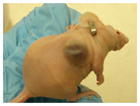
|
COLON, BREAST, PROSTATE: Surgical orthotopic implantation: intact fragments of human cancer, including tumors taken directly from the patient, transplanted into the corresponding organ of immunodeficient rodents16 | Orthotopic xenograft model in which hepatoma 129 cells originating from C3H mice are injected into fibrotic livers of mice pretreated with TAA and EtOH45 | Mouse HCC cell line derived from GEM tumor with specific molecular pathway dysregulated, with immunofluorescent marker, injected into fibrotic liver of immune-competent mice |
| Transgenic GEM | Constitutive Transgenic |
PANCREATIC: KrasG12V and chronic pancreatitis169; Trp53R172H and KrasG12D double transgenic driven by insulin promoter170: two models of invasive and metastatic pancreatic cancer | Mouse C-myc/Human E2F-1 overexpression driven by albumin promoter171: HCC at 6–8 months | Double transgenic overexpressing profibrotic gene combined with liver-specific oncogene |
| Dominant Negative Transgenic |
PITUITARY: Rb and p27Kip1 Cdk inhibitor tissue specific knockout mice develop pituitary tumors with different phenotypes172 | Mdr-2 knockout mice are unable to secrete phospholipids into bile, and develop cholangitis and HCC at 6–12 months166 | Double transgenic liver-specific E-cadherin knockout and β-catenin overexpression | |
| Inducible Transgenic |
MELANOMA: Double transgenic combining Tet-induced overexpression of mutated HrasV12G and Ink4a knockout173 | Tet-inducible Met expression under albumin promoter: 60% HCC at 12 months; tumors regressed when transgene (Tg) was inactivated91 | Tet-induced, liver-specific overexpression of known oncogene in fibrotic mice | |
| Endogenous GEM | Conditional Gene Targeting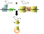
|
PROSTATE: Double transgenic Cre-mediated PTEN−/− homozygous loss and p19Arf +/−: cooperativity in cancer development174 | Cre-mediated liver specific PTEN−/− knockout: 66% HCC at 8 months108 | Cre-mediated, liver-specific knockout of known tumor suppressor gene in fibrotic mice |
2.1. Xenograft models
The demonstration that concentrated cancer cells grown in vitro could form tumors when implanted subcutaneously into an immunocompromised mouse was first established in 1969 [5]. This xenograft model has since demonstrated several advantages that explain its persistence as the mainstay of pre-clinical studies of anti-neoplastic drugs in vivo: the tumors are rapidly and easily induced, and their subcutaneous location enables direct measurement of tumor growth. More recently, however, several critical differences between xenograft- and patient-derived specimens have become apparent, as discussed below. In addition, cancer is now appreciated as a complex disease dependent upon the interaction between transformed cells harboring oncogenic mutations, referred to as the ‘cell autonomous compartment’, and their surrounding tumor environment, the ‘non-cell autonomous constituents’ made up of normal cells, stromal cells, and immune cells [4], features that are not part of the xenograft approach.
Mouse models of cancer were first introduced over 60 years ago. Shortly after its inception in 1955, the Developmental Therapeutics Program at the National Cancer Institute (NCI) adopted the use of three transplanted rodent models of sarcoma, carcinoma, and leukemia, for the purposes of selecting agents for clinical use in cancer patients. Thousands of molecules were tested in mice bearing murine leukemias during the first decades of modern cancer drug development, circa 1945–1969 [6]. This tumor panel was later expanded to include human tumor xenografts, with the intention to study drug activity against solid tumors [7]. In 1990, the NCI focused on the development of in vitro assays in 60 different cell lines in order to screen pharmaceutical agents for their potency and their selective activity against either a particular disease category or specific cell line [8,9], the most promising of which were to be subsequently evaluated in the nude mouse xenograft model.
The validity of xenografts as a predictive indicator of probable clinical activity is limited, with the most success seen in cytotoxic agents. A retrospective analysis performed by the NCI for 39 compounds in which both xenograft testing and Phase II clinical data were available showing that less than 50% of agents with activity in more than one-third of xenografts showed clinical activity (p = 0.04) [6]. The same study demonstrated that activity in a particular histology in a tumor model did not closely correlate with activity in the same human cancer histology [10], with the exception of non-small cell lung and ovarian cancer [11].
There are several variables inherent to the xenograft experiments which may impact on the divergent outcomes compared to human disease, including growth properties and size at initiation of treatment of xenograft tumor, ectopic versus orthotopic location of tumor, local versus metastatic disease [12], tolerance for high doses of chemotherapeutic agents in mice [13], and variability in selected endpoints. These variables can be minimized if given due consideration in the design of preclinical cancer drug experiments. However, the greatest discrepancies between success of cancer therapies in xenograft models and in human clinical trials are likely due to critical differences in both the tumor cells and their microenvironment. Natural tumor progression is a micro-evolutionary process during which increasingly aggressive clones, generated through genetic instability, emerge from an initially monoclonal lesion. Autochthonous tumors, those that evolve in situ from normal cells, tend to have a diminished genetic heterogeneity compared to tumor xenografts, although selective pressures of cell culture or tissue explantation can cause a rapid expansion of a certain clonal constituent of polyclonal tumors [14,15].
One solution to this disparity between cancer cell lines and human tumors is surgical orthotopic implantation, in which intact fragments of human cancer taken directly from the patient are transplanted into the corresponding organ of immunodeficient rodents, as reviewed by Hoffman [16]. This technique has been applied to breast, lung, and prostate cancer among others.
Additional advances have been made in the xenograft model through the addition of mesenchymal stem cells to weakly metastatic cancer cell lines, which enhances the ability of the cell lines to form tumors and to metastasize [17]. Wu et al. were able to isolate a side population (SP) from 29 sarcomas which preferentially formed tumors when grafted into immunodeficient mice; only cells from tumors that developed from the SP cells had the ability to initiate tumor formation upon serial transplantation [18].
Our deepening appreciation of the non-cell autonomous constituents of the tumor microenvironment, including the stroma and immune cells relevant to liver pathology in particular, provides further evidence that the xenograft model is more appropriately termed animal culture, as suggested by Tuveson and Frese [4].
2.2. Genetically engineered mouse models (GEM)
The most sophisticated animal models of human cancer are those that have been genetically engineered to mimic the pathophysiological and molecular features of human malignancies [4]. Such models enable the investigation of a range of discrete molecular stages that occur during tumor progression both within tumor cells and within their microenvironment; additionally, mice harboring multiple mutations provide information regarding pathway cooperativity and dependency in vivo [19].
Despite these strengths, there are a number of important limitations in mouse models of cancer, such as variation in basic cellular processes, as well as in telomere length and telomerase expression [20,21]. It is also well documented that identical genetic lesions can produce different pathologies in mice than in humans [22]. GEM can be categorized as either transgenic or endogenous models.
2.3. Transgenic models
Transgenic mice are those that are engineered to express either oncogenes or dominant-negative tumor suppressor genes in a non-physiologic manner due to ectopic promoter and enhancer elements [4,19]. Microinjection of recombinant DNA directly into the pronucleus of a fertilized mouse egg is the classic method for generating transgenic mice [23], but transgenic mice can also be produced through gene targeting (“knock-in”) and lentiviral transduction in embryonic stem cells.
Constitutive expression of cellular and viral oncogenes and germline disruptions of tumor suppressor genes were the first approaches used to create strains of cancer-prone mice [19,24]. The cDNA constructs can contain promoter elements designed to restrict tissue tropism, so although the effect of the oncogenic gain will be constitutive, its expression can be limited to specific tissues by the use of tissue-specific promoters [19], for example the albumin promoter in liver transgenic models.
Germline tumor suppressor cell mutant mice were initially developed to parallel human inherited cancer predisposition syndromes. However, although many of these heterozygous mice were tumor-prone and demonstrated loss of the wild-type allele in their tumors, few of them developed the clinical features of the cognate human syndrome. For example, loss of the retinoblastoma gene product Rb in humans leads to retinoblastomas, osteosarcomas, and small cell lung cancer; whereas Rb heterozygote mice develop thyroid and pituitary tumors but no retinoblastomas [25]. Rb heterozygotes are able to compensate for loss of Rb, a finding that highlights the existence of shared and predictable cellular process within both species [20,26]. So, although identical genetic lesions may not perfectly recapitulate the human disease in mice, there is no doubt that these genetically engineered mice are valuable tools for understanding the underlying biological mechanisms of tumorigenesis [22]. Their ability to recapitulate the genetic features of amplified proto-oncogenes, such as c-myc [27], has contributed greatly to our understanding of cancer biology.
There are, however, additional weaknesses of these models that have spurred the development of more advanced methods. For example, because the genes affected may be vital to normal development, over-expression or ablation may lead to embryonic lethality or infertility [24]. Promoter fragments typically represent the minimal sequence required for tissue-specific expression and do not necessarily allow the same control conferred by endogenous regulatory elements [28]; for example, a typical transgene would not include all transcription factor and microRNA binding sites [4,29]. And, although the DNA fragments are thought to associate by homologous recombination before integration and in most cases insert at a single chromosome site [23], there is little control over site of integration and copy number [22]. This can result in pronounced variability of expression patterns, as the exogenous gene can affect genes near its insertion site or can be affected by endogenous control elements [22,30–32]. Also, although conventional mouse mutants may be useful for modeling familial forms of cancer, they do not mimic sporadic tumorigenesis because the initiating mutation is present in all cells of the body, including those that constitute the tumor microenvironment [33].
2.4. Inducible systems of oncogene expression
Bujard and colleagues developed a strategy for temporally controlled and reversible transgene expression, using a tetracycline (tet-) inducible system [34]. These drug- or ligand-inducible systems involve the use of a chimeric transcriptional activator that reversibly activates a target gene in response to the administration of the inducing agent.
The Escherichia coli tetracycline resistance operon has been applied widely to generate cell lines and murine models with tightly regulated gene expression in response to tetracycline [35]. The tet transactivator functions either as a constitutive repressor that is inducibly inhibited by ligand to allow expression from the tet operon (tTA), or it acts as an inducible activator of the tet operon upon ligand addition (rtTA) [19]. This system has been particularly useful to study the concept of oncogene addiction; nearly all oncogenes tested thus far seem to be required not only for tumor initiation, but also for tumor maintenance [33].
2.5. Endogenous GEM: knock-out models
Endogenous GEM are those that lose the expression of tumor suppressor genes (TSG) or that express dominant-negative tumor suppressor genes or oncogenes from their native promoters [4]. The original ‘knockout’ mouse model entailed disruption of an allele in endogenous embryonic stem cells using a targeting vector. Biallelic disruption of TSG often results in embryonic lethality, but heterozygous mice can be used to determine the tumorigenic potential of the genes, such as the retinoblastoma tumor suppressor gene (Rb) [25]. These germline mutations are present throughout the mouse and are constitutively expressed, unlike the sporadic mutations occurring in human tumors that are surrounded by normal tissue.
2.6. Endogenous GEM: conditional gene targeting
As reviewed by Maddison et al. [22], model systems have now been developed which allow both spatial and temporal control of gene expression. These are predominantly dependent on the creation of bi-transgenic mice: those carrying a tissue-specific, inducible transactivator gene are crossed to mice carrying the allele of interest which has been engineered to be controlled by the transactivator. Offspring that carry both transgenic elements are treated with the inducer to express the transactivator gene in a specific tissue, which then acts on the desired allele. This system requires the exogenous delivery of the cre gene (usually by an adeno- or retrovirus), and the induction is irreversible.
Conditional inactivation of tumor suppressor genes relies on the ability of a viral or prokaryotic site-specific recombinase to recognize a pair of target DNA sequences and catalyze recombination at these sites, which results in either deletion or inversion of the intervening DNA sequence [19]. A commonly used tool is the Cre-Lox system, wherein Cre (Causes recombination) recombinase, isolated from bacteriophage P1, catalyses site-specific recombination between defined 34 bp Lox P sites (Locus χ of crossover P1) [36,37]. If gene χ is placed between two Lox P sites and then exposed to Cre, it will be excised, or ‘floxed out’. An alternative system to Cre-Lox uses the FLP recombinase, which recognizes the 48 bp Frt site [38]. Transgenic mice that express recombinase from a specific promoter are bred to mice carrying conditional tumor suppressor gene mutations, so that the TSG can be bi-allelically inactivated to allow the generation of organ- and cell-lineage-specific tumors models [19].
Conditional activation of oncogenes is created by the insertion of a LoxP flanked transcriptional silencing element between the promoter and the mutant oncogene-encoding sequence. Conditional oncogenes are constructed using classic transgenic technology, but expression of the oncogene is only activated by the recombinase-mediated removal of the transcriptional silencer. This allows for tissue-specific oncogene expression [39].
This second generation of GEM, which more faithfully recapitulates sporadic tumor formation by the induction of somatic mutations in a time- and tissue-specific fashion, has provided great insight into the contribution of genes in the initiation, progression, and treatment of cancer. We will now discuss how each of these systems has been used to further our understanding of liver cancer.
3. Experimental models of hepatocellular cancer
Hepatocellular carcinoma universally arises upon a background of inflammation and fibrosis. Creation of animal models of HCC presents a particular experimental challenge because of the diffculty in modeling chronic inflammation without using carcinogens to induce liver injury, and because of the heterogeneity of molecular pathways that are dysregulated during this transition from cirrhosis to cancer.
HCC is preceded in both rodents and humans by the development of premalignant lesions including foci of altered hepatocytes and dysplastic nodules, which exhibit a higher risk of malignant evolution than normal cells [40,41]. Various genetic alterations and exposures to chemical carcinogens have been studied in animals in order to recapitulate the phenotypic, biological, and molecular events that occur during this transformation.
3.1. Xenograft models of HCC
In a recent attempt to characterize primary human xenografts in liver cancer, seven different primary HCC cell lines were injected into SCID mice. The mice were then treated with common chemotherapeutic agents such as cisplatin and gefitinib. There were significant differences in tumor growth inhibition between xenografts, which reinforced the concern for high internal variability of this model in human cancer. Interestingly, the study concluded that most of the chemotherapeutic agents currently used in the treatment of HCC have little or no anti-neoplastic activity in these models [42].
Ma et al. have examined HCC cells expressing CD133 [43], which exhibit stem cell properties and are chemoresistant: purified CD133(+) HCC cells isolated from human HCC cell lines and harvested from xenograft mouse models survived chemotherapy in increased proportions relative to most tumor cells which lack the CD133 phenotype [44]. The inclusion of stem cell-enriched HCC cell lines will likely enhance future pre-clinical studies in HCC therapeutics (see Table 1).
A group of investigators at the University Hospital Bonn created an orthotopic xenograft model in which hepatoma 129 cells originating from C3H mice were injected into fibrotic livers of mice pretreated with thi-oacetamide by intraperitoneal injection and alcohol per oral [45]. They found that tumors in fibrotic livers grew significantly larger and more rapidly than those in normal livers, and were able to metastasize and form satellite nodules. Gene expression analysis revealed greater intratumoral expression of vascular endothelial growth factor (VEGF) and its receptor (VEGFR), and of MMP-2 and MMP-9 in the fibrotic liver tumors. This useful model provides a unique tool for testing drug e3-cacy in orthotopic hepatoma xenograft within the context of liver fibrosis.
3.2. Viral models of HCC
Infection causing latent or chronic viral hepatitis is the most common aetiology of HCC, comprising 80% of cases worldwide. Hepatitis B virus (HBV) is endemic in China, Southeast Asia, and sub-Saharan Africa; there, vertical transmission of the virus results in high rates of HCC. Hepatitis C (HCV) viral infection is more prevalent in the United States and Europe than either HBV or HIV [46]. The woodchuck hepatitis virus (WHV) induces a liver inflammation, injury and repair process in woodchucks similar to those of HBV-positive patients and has therefore proven to be a useful model of the disease.
3.2.1. Hepatitis B virus
HBV is a DNA virus that causes acute and chronic hepatocyte injury, inflammation, and HCC. During prolonged infection, viral DNA sequences integrate into the host cell genome, where they and the flanking cellular sequences are commonly rearranged [47], a phenomenon that can activate an adjacent cellular oncogene. In addition, viral infection can induce hepatocyte injury mediated by the antiviral cellular immune response and, to a lesser extent, by direct injury to the cells. Although most cases of HBV-associated HCC arise in a background of inflammation and fibrosis, the virus is notorious for also causing HCC in the absence of cirrhosis, most likely by integrating into the host chromosome and thereby promoting transcriptional transactivation of mitogenic factors.
The HBV virus is a circular DNA molecule containing four open reading frames encoding four HB viral proteins: preS/S, preC/C, P and X protein (HBx). The most common viral marker in HCC is the integration of HBV genomic DNA encoding HBx. In 1994, Koike et al. published their description of a transgenic mouse model demonstrating that high levels of HBx expression were sufficient to generate HCC in 84% of male transgenic mice at age 13–24 months [48] (see Table 2). Analysis of proliferation and DNA content in these mice suggested that the continued expression of HBx gene initiated tumor formation by inducing DNA synthesis and placing large numbers of hepatocytes subjective to secondary events for transformation [48]. Yu et al. also confirmed the development of HCC in HBx transgenic mice [49]. Although another research group did not see spontaneous HCC development, those HBx transgenic mice were more susceptible to chemical carcinogenesis than control mice [50]. The reason for this discrepancy is unclear, but the difference in genotype of HBV should be noted: HCC tumors arose in genotype C HBx transgenic mice but not in other genotypes [51].
Table 2.
Genetically engineered models of hepatocellular carcinoma
| Gene | Type of mutation or tissue promoter/construct | Phenotype (+/− and −/−) | Chemically induced/metastasis | References |
|---|---|---|---|---|
| Viral models | ||||
| Hepatitis B virus large envelope protein | BgIII-A fragment of HBV encoding large envelope protein under control of albumin promoter and enhancer | Focal necrosis, inflammation, and subsequent HCC in 72% males | No metastases; rare local invasion | [47,52] |
| Hepatitis B virus X protein | EcoRI–BglII fragment of HBV including the X gene under its own promoter and enhancer | HCC in 84% after 13–24 months in mice with high HBx expression | Lung metastasis | [48,175,176] |
| Hepatitis C virus | HCV core-E1–E2 transgenic under albumin promoter and HCV core transgenic under HBV X promoter | No DEN: No HCC in either strain by 21 months. +DEN: 100% HCC at 32 weeks; HCV core-E1–E2 with largest tumors (p = 0.008) | DEN injected weekly × 6 weeks | [56] |
| Hepatitis C virus | HCV core under HBV X promoter; HCV E1–E2 under HBV X promoter | Core transgenics: 32% HCCs in male mice at 16–23 months; E1– E2 transgenics: no HCC. No evidence hepatitis | None reported | [56,59] |
| Hepatitis C virus | HCV core-E1–E2 transgenic under albumin promoter and the entire HCV transgenic under albumin promoter | HCC in core-E1–E2 transgenic and entire HCV transgenic after 13 months | None reported | [60] |
| Cell cycle models | ||||
| p53 germline knockout and liver-specific viral receptor TVA, injected with PyMT oncogene | p53 germline knockout [177] crossed with mice expressing viral receptor TVA under albumin promoter (Alb-TVA), injected at age 3 days intrahepatically with mouse polyoma virus middle T antigen | HCC in 42% of p53 null mice, in 37% of p53+/−, and in 66% of p53+/+ mice expressing TVA injected with PyMT at 4 months. No TVA-negative littermates developed HCC | Metastases in p53 null mice (6/16); less in p53+/− (1/14) | [67,177] |
| Trp53 and INK4a/ARF conditional mutant mice, injected with PyMT oncogene | Albumin Cre mice crossed with Trp53 conditional mutant and INK4a/ARF conditional mutant, injected at age 3 days intrahepatically with mouse polyoma virus middle T antigen | >90% HCC in combined Trp53, INK4a/ARF null mice injected with PyMT compared to single null gene | Metastases in Trp53 null mice (30%) and in combined Trp53, INK4a/ARF mice (63%) at 6 months | [68] |
| P53 conditional expression | Hepatoblasts transduced with oncogenic ras (Hras V12) and a tet-responsive P53 miRNA design short hairpin RNA | Complete tumor regressions when endogenous p53 reactivated in p53-deficient tumors | None reported | [69] |
| c-myc | c-myc over-expression under albumin enhancer/promoter [74,90,178,179]; under α1 antitrypsin promoter [180,181] | 15 weeks: polyploidy cells, dysplasia >60% [179]; 15 mos: 91% adenomas [74,178]; 54% HCC [178,180,181]; | None reported | [74,90,178–181] |
| c-myc and E2F-1 | Mouse c-myc and human E2F-1 over-expression under albumin promoter | 6–8 mos: 100% HCC [171,178] | None reported | [90,171,178,179] |
| c-myc and TGFα | c-myc over-expression under albumin enhancer/promoter; TGFα over-expression under metallothionein 1 promoter | 4 mos: 70% dysplastic nodules; 18% HCC [90] | Zinc in H2O accelerated nodule formation by 6–8 weeks | [90,182] |
| SV40 T-antigen conditional and inducible expression | SV40 T-antigen expression under albumin enhancer/promoter [74]; under major urinary protein enhancer/promoter [183]; under metallothionein 1 promoter [184]; under α1 antitrypsin promoter [185]; under antithrombin III promoter [186]; tetracycline-inducible expression: mice expressing tTa under albumin promoter crossed with mice expressing T antigen under tTa promoter [75] | 3–7 mos: adenomas and HCC [74]; 10–12 weeks: HCC[185]; after 4–6 weeks: 100% HCC [186] | Lung metastases [186] | [74,75,181,183–186] |
| E2F-1 | E2F-1 over-expression under control of albumin enhancer/promoter | 10 mos: 100% adenomas and dysplastic nodules; 12 mos: 33% HCC | None reported | [71,90,179] |
| Telomere dysfunction models | ||||
| mTERT−/− and p53+/− or WT | Germline mTERT and p53 knockout over several generations and CCl4 liver injury | 50 weeks: 100% HCC in p53+/− both generations (G0 and G3/G4); 44% in wild-type G0 versus 9% HCC in wild-type G3/G4 | CCl4 by IP injection 3×/week × 4 months | [66] |
| Pathway specific models | ||||
| Wnt/β-catenin | ||||
| Activating mutation in β-catenin: truncated NH2 terminal transgenic | EAB/9K/Δ N131 β-catenin construct under control of liver-specific enhancer of aldorase B gene (expressed throughout embryonic and post-natal development) | Death at 3 weeks from hepatomegaly; no dysplastic foci in liver | N/A | [127] |
| Activating mutation in β-catenin: exon 3 conditional knockout | Catnblox(ex3) knockout and fatty acid binding protein Fabpl-cre transgenic | Death at 5 weeks from liver damage/mitochondrial swelling. No dysplastic foci in liver; +intestinal polyps | N/A | [128] |
| Activating mutation in β-catenin: exon 3 conditional knockout | Catnblox(ex3) knockout injected with recombinant adenovirus expressing Cre from human CMV promoter | High multiplicity injection (109 pfu/mouse): death at 3 weeks with hepatomegaly/mitochondrial swelling. Low multiplicity injection (107–8 pfu/mouse): No dysplastic foci in liver >6 mos | N/A | [128] |
| β-catenin exon 3 knockout and activated H-ras (H-rasG12V) double-transgenic conditional | Catnblox(ex3) knockout and H-ras (Tglox(pA)H-ras*) double-transgenic with recombinant adenovirus expressing Cre from human CMV promoter | Low multiplicity infection (108 pfu/mouse): 100% HCC at 6 months | Intrahepatic invasion | [131] |
| APC knockout liver-specific | ApcΔex14 knockout (−/−) injected in tails with recombinant adenovirus expressing Cre (injections infected primarily and massively the liver) | High multiplicity infection (109 pfu/mouse): Death within 2 months and hepatomegaly. Lower multiplicity infection (0.5 × 10⁹ pfu/mouse): 67% HCC at 9 months. Apc+/− had no liver abnormalities | None reported | [130] |
| β-catenin wild-type | β-catenin over-expression under control of albumin enhancer/promoter | Hepatomegaly (15% increased liver/body weight ratio); no dysplastic nodules at 24 months | N/A | [129] |
| PI3K/Akt pathway | ||||
| PTEN−/− | Albumin cre/PTENlox/lox | Steatohepatitis; Adenomas at 44 weeks and 66% HCC at 78 weeks [108]; HCC in 66% of males at 44 weeks and in 83% of males and 50% of females at 78 weeks [109] | Lung metastases | [108,109] |
| Insulin growth factor pathway | ||||
| IGF2 transgenic | IGF2 over-expression under control of urinary protein promoter | HCC in <10% at 18–24 months; also lymphomas, sarcomas, and thyroid carcinomas | None reported | [187] |
| IGF2 knockout and TGFα transgenic | TGFα over-expression under metallothionein 1 promoter [86] crossed with IGF2 heterozygous knockout mice (paternal null allele; maternal wild-type, normally imprinted) | (1) IGF2wt/wt: no HCC; 4% adenoma; (2) IGF2+/−: dwarves, normal liver phenotype; (3) TGFα × IGF2wt/wt and (4) TGFα + IGF2wt/−: 100% HCC at 18 months | None reported. Zinc in drinking water starting at age 10 months | [188] |
| Epidermal growth factor pathway | ||||
| EGF transgenic | Double-transgenic of the liver construct Alb-DS4 that encodes autocrine growth factor IgEGF crossed with AAT-myc mice | EGF transgenic (Alb-DS4): mortality from HCC by age 7.1 months; EGF/myc double- transgenic: accelerated mortality to 4.4 months | [115] | |
| Ras signaling | ||||
| H-ras | Mutant c-H-ras overexpression under albumin promoter | Hepatomegaly, lung tumors [74] | None reported | [74] |
| HGF/c-Met and TGF-α | ||||
| HGF transgenic | Mouse HGF expression driven by metallothionein promoter [95]; by albumin promoter[189] | Hepatomegaly; >17 months: adenomas and rare HCCs [95]; rapid recovery after partial hepatectomy, no dysplasia [189] | Most animals not given zinc because transgene expression adequate | [95,189] |
| HGF overexpression +/− β-catenin conditional knockout | Hydrodynamic injection of plasmid containing HGF under CMV promoter (pCMV-HGF) into wild-type and into AFP-enhancer albumin promoter-Cre floxed β-catenin knockout mice | HGF over-expression: hepatomegaly and increased Wnt/β-catenin signaling; no dysplastic nodules. HGF over- expression in β-catenin knockout: no alterations in liver | N/A | [96] |
| HGF + c-myc | Double-transgenic mouse c-myc driven by albumin promoter/enhancer and human HGF driven by albumin regulatory elements | Inhibition of hepatocarcinogenesis by HGF in c-myc transgenic mice: 0% HCC in HGF/c-myc versus 60% HCC at 16 months in c-myc single transgenic, even with addition of phenobarbital | Phenobarbital | [97] |
| HGF + TGF-α | Double-transgenic mouse TGFα over-expression under metallothionein 1 promoter and human HGF driven by albumin promoter | Increased proliferation and c-myc expression in HGF over- expressing mice. Diminished hepatocarcinogenesis by HGF in TGFα transgenic mice: 33% (3/9 mice) HCC in HGF/TGFα versus 60% (6/10) in TGFα single transgenic | None reported | [98] |
| Met transgenic | Tetracycline-inducible expressing human Met under liver-specific promoter crossed with mice expressing tetracycline transactivator under liver-specific liver activating protein (MET-TRE/LAP-tTA) [91,99] | 12 months: 60% HCC; tumors regressed when transgene was inactivated [91]; by 4 months, adenomas and HCC [99]. +Recurrence of HCC in mice whose original tumors had regressed on Doxycycline | None reported | [91,99] |
| Met and β-catenin | Transposable vectors containing wild-type human MET, constitutively- activated mutated form of β-catenin (ΔN90-CTNNB1), and dominant-negative TCF- 1 (DNHNF1), by hydrodynamic transfection into livers | Combination MET and ΔN90- CTNNB1: 74% HCC within 1 month (no adenomas); combination MET and DNHNF1: 50% hepatic adenomas within 1 month; each one individually, no tumors | Death within 3 months | [99] |
| c-Met conditional knockout | c-Met conditional liver- specific knockout (MetLivKO) using floxed Met (c-metfl/f)l and Cre driven by albumin promoter (AlbCre−/−) | 100% HCC in MetLivKO at 6 months versus 44% in control; greater number and size of tumors in MetLivKO; protumorigenic effects of c-Met deficiency reversed by early administration of antioxidants | N-nitrosodiethylamine | [94] |
| TGFα | TGFα over-expression under metallothionein 1 promoter [86,87,90] | 10–15 mos: 50% HCC [87];100% HCC [86] +mammary/pancreatic hyperplasia [86,88] | Zinc in H2O increased tumor formation; no metastases | [86–88] |
Chisari et al. described a transgenic model that overproduces the hepatitis B virus large envelope polypeptide and accumulates toxic quantities of hepatitis B surface antigen (HBsAg) [52]. This hepatocellular injury initiates a programmed response within the liver, characterized by inflammation, regenerative hyperplasia, transcriptional deregulation, aneuploidy and eventually HCC. Inappropriate expression of a single structural viral gene was thereby shown to be sufficient to cause malignant transformation. The process of oncogenesis seen in this model also supports the theory that severe, prolonged cellular injury can induce a proliferative response that fosters secondary genetic events that lead to unrestrained growth [47]. However, the level of viral protein expression in this model may well surpass the expression in human infection.
The transgenic mouse expressing PreS, S and X proteins (Tg (HBV Alb-1) Bri44) described by Chisari et al. [52] has also been studied more extensively for its stepwise accumulation of liver disease. Gene expression in this model generates hepatocyte damage and inflammation early, generating dysplastic nodules by age 9 months, and macroscopic HCC nodules by age 18 months [53].
3.2.2. Hepatitis C virus
Hepatitis C virus (HCV) infects 170 million people worldwide, and the recent increase in HCC in the United States has been attributed to an increase predominantly among patients with chronic HCV infection. HCV does not cause insertional mutagenesis, but rather is thought to produce HCC through the cumulative effects of chronic infection, injury and repair. Most cases of HCC occur after several decades of infection with HCV and in a microenvironment of cirrhosis [54].
Several models have attempted to emulate HCV viral infection in hepatocytes in order to better understand its oncogenicity. Transgenic mice encoding the core, E1 and E2 structural proteins under control of the albumin promoter did not develop hepatic disease [55], although when the same strains were exposed to diethylnitrosamine (DEN), there were significantly larger HCC tumors in core-E1–E2 transgenic mice relative to the core and non-transgenic strains [56]. Koike et al. described two different mouse strains expressing HCV core protein under control of the HBV promoter; these mice developed steatosis after several months [57] and HCC in 32% of animals after 16–23 months [58,59]. The same study found no adenomas or carcinomas in transgenic mice over-expressing HCV envelope genes. Lerat et al. also described the development of HCC in mice transgenic for the entire HCV genome or core-E1–E2 structural genes under the control of albumin promoter [60].
The mechanism by which HCV core protein promotes oncogenesis is unclear. HCV core transgenic mice have been studied for their gene expression patterns, revealing that interleukin-1 (IL-1) and tumor necrosis factor (TNF) are transcriptionally activated in these models. Reactive oxygen species (ROS) are produced in HCV core transgenic mice even in the absence of hepatitis and inflammation [61]. Alcohol can act synergistically to produce ROS in HCV-core protein transgenic mice [62]. Clinically, heavy alcohol use is known to enhance the development of cirrhosis and HCC [60] in patients chronically infected with HCV; thus, production of ROS may be the common instigator.
3.2.3. Woodchuck hepatitis virus
Woodchucks develop cirrhosis and HCC from chronic Woodchuck Hepatitis Virus (WHV) infection. During the course of infection, WHV DNA is stably integrated into the DNA of 1–5% of hepatocytes [63], and causes HCC within the first 2–4 years of life [64]. Over 50% of these HCCs contain integrations of WHV DNA within, or immediately adjacent to, a unique and functional N-myc 2 retroposon, and are associated with increased IGF-2 expression [65].
3.3. Experimental models recapitulating molecular events of hepatocarcinogenesis
3.3.1. Cell cycling pathways: p53, Rb, E2F, SV40 T antigen
Cancer is a disease of the cell cycle in the majority of cases, as most tumors contain defects in cell cycle machinery. Fundamental to our understanding of cancer biology have been models simulating loss of tumor suppressors p53 and Retinoblastoma (Rb), key regulators of cell cycling and frequent targets of carcinogens. There is a large body of evidence indicating a pivotal role for cell cycle deregulation during hepatocarcinogenesis [41].
Tumor suppressor p53 acts to restrict proliferation in response to DNA damage or deregulation of mitogenic oncogenes, by leading to the induction of various cell cycle checkpoints, to apoptosis, or to cellular senescence. p53 heterozygous mutant mice appear to be susceptible to HCC formation in the context of liver injury, but only in the absence of intact telomerase [66].
Trp53 knockout mice develop larger, more invasive tumors than wild-type mice when mouse polyoma virus middle T antigen (PyMT) viral oncogene is introduced into the liver under an albumin promoter [67]. Liver-specific knockout of Trp53 when combined with liver-specific PyMT expression also results in an invasive, metastatic phenotype. Concomitant loss of Ink4a/Arf tumor suppressor locus accelerates this process [68].
Lowe and colleagues assessed the extent to which p53 loss is required for maintaining established tumors [69]. To do so, they first transduced hepatoblasts in vitro with oncogenic ras (HrasV12) and a tet-responsive p53 shRNA (miR30 design short hairpin RNA), and then injected the cells into the spleen of nude mice. Next, they used RNA interference (RNAi) to conditionally regulate p53 expression in the nodules that had formed by transduced hepatoblasts seeding in the liver. The authors concluded that p53 loss can be required for the maintenance of aggressive carcinomas, and that the cellular senescence program can act together with the innate immune system to potently limit tumor growth.
The retinoblastoma (Rb) pathway plays its role in cell cycle regulation by guarding and triggering DNA replication and cell cycle division in late G1. Rb binds members of the E2F family, and in doing so represses transcription of E2F regulated genes, which mediate DNA synthesis and cell cycle regulation [70]. After noting upregulation of E2F in liver tumors from their c-myc/TGF-α double-transgenic mice, Conner et al. generated E2F transgenic mice under control of the albumin enhancer/promoter [71]. All of these mice formed adenomas after 10 months, and a minority developed HCC (2/6). When crossed with c-myc transgenic mice, HCC development was accelerated, with 100% tumor formation within 6–8 months. Further investigation of this model revealed activation of the Wnt/β-catenin pathway in a majority of the tumors, as demonstrated by accumulation of nuclear β-catenin; this occurred in the absence of mutations of β-catenin [72].
SV40 (Simian Vacuolating Virus 40) large T antigen (TAg) is an oncoprotein derived from the polyomavirus SV40 which is capable of transforming a variety of cell types. The transforming activity of TAg is due mainly to its perturbation of the retinoblastoma (pRB), p53 and p105 tumor suppressor proteins. This causes the cells to leave G1 phase and enter into S phase, which permits DNA replication of both the cell and the viral genome [73]. In addition, TAg binds to several other cellular factors, including the transcriptional co-activators p300 and CBP, which may contribute to its transforming capacity. SV40 T-antigen expression under the albumin enhancer/promoters provoked the appearance of adenomas and HCC within 3–7 months [74]. A tetracycline-inducible binary transgenic mouse model of SV40 was found to develop hepatic neoplasia in 60% of cases (3/5); no neoplasia was observed in mice with suppression of transgene expression by tetracycline administration [75].
3.3.2. Telomere dysfunction
Telomeres are regions of DNA near the ends of eukaryotic chromosomes that act to prevent loss of genetic information during chromosomal replication. They are synthesized and maintained by telomerase, part of a group of enzymes called TERT (telomerase reverse transcriptases). Because of cell division mechanisms and because telomerase expression is repressed in most human cells (with the exception of stem cells and some leukocytes), telomere length decreases with each cell division. Once telomeres reach a critically short length, they unfold; this uncapping is detected and the cell undergoes senescence (the “Hayflick limit”) [76]. Neutralization of p53 or Rb function results in continued telomere attrition, culminating in chromosomal instability and cell death [77]. Low levels of telomerase are associated with aging and tumorigenesis in some tumors such as colorectal cancer [78] but levels are typically increased in HCC [79,80].
Telomere attrition has been documented in hepatocytes from cirrhotic patients [81,82]. It is thought that repeated rounds of hepatocyte injury and regeneration may promote telomere shortening, which would ultimately lead to chromosomal instability (CIN), a common feature of HCC. Indeed a correlation between CIN, telomere shortening, and HCC was demonstrated in a series of 39 patients with HCC by analysis of liver biopsies for ploidy and telomere length [83].
In mice, reduction in telomere length is not observed, probably due to long initial telomere length and active telomerase expression [84]. However, in p53-mutant mice, deficiency of telomerase promotes formation of non-reciprocal translocations and epithelial cancers [85]. The cooperative roles of telomerase-induced chromosomal instability and attenuated p53 function in the liver was illustrated by a study which showed enhanced HCC formation in p53-mutated telomerase knockout mice (mTERT−/−). In the setting of intact telomeres, however, p53 mutation had no effect on tumor formation [66].
3.3.3. Growth factor signaling pathways
3.3.3.1. TGF-α and c-myc
Transforming Growth Factor (TGF)-α binds and activates EGFR and is mitogenic toward hepatocytes. In most organs, metallonein-driven over-expression of TGF-α causes epithelial hyperplasia [41]. In liver and breast tissue, the phenotype extends to neoplastic transformation. Tumor incidence is 100% in susceptible mice strains after a substantial latency [86–88]. Gefitinib, an EGFR inhibitor, significantly reduces HCC development in rats with cirrhosis induced by DEN administration [89].
Co-expression of TGF-α and c-myc can occur in HCC. Liver-specific c-myc over-expression induces persistent hepatocyte proliferation and eventual HCC. When c-myc and TGF-α are co-expressed, this process is accelerated [90].
3.3.3.2. Hepatocyte growth factor and c-Met pathway
When stimulated by its ligand, hepatocyte growth factor (HGF) elicits multiple biological responses including proliferation, migration, invasion, and morphogenesis [91]. Over-expression, amplification, and mutation of the MET proto-oncogene which encodes protein tyrosine kinase receptor Met have been demonstrated in human HCC samples [92,93]. Nevertheless, experimental mouse models of HCC have revealed that the net outcome of HGF/c-Met activation could be either stimulation or inhibition of hepatocarcinogenesis [94]. Transgenic mice over-expressing HGF driven by the metallothionein promoter developed HCC [95], but when HGF expression was driven by the CMV promoter, mice developed hepatomegaly but not dysplasia [96]. Inhibition of hepatocarcinogenesis by HGF in c-myc transgenic mice was demonstrated by Thorgeirsson et al. in 1996: none of the liver-specific HGF/c-myc over-expressing mice developed HCC and only 30% developed adenomas, versus HCC in 60% of the c-myc single transgenic, even with addition of phenobarbital [97]. Similarly, HGF co-expression inhibited tumor formation in TGF-α transgenic mice [98].
The paradoxical effects of HGF ligand expression are mirrored in Met receptor expression. Bishop and colleagues demonstrated that over-expression of wild-type Met in hepatocytes of transgenic mice leads to the development of HCC [91]. Interestingly, these mice were found to have frequent activating mutations of β-catenin, and it was subsequently discovered that there was a correlation between MET activation and β-catenin mutations in human HCCs. Spurred by these findings, vectors of human MET and β-catenin with activating mutations were hydrodynamically cotransfected: these mice developed larger HCCs with short latency periods, confirming a cooperative relationship between MET over-expression and β-catenin mutations [99].
Recently, however, Takami et al. reported that loss of c-Met signaling enhanced rather than suppressed the early stages of chemical hepatocarcinogenesis [94]: C-met conditional knockout (MetLivKO) mice treated with N-nitrosodiethylamine developed significantly more and bigger tumors and with a shorter latency compared with control mice. These knockout mice had increased oxidative stress demonstrated signs of was reversed by administration of antioxidant N-acetyl-L-cysteine. The authors concluded that intact HGF/c-Met signaling is essential for maintaining normal redox homeostasis in the liver. Further studies will be needed before definitive conclusions can be drawn regarding the role of HGF/c-Met signaling in HCC.
3.3.3.3. PTEN/Akt/mTOR signaling pathway
The serine/threonine kinase Akt (PKB) was first isolated as an oncogene transduced by the acute transforming retrovirus [100,101]. Its role in human cancer was established shortly thereafter by demonstration of its frequent amplification and over-expression in various cancers, including breast and ovarian [102]. Akt acts as a cytoplasmic central regulator of numerous signals related to cell cycling (Cyclin D1), cell survival (Mdm2/p53), cardiovascular homeostasis (eNOS), and cell growth (mTOR), among others [103]. PTEN is a negative regulator of the pathway and its loss activates Akt.
Tissue-specific knockout models of PTEN in pancreas develop tumors with high penetrance [104]. Transgenic animals over-expressing Akt develop a hyperplasic but not malignant phenotype, typically requiring a second hit to generate cancer [105,106]. Notably, mTOR inhibition can reverse these phenotypes, suggesting the presence of an mTOR-dependent survival signal downstream of Akt [107]. Liver-specific deletion of PTEN results in hepatomegaly and steatohepatitis by 10 weeks and HCC in a majority of male mice by 20 months [108,109].
3.3.3.4. IGF and EGF signaling pathway
The insulin growth factor (IGF1 and IGF2) signaling pathways regulate cell growth, differentiation and survival, and play a central role in embryogenesis and regulation of lifespan. IGF-2 possesses both mitogenic and metabolic properties; 16–40% of human HCCs demonstrate over-expression of IGF-2 [110].
The coordinated expression of IGF-2 and its receptor suggests a role for IGF-2R in regulation of extracellular IGF-2 concentration; alterations in the expression of IGF-2R in human tumors suggest it may act as a tumor suppressor gene [111].
Transcriptional activation of IGF2 has been demonstrated in HCCs arising in HBV-associated human samples [112] and in HBV transgenic mice [41]. To investigate whether IGF-2 has a promoter role in a slowly developing HCC model, TGFα transgenic mice were crossed with IGF-2 hemizygous knockout mice containing either only one maternal allele or two alleles. Imprinting usually blocks IGF-2 expression from the maternal allele in liver. However, IGF-2 re-expression occurred in all 4 of these models, and was chronologically associated with late stages of progression toward HCC [113].
Epidermal growth factor (EGF) is a potent mitogen to hepatocytes. Unlike in other malignancies, the EGF receptor is rarely mutated in HCC, and several reports suggest an EGF-mediated autocrine growth stimulation of hepatoma cells [114]. This was further supported by the accelerated liver tumor formation after constitutive over-expression of a secretable form of EGF (IgEGF). All double-transgenic mice with liver-specific IgEGF over-expression in cooperation with AAT-myc died by 4.4 months from HCC, whereas only 44% of ATT-myc mice had developed HCC by age 14 months [115].
3.3.3.5. Wnt/β-catenin pathway
A key pathway implicated in hepatic tumorigenesis is the canonical Wnt pathway, in which β-catenin acts as a co-activator of the TCF/LEF family of transcription factors and regulates the expression of several genes related to cell proliferation and apoptosis. The Wnt/β-catenin signaling pathway normally functions in cellular differentiation, proliferation, and apoptosis, and has a fundamental role in embryogenesis. Liver development in xenopus, zebra-fish, and mouse embryogenesis has been shown to be dependent on functional Wnt signaling [116,117].
There is general agreement that Wnt signaling is upregulated in a subset of HCCs [118]. Mutations of genes encoding several components of the Wnt pathway have been described, including β-catenin (19–44%), AXIN1 and AXIN2 (5–14% and 3–10%) [119–123]. The mutations of β-catenin identified in HCC are located in exon 3 of the CTNNB1 gene, the phosphorylation site for GSK3α/β. In addition, immunohistological studies have demonstrated abnormal cytoplasmic and nuclear accumulation of β-catenin in 17–40% of human HCCs [124,125]. In addition to accumulated mutations, stimulation of proliferation in liver cancer cell lines transfected with Hepatitis C core viral protein is at least partially mediated by upregulation of Wnt-1 protein expression [126]. This correlation between HCV and the Wnt pathway needs to be verified by in vivo studies.
Although mutations in β-catenin are thought to be tumorigenic in human HCCs, transgenic mouse models over-expressing either a stable mutant form of β-catenin [127,128] or a constitutively activated, non-mutated form of β-catenin exhibit hepatomegaly, but no HCC [127–129]. Surprisingly, although mutations in the tumor suppressor APC are very rarely seen in HCC and patients with germline APC mutations do not typically develop HCC, it has been found that liver-targeted loss of APC in mice can lead to HCC through activation of β-catenin signaling [130].
It seems that a second hit from an additional mutation is required to generate tumors in β-catenin transgenic mice. Simultaneous co-expression of a Wnt-activating β-catenin mutation (Catnblox(ex3)) and mutation in H-ras introduced by adenovirus-mediated Cre expression resulted in HCC in 100% of the double-transgenic progeny [131]. The interplay between the growth factor signaling pathways and the Wnt/β-catenin pathway was amply illustrated in the simultaneous over-expression of HGF and β-catenin knockout mouse model generated by Monga and colleagues [96]: the proliferative effects of HGF over-expression were mediated by β-catenin stabilization, and were negated in β-catenin null mice.
3.3.4. Other HCC models
3.3.4.1. Fibroblast growth factor in muscle
While most mouse models of HCC express growth factors and oncogenes under liver-specific promoters, liver-specific expression is not a requirement for development of HCC. For example, a transgenic model over-expressing fibroblast growth factor 19 (FGF19) in skeletal muscle develops HCC in 53% of mice by age 10–12 months [132]. Interestingly, unlike the vast majority of both human tumors and murine models, these tumors are more common in female progeny. Hepatocellular proliferation was significantly increased in these mice and in non-transgenic mice injected with FGF19 protein. Furthermore, immunostaining for β-catenin revealed nuclear staining in 4/4 female mouse tumors, and subsequent sequencing of the GSK3β phosphorylation site of β-catenin revealed mutations in 16%, which implicates activation of the Wnt/β-catenin signaling pathway as a potential mechanism for hepatocellular transformation in this model.
3.3.4.2. Urokinase-type plasminogen activator
Not all genetically modified models of HCC arise from predicted oncogene over-expression, tumor suppressor loss, or liver injury. In a transgenic model over-expressing the urokinase-type plasminogen activator (uPA) transgene under the albumin promoter, for example, most mice died from liver hemorrhage within 4 days of birth; in the two transgenic lineages developed from surviving founder mice, there was a surprising 100% incidence of HCC at 8–20 months of age. Moreover, the surviving mice regained normal clotting function, and their livers were repopulated by clonal, regenerative nodules that no longer expressed the transgene. Tumor progenitor cells were found to contain transgene-deleting chromosomal rearrangements which likely extended into flanking DNA. Therefore, the initiating event in this HCC model was likely extensive DNA rearrangements occurring during rapid regeneration [133].
3.4. Chemically-induced fibrosis and hepatocarcinogenesis
Cirrhosis is a major cause of mortality as both a precursor to malignancy and a cause for liver failure. As a disease with clear environmental non-hereditary components to its aetiology, liver fibrosis and cancer is well suited for modeling using chemical induction. Experimental models of liver disease can be categorized as cholestatic, nutritional, alcoholic, immunological, and toxic, and have been reviewed elsewhere [134].
Briefly, several hepatotoxic agents have been used both in the induction of generalized liver disease and HCC (see Table 3). Chemical models of hepatocarcinogenesis often involve initiation by a carcinogen followed by a growth stimulus promoter to induce clonal expansion of initiated cells, such as partial hepatectomy (Solt–Farber method [135]) or phenobarbital [136]. Alternatively, rodents are subjected to repeated administration of carcinogens such as DEN, DMN, or CCl4 over a prolonged period [136]. Most initiated cells accrue damage and ultimately undergo apoptosis, but the small number that respond to promoters evolve into dysplastic foci and later to dysplastic nodules. These foci and nodules can disappear following the removal of promoters in a process termed remodeling, which typically involves apoptosis of the preneoplastic cells [41]. Nodules which have acquired the capacity for autonomous growth progress to neoplastic nodules and HCC, an irreversible process involving the accumulation of genomic damage [137].
Table 3.
Toxic models of liver fibrosis and HCC
| Diet or chemical | Mechanism of action | Phenotype | References |
|---|---|---|---|
| Choline-deficient and ethionine (CDE) diet | Oxidative DNA damage, DNA strand breaks and chromosomal instability [41] | 30–35 weeks: 100% HCC | [135,190–192] |
| Ciprofibrate | Synthetic peroxisome proliferators, non-genotoxic carcinogen | 60 weeks: 100% HCC [193] | [90,193,194] |
| Diethylnitrosamine (DENA) | Genotoxic hepatocarcinogen | 100% HCC in males, 30% in females. Extensive chromosomal damage | [90,168,194,195] |
| Thioacetamide (TAA) | Metabolites induce oxidative stress | 100% HCC | [134,146] |
| 2-Acetylaminoflouren (2-AAF) | Genotoxic | Used primarily as promoter in initiation/promotion protocols | [194,196] |
| Phenobarbital | Non-genotoxic | Used as promoter in initiation/promotion protocols; increases HCC by 500%. Can inhibit tumor formation in mice given DEN. Associated with β-catenin activation [197] | [197,198] |
The most commonly employed model for liver disease is carbon tetrachloride (CCl4) administered in drinking water, in inhaled gases, or by intraperitoneal injection. The reactive metabolite trichloromethyl radical is produced during the oxidative metabolism of CCl4 by cytochrome p450, and causes liver damage by eliciting production of reactive oxygen intermediates and by peroxidative degradation of membrane phospholipids [138]. Compounds like phenobarbital, ethanol, and acetone induce microsomal cytochrome p450 and therefore potentiate the hepatotoxicity of CCl4, as does hypoxia; therefore, hepatocellular injury and necrosis are predominantly seen in the centrolobular zone where the oxygen tension is low [134,138].
Dimethylnitrosamine (DMN) is a carcinogenic agent which causes liver injury by covalent binding and methylation of nucleic acids and proteins in hepatocytes [139]. Animals administered DMN either per oral or by intraperitoneal injection develop cirrhosis within 3–4 weeks, and can continue to have stable or progressive disease for several months after discontinuation of the agent [140].
Diethylnitrosamine (DEN) induces pericentral foci of small dysplastic hepatocytes and acts by ethylating nucleophilic sites in DNA [141,142], causing cirrhosis and multifocal HCC within 18 weeks [89,143]. Frequent β-catenin mutations have been found in HCCs induced by DENA in mice [144], and when combined with a methyl-deficient diet, DEN administration generates p53 mutation or rearrangement in rats [145].
Thioacetamide (TAA) in drinking water (0.03%) or by intraperitoneal injection induces fibrosis in rats and mice over a period of 2–3 months, which may be secondary to the oxidant properties of TAA and the induction of hepatic oxidative stress [134,146,147]. Acute liver injury and subsequent fibrosis can be created by administration of D-galactosamine (GalN), a hepatotoxin that induces liver damage by depleting uridine nucleotides and therefore diminishing RNA and protein synthesis [148].
Cholestatic cirrhosis has been induced by extrahepatic bile duct ligation (BDL) in rats, rabbits, dogs, and monkeys. Histologically, the BDL model is characterized by infiltration of connective tissue in the portal zone and proliferation of bile duct epithelial cells and hepatocytes. This methods allows rapid four-week induction of cirrhosis, and the mortality is high [134].
Choline-, methionine-deficient diets administered over 3–12 week periods induce cirrhosis and HCC in rats and mice, even when followed by an adequate diet [41]. Injury in these diets is most likely attributable to depletion of hepatic antioxidant mechanisms, such as reduced glutathione, which leads to oxidative DNA damage, inflammation and fibrosis [41,149]. Histologic changes seen in rodents fed this diet include periportal fatty liver, focal hepatocyte necrosis, oval cell proliferation, infrequent cirrhosis [150] and HCC [151]. The variation in animal susceptibility to choline deficiency is a disadvantage to this model [134].
3.5. Models of liver fibrosis and HCC: creating a tumor environment
The tumor microenvironment is emerging as a fundamental determinant of oncogenesis and metastasis. The liver presents an ideal organ in which to study the interaction between tumors and their microenvironment, as hepatocellular carcinoma (HCC) develops in a background of liver fibrosis in about 90% of cases. While the notion that the tumor microenvironment may help instigate tumor formation is gaining acceptance, the manner in which this occurs remains a mystery. In addition to the traditional toxic method of inducing fibrosis in rodents, there are numerous transgenic models that have been designed to recapitulate the phenotype of chronic inflammation leading to fibrosis and HCC seen in humans (see Table 4).
Table 4.
Genetically modified models of liver fibrosis, inflammation, and HCC
| Gene | Type of mutation or tissue promotor/construct | Phenotype | Dysplasia or HCC | References |
|---|---|---|---|---|
| TGF-β | Porcine TGF-β over-expression under albumin promoter | Early death due to extra- intestinal manifestations [153]; mild fibrosis [155,156] | 100% HCC in transgenic mice treated with TAA [156] | [153,155,156] |
| TGF-β inducible transgenic | Fusion CRP/TGF-β1 under CRP promotor, induced by LPS injection | Collagen deposition at age 6 weeks | None reported | [154] |
| ELF+/− knockout PDGF-B | ELF+/− knockout mice PDGF-B over-expression using Cre-LoxP under albumin promoter; made Tamoxifene- inducible by breeding with mice expressing Cre under transthyretin receptor promoter | Steatosis 100% liver fibrosis at age 4–6 weeks | 40% HCC at >15 months None reported | [157,199] [159] |
| PDGF-C | Human PDGF-C expression driven by albumin promoter | Fibrosis and steatosis | 80% HCC at 12 months | [160] |
| IL-6 knockout | IL-6 knockout (IL-6−/−) | Hepatocyte necrosis and compensatory proliferation both decreased in IL-6−/− mice | <10% HCC in IL-6−/− mice compared to 100% HCC at 8 months in male WT mice; 13% HCC in female WT mice | [162,163] |
| MyD88 knockout | MyD88−/− | Diminished production of IL-6 in MyD88−/− mice | Suppression of DEN-induced HCC: MyD88−/− mice had fewer smaller HCCs than WT mice | [162,163] |
| Alpha-1-antitrypsin (AAT) | Transgenic mice using AAT Z genomic clones | High copy Z lineage: AAT accumulation in endoplasmic reticulum; hepatitis and HCC | 82% HCC at 16–18 months | [164] |
| Mdr-2 | Mdr-2 gene knockout | Early: non-suppurative inflammatory cholangitis | HCC at 6–12 months with +lung metastasis [166] | [165,166] |
| Acox1−/− | Fatty acyl-CoA oxidase null (AOX−/−) [167] | Steatohepatitis followed by regeneration | 100% HCC at 15 months [167] | [167] |
Stellate cell transactivation is a hallmark of hepatic fibrogenesis. Many genetic models of liver fibrosis have focused on the over-expression of TGF-β, a major fibrogenic factor that drives matrix deposition from activated stellate cells [152]. Sanderson et al. generated transgenic mice containing a fusion gene (Alb/TGF-β1) under the control of the regulatory elements of the mouse albumin gene; these mice developed mild fibrosis by 12 weeks, and rarely developed cirrhosis [153]. Similar mild to moderately fibrotic phenotypes have been demonstrated by other investigators [154,155]. When exposed to thioacetamide, TGF-β1-over-expressing transgenic mice develop fibrosis at an accelerated rate [155], and develop HCC more frequently than wild-type mice (9/9 versus 4/10 mice at 9 months) [156].
Intracellular signaling from TGF-β occurs through signaling members TGF-β receptor type II (TBRII), SMAD2, SMAD4, and SMAD adaptor, which are tumor suppressors in gastrointestinal cancers. None of the SMAD mutant models have developed HCC, however. SMAD function is dependent upon adaptor proteins such as embryonic liver fodrin (ELF), a β-spectrin protein. ELF associates with SMAD3, SMAD 4, and the TGF-β receptor complex, and ultimately leads to their translocation to the nucleus. Mishra et al. report that ELF+/− knockout mice develop steatosis and spontaneous HCC. Loss of ELF in these mice results in cell cycle disruption with significant increases in Cdk4, cyclin D1 and pRb hyperphosphorylation [157].
In addition to TGF-β, activated stellate cells produce a number of other profibrotic cytokines such as platelet derived growth factor (PDGF). Induction of PDGF receptor mRNA is one of the earliest events in stellate cell activation, and its over-expression has been linked to fibrosis [158]. Kanzler’s group developed a model in which the PDGF-B ligand is inducibly over-expressed in the liver. They found that PDGF-B expression caused hepatic stellate cell activation and collagen deposition [159]. Campbell et al. have described a PDGF-C transgenic model expressing human PDGF-C driven by the albumin promoter. These mice develop fibrosis and steatosis, and 80% develop HCC by 12 months of age [160]. Interestingly, no cirrhosis or regenerating nodules were observed in either of these models.
Interleukin-6 (IL-6) is the cytokine largely responsible for hepatic response to infections and inflammation. IL-6 serum concentrations are increased in patients with HBV and HCV infections and with HCC [161]. Naugler et al. induced liver disease with DEN in IL-6 knockout (IL-6−/−) mice to determine whether gender bias in IL-6 production accounts for the sex difference seen in HCC development in both humans and in rodent models [162]. The carcinogenic effects of DEN were suppressed in IL-6−/− male mice: <10% developed HCC by 8 months of age, compared to 100% in wild-type male mice. No difference was seen in IL-6−/− versus WT female mice. Estrogens inhibit IL-6 promoter activity by decreasing activity of the transcription factors NF-κB and C/EBPβ, a process dependent on IKKβ and toll-like receptor (TLR) adaptor Myd-88. In the same study, Myd-88 was found to be required for IL-6 induction by necrotic hepatocyte debris, and Myd-88 knockout (Myd-88−/−) male mice developed fewer and smaller HCCs in response to injury by DEN than did WT male mice. The results of this experiment provide a potential explanation for the gender differences in the incidence of liver cancer, which ranges between 2:1 and 4:1 male to female ratio [163].
Alpha-1-antitrypsin (AAT)-deficient transgenic mice express the transport-impaired Z variant of the human disease. These mice accumulate AAT and form foci of hyperplasia surrounded by inflammatory infiltrates [41], developing hepatitis, adenomas after 12 months, and HCC after 16–20 months [164].
The Mdr-2 gene encodes a protein involved in transport of phosphatidylcholine into the bile. Mdr-2 knockout mice accumulate toxic bile salts in their intrahepatic biliary system, which causes a non-suppurative inflammatory cholangitis and ductular proliferation and eventually nodules and HCC at 6–12 months [165,166]. A similar pathogenesis occurs in acyl-CoA oxidase (AOX) knockout mice, which develop steatohepatitis followed by a complete liver regeneration; this sequence of inflammation followed by proliferation results in the formation of HCCs by the age of 15 months [167].
4. Integrating functional genomics in HCC: from mice to humans
The progression from dysplastic foci to HCC involves the accumulation of genetic changes which can be monitored with cytogenetic studies that show karyotypic alterations in various chromosomes [137]. This type of chromosomal gains and losses are particularly numerous in lesions from rodents subjected to the carcinogen initiator–promoter protocol, or in SV40/T antigen transgenic mice. Various genes involved in hepatocarcinogenesis such as c-H-ras, met, HGF, myc, and p53 are located on rat chromosomes exhibiting frequent aberrations [41].
Thorgeirrson et al. applied a genome-wide microarray analysis to three transgenic mouse models of HCC, and found that although gene expression profiles in tumors derived from the three transgenic lines were highly similar, it was possible to identify oncogene-specific gene expression signatures at an early dysplastic stage of hepatocarcinogenesis [168]. In a related study, gene expression patterns of HCC tumors from seven different mouse models and 91 human HCCs from prede-fined subclasses were measured to compare the molecular features of mouse and human HCCs [90]. The authors found that gene expression patterns in tumors from Myc, E2f1 and Myc/E2f1 transgenic mice were similar to those of the better survival group of human HCC, whereas the expression patterns in HCCs from Myc/Tgfα transgenic mice and from DEN-treated mice were most similar to those of the poorer survival group of human HCC. Gene expression patterns in HCC from Acox1−/− mice and in ciprofibrate-induced HCCs were least similar to those observed in human HCCs. This study supports the notion that comparison of gene expression between the two species can be used to identify the mouse models of HCC that most closely mimic the tumors in humans.
5. Conclusion
We have described both traditional models of carcinogenesis in which the expression of oncogenes and tumor suppressor genes is genetically altered to produce HCC, and other models in which tumor formation is dependent on inflammation. The natural history of HCC development in humans, combined with the evidence that genetic mutations alone sometimes do not generate tumors unless initiated by a proinflammatory agent, underscore the need to develop new models in which HCCs develop spontaneously in an environment of fibrosis, in order to best recapitulate the human disease process. In addition, integrative functional genomic studies have suggested that human HCCs can be classified into subgroups based on molecular pathway activation. Comparison of gene expression between mouse models and human HCC may allow us to create mouse models in future which recapitulate the various subgroups, which would make ideal models for preclinical studies.
Acknowledgments
We dedicate this work to our friend and colleague Eric Lemmer, M.D., Ph.D., whose presence at its inception was highly inspirational, and whose absence today we still lament.
Abbreviations
- HCC
hepatocellular carcinoma
- HCV
hepatitis C virus
- TKR
tyrosine kinase receptor
- HBV
hepatitis B virus
- TSG
tumor suppressor gene
- TSP
tissue specific promoter
- Tg
transgene
Footnotes
P. Newell is a recipient of an American Liver Foundation (ALF) Postdoctoral Research Fellowship Award (2007). A. Villanueva is supported by grants from the Fundacíon Caixa Galicia and the National Cancer Center. S. Friedman is a Professor of Medicine and Chief of the Division of Liver Diseases, supported by NIH Grant number DK37340. K. Koike is Chairman of Department of Infectious Diseases, University of Tokyo, supported by grant from the Ministry of Health, Labor and Welfare, and Ministry of Education, Science, Sports and Culture of Japan. J.M. Llovet is Director of the HCC Program in Mount Sinai and Professor of Research-ICREA in the Hospital Clínic Barcelona, supported by National Institute of Health-NIDDK grant 1R01DK076986-01, National Institute of Health-I+D Program (Spain) grant number SAF-2007-61898. The authors declare that they do not have anything to disclose regarding funding from industries or conflict of interest with respect to this manuscript.
References
- 1.El-Serag HB, Rudolph KL. Hepatocellular carcinoma: epidemiology and molecular carcinogenesis. Gastroenterology. 2007;132:2557–2576. doi: 10.1053/j.gastro.2007.04.061. [DOI] [PubMed] [Google Scholar]
- 2.Farazi PA, DePinho RA. Hepatocellular carcinoma pathogenesis: from genes to environment. Nat Rev Cancer. 2006;6:674–687. doi: 10.1038/nrc1934. [DOI] [PubMed] [Google Scholar]
- 3.Chiang D. Focal VEGFA gains and molecular classification of hepatocellular carcinomas. Hepatology. 2007;46:530A. [Google Scholar]
- 4.Frese KK, Tuveson DA. Maximizing mouse cancer models. Nat Rev Cancer. 2007;7:654–658. doi: 10.1038/nrc2192. [DOI] [PubMed] [Google Scholar]
- 5.Rygaard J, Povlsen CO. Heterotransplantation of a human malignant tumour to “Nude” mice. Acta Pathol Microbiol Scand. 1969;77:758–760. doi: 10.1111/j.1699-0463.1969.tb04520.x. [DOI] [PubMed] [Google Scholar]
- 6.Kelland LR. Of mice and men: values and liabilities of the athymic nude mouse model in anticancer drug development. Eur J Cancer. 2004;40:827–836. doi: 10.1016/j.ejca.2003.11.028. [DOI] [PubMed] [Google Scholar]
- 7.Venditti JM. Preclinical drug development: rationale and methods. Semin Oncol. 1981;8:349–361. [PubMed] [Google Scholar]
- 8.Alley MC, Scudiero DA, Monks A, Hursey ML, Czerwinski MJ, Fine DL, et al. Feasibility of drug screening with panels of human tumor cell lines using a microculture tetrazolium assay. Cancer Res. 1988;48:589–601. [PubMed] [Google Scholar]
- 9.Monks A, Scudiero D, Skehan P, Shoemaker R, Paull K, Vistica D, et al. Feasibility of a high-flux anticancer drug screen using a diverse panel of cultured human tumor cell lines. J Natl Cancer Inst. 1991;83:757–766. doi: 10.1093/jnci/83.11.757. [DOI] [PubMed] [Google Scholar]
- 10.Johnson JI, Decker S, Zaharevitz D, Rubinstein LV, Venditti JM, Schepartz S, et al. Relationships between drug activity in NCI preclinical in vitro and in vivo models and early clinical trials. Br J Cancer. 2001;84:1424–1431. doi: 10.1054/bjoc.2001.1796. [DOI] [PMC free article] [PubMed] [Google Scholar]
- 11.Voskoglou-Nomikos T, Pater JL, Seymour L. Clinical predictive value of the in vitro cell line, human xenograft, and mouse allograft preclinical cancer models. Clin Cancer Res. 2003;9:4227–4239. [PubMed] [Google Scholar]
- 12.Kerbel RS. Human tumor xenografts as predictive preclinical models for anticancer drug activity in humans: better than commonly perceived-but they can be improved. Cancer Biol Ther. 2003;2:S134–S139. [PubMed] [Google Scholar]
- 13.Inaba M, Kobayashi T, Tashiro T, Sakurai Y. Pharmacokinetic approach to rational therapeutic doses for human tumor-bearing nude mice. Jpn J Cancer Res. 1988;79:509–516. doi: 10.1111/j.1349-7006.1988.tb01620.x. [DOI] [PMC free article] [PubMed] [Google Scholar]
- 14.De Both NJ, Vermey M, Groen N, Dinjens WN, Bosman FT. Clonal growth of colorectal-carcinoma cell lines transplanted to nude mice. Int J Cancer. 1997;72:1137–1141. doi: 10.1002/(sici)1097-0215(19970917)72:6<1137::aid-ijc32>3.0.co;2-z. [DOI] [PubMed] [Google Scholar]
- 15.Staroselsky AN, Radinsky R, Fidler IJ, Pathak S, Chernajovsky Y, Frost P. The use of molecular genetic markers to demonstrate the effect of organ environment on clonal dominance in a human renal-cell carcinoma grown in nude mice. Int J Cancer. 1992;51:130–138. doi: 10.1002/ijc.2910510123. [DOI] [PubMed] [Google Scholar]
- 16.Hoffman RM. Orthotopic metastatic (MetaMouse) models for discovery and development of novel chemotherapy. Methods Mol Med. 2005;111:297–322. doi: 10.1385/1-59259-889-7:297. [DOI] [PubMed] [Google Scholar]
- 17.Karnoub AE, Dash AB, Vo AP, Sullivan A, Brooks MW, Bell GW, et al. Mesenchymal stem cells within tumour stroma promote breast cancer metastasis. Nature. 2007;449:557–563. doi: 10.1038/nature06188. [DOI] [PubMed] [Google Scholar]
- 18.Wu C, Wei Q, Utomo V, Nadesan P, Whetstone H, Kandel R, et al. Side population cells isolated from mesenchymal neoplasms have tumor initiating potential. Cancer Res. 2007;67:8216–8222. doi: 10.1158/0008-5472.CAN-07-0999. [DOI] [PubMed] [Google Scholar]
- 19.Tuveson DA, Jacks T. Technologically advanced cancer modeling in mice. Curr Opin Genet Dev. 2002;12:105–110. doi: 10.1016/s0959-437x(01)00272-6. [DOI] [PubMed] [Google Scholar]
- 20.Rangarajan A, Weinberg RA. Opinion: comparative biology of mouse versus human cells: modelling human cancer in mice. Nat Rev Cancer. 2003;3:952–959. doi: 10.1038/nrc1235. [DOI] [PubMed] [Google Scholar]
- 21.Blasco MA, Lee HW, Hande MP, Samper E, Lansdorp PM, DePinho RA, et al. Telomere shortening and tumor formation by mouse cells lacking telomerase RNA. Cell. 1997;91:25–34. doi: 10.1016/s0092-8674(01)80006-4. [DOI] [PubMed] [Google Scholar]
- 22.Maddison K, Clarke AR. New approaches for modelling cancer mechanisms in the mouse. J Pathol. 2005;205:181–193. doi: 10.1002/path.1698. [DOI] [PubMed] [Google Scholar]
- 23.Jaenisch R. Transgenic animals. Science. 1988;240:1468–1474. doi: 10.1126/science.3287623. [DOI] [PubMed] [Google Scholar]
- 24.Macleod KF, Jacks T. Insights into cancer from transgenic mouse models. J Pathol. 1999;187:43–60. doi: 10.1002/(SICI)1096-9896(199901)187:1<43::AID-PATH246>3.0.CO;2-P. [DOI] [PubMed] [Google Scholar]
- 25.Jacks T, Fazeli A, Schmitt EM, Bronson RT, Goodell MA, Weinberg RA. Effects of an Rb mutation in the mouse. Nature. 1992;359:295–300. doi: 10.1038/359295a0. [DOI] [PubMed] [Google Scholar]
- 26.Robanus-Maandag E, Dekker M, van der Valk M, Carrozza ML, Jeanny JC, Dannenberg JH, et al. p107 is a suppressor of retinoblastoma development in pRb-deficient mice. Genes Dev. 1998;12:1599–1609. doi: 10.1101/gad.12.11.1599. [DOI] [PMC free article] [PubMed] [Google Scholar]
- 27.Adams JM, Harris AW, Pinkert CA, Corcoran LM, Alexander WS, Cory S, et al. The c-myc oncogene driven by immunoglobulin enhancers induces lymphoid malignancy in transgenic mice. Nature. 1985;318:533–538. doi: 10.1038/318533a0. [DOI] [PubMed] [Google Scholar]
- 28.Gannon M, Gamer LW, Wright CV. Regulatory regions driving developmental and tissue-specific expression of the essential pancreatic gene pdx1. Dev Biol. 2001;238:185–201. doi: 10.1006/dbio.2001.0359. [DOI] [PubMed] [Google Scholar]
- 29.Mattick JS, Makunin IV. Non-coding RNA. Hum Mol Genet. 2006;15:R17–R29. doi: 10.1093/hmg/ddl046. [DOI] [PubMed] [Google Scholar]
- 30.Palmiter RD, Brinster RL. Germ-line transformation of mice. Annu Rev Genet. 1986;20:465–499. doi: 10.1146/annurev.ge.20.120186.002341. [DOI] [PMC free article] [PubMed] [Google Scholar]
- 31.Dorer DR. Do transgene arrays form heterochromatin in vertebrates? Transgenic Res. 1997;6:3–10. doi: 10.1023/a:1018460413680. [DOI] [PubMed] [Google Scholar]
- 32.Politi K, Kljuic A, Szabolcs M, Fisher P, Ludwig T, Efstratiadis A. ‘Designer’ tumors in mice. Oncogene. 2004;23:1558–1565. doi: 10.1038/sj.onc.1207275. [DOI] [PubMed] [Google Scholar]
- 33.Jonkers J, Berns A. Conditional mouse models of sporadic cancer. Nat Rev Cancer. 2002;2:251–265. doi: 10.1038/nrc777. [DOI] [PubMed] [Google Scholar]
- 34.Baron U, Bujard H. Tet repressor-based system for regulated gene expression in eukaryotic cells: principles and advances. Methods Enzymol. 2000;327:401–421. doi: 10.1016/s0076-6879(00)27292-3. [DOI] [PubMed] [Google Scholar]
- 35.Gossen M, Freundlieb S, Bender G, Muller G, Hillen W, Bujard H. Transcriptional activation by tetracyclines in mammalian cells. Science. 1995;268:1766–1769. doi: 10.1126/science.7792603. [DOI] [PubMed] [Google Scholar]
- 36.Le Y, Sauer B. Conditional gene knockout using Cre recombinase. Mol Biotechnol. 2001;17:269–275. doi: 10.1385/MB:17:3:269. [DOI] [PubMed] [Google Scholar]
- 37.Branda CS, Dymecki SM. Talking about a revolution: The impact of site-specific recombinases on genetic analyses in mice. Dev Cell. 2004;6:7–28. doi: 10.1016/s1534-5807(03)00399-x. [DOI] [PubMed] [Google Scholar]
- 38.Sadowski PD. The Flp recombinase of the 2-microns plasmid of Saccharomyces cerevisiae. Prog Nucleic Acid Res Mol Biol. 1995;51:53–91. [PubMed] [Google Scholar]
- 39.Lakso M, Sauer B, Mosinger B, Jr, Lee EJ, Manning RW, Yu SH, et al. Targeted oncogene activation by site-specific recombination in transgenic mice. Proc Natl Acad Sci USA. 1992;89:6232–6236. doi: 10.1073/pnas.89.14.6232. [DOI] [PMC free article] [PubMed] [Google Scholar]
- 40.Feo F, De Miglio MR, Simile MM, Muroni MR, Calvisi DF, Frau M, et al. Hepatocellular carcinoma as a complex polygenic disease. Interpretive analysis of recent developments on genetic predisposition. Biochim Biophys Acta. 2006;1765:126–147. doi: 10.1016/j.bbcan.2005.08.007. [DOI] [PubMed] [Google Scholar]
- 41.Feo F, Pascale R, Calvisi D. Models for liver cancer. In: Alison M, editor. The cancer handbook. John Wiley & Sons; 2007. pp. 1–16. [Google Scholar]
- 42.Huynh H, Soo KC, Chow PK, Panasci L, Tran E. Xenografts of human hepatocellular carcinoma: a useful model for testing drugs. Clin Cancer Res. 2006;12:4306–4314. doi: 10.1158/1078-0432.CCR-05-2568. [DOI] [PubMed] [Google Scholar]
- 43.Ma S, Chan KW, Hu L, Lee TK, Wo JY, Ng IO, et al. Identification and characterization of tumorigenic liver cancer stem/progenitor cells. Gastroenterology. 2007;132:2542–2556. doi: 10.1053/j.gastro.2007.04.025. [DOI] [PubMed] [Google Scholar]
- 44.Ma S, Lee TK, Zheng BJ, Chan KW, Guan XY. CD133(+) HCC cancer stem cells confer chemoresistance by preferential expression of the Akt/PKB survival pathway. Oncogene. 2008;27:1749–1758. doi: 10.1038/sj.onc.1210811. [DOI] [PubMed] [Google Scholar]
- 45.Kornek M, Raskopf E, Tolba R, Becker U, Klockner M, Sauerbruch T, et al. Accelerated orthotopic HCC growth is linked to increased expression of pro-angiogenic and pro-metastatic factors in murine liver fibrosis. Liver Int. 2008;28:509–518. doi: 10.1111/j.1478-3231.2008.01670.x. [DOI] [PubMed] [Google Scholar]
- 46.Rustgi VK. The epidemiology of hepatitis C infection in the United States. J Gastroenterol. 2007;42:513–521. doi: 10.1007/s00535-007-2064-6. [DOI] [PubMed] [Google Scholar]
- 47.Chisari FV, Klopchin K, Moriyama T, Pasquinelli C, Dunsford HA, Sell S, et al. Molecular pathogenesis of hepatocellular carcinoma in hepatitis B virus transgenic mice. Cell. 1989;59:1145–1156. doi: 10.1016/0092-8674(89)90770-8. [DOI] [PubMed] [Google Scholar]
- 48.Koike K, Moriya K, Iino S, Yotsuyanagi H, Endo Y, Miyamura T, et al. High-level expression of hepatitis B virus HBx gene and hepatocarcinogenesis in transgenic mice. Hepatology. 1994;19:810–819. [PubMed] [Google Scholar]
- 49.Yu DY, Moon HB, Son JK, Jeong S, Yu SL, Yoon H, et al. Incidence of hepatocellular carcinoma in transgenic mice expressing the hepatitis B virus X-protein. J Hepatol. 1999;31:123–132. doi: 10.1016/s0168-8278(99)80172-x. [DOI] [PubMed] [Google Scholar]
- 50.Slagle BL, Lee TH, Medina D, Finegold MJ, Butel JS. Increased sensitivity to the hepatocarcinogen diethylnitrosamine in transgenic mice carrying the hepatitis B virus X gene. Mol Carcinog. 1996;15:261–269. doi: 10.1002/(SICI)1098-2744(199604)15:4<261::AID-MC3>3.0.CO;2-J. [DOI] [PubMed] [Google Scholar]
- 51.Koike K. Transgenic mouse models of viral hepatitis: insight into viral hepatocarcinogenesis. Viral Hepatitis Rev. 1999;5:177–203. [Google Scholar]
- 52.Chisari FV, Filippi P, Buras J, McLachlan A, Popper H, Pinkert CA, et al. Structural and pathological effects of synthesis of hepatitis B virus large envelope polypeptide in transgenic mice. Proc Natl Acad Sci USA. 1987;84:6909–6913. doi: 10.1073/pnas.84.19.6909. [DOI] [PMC free article] [PubMed] [Google Scholar]
- 53.Dunsford HA, Sell S, Chisari FV. Hepatocarcinogenesis due to chronic liver cell injury in hepatitis B virus transgenic mice. Cancer Res. 1990;50:3400–3407. [PubMed] [Google Scholar]
- 54.Liang TJ, Heller T. Pathogenesis of hepatitis C-associated hepatocellular carcinoma. Gastroenterology. 2004;127:S62–S71. doi: 10.1053/j.gastro.2004.09.017. [DOI] [PubMed] [Google Scholar]
- 55.Kawamura T, Furusaka A, Koziel MJ, Chung RT, Wang TC, Schmidt EV, et al. Transgenic expression of hepatitis C virus structural proteins in the mouse. Hepatology. 1997;25:1014–1021. doi: 10.1002/hep.510250437. [DOI] [PubMed] [Google Scholar]
- 56.Kamegaya Y, Hiasa Y, Zukerberg L, Fowler N, Blackard JT, Lin W, et al. Hepatitis C virus acts as a tumor accelerator by blocking apoptosis in a mouse model of hepatocarcinogenesis. Hepatology. 2005;41:660–667. doi: 10.1002/hep.20621. [DOI] [PubMed] [Google Scholar]
- 57.Moriya K, Yotsuyanagi H, Shintani Y, Fujie H, Ishibashi K, Matsuura Y, et al. Hepatitis C virus core protein induces hepatic steatosis in transgenic mice. J Gen Virol. 1997;78:1527–1531. doi: 10.1099/0022-1317-78-7-1527. [DOI] [PubMed] [Google Scholar]
- 58.Moriya K, Fujie H, Shintani Y, Yotsuyanagi H, Tsutsumi T, Ishibashi K, et al. The core protein of hepatitis C virus induces hepatocellular carcinoma in transgenic mice. Nat Med. 1998;4:1065–1067. doi: 10.1038/2053. [DOI] [PubMed] [Google Scholar]
- 59.Koike K, Moriya K, Kimura S. Role of hepatitis C virus in the development of hepatocellular carcinoma: transgenic approach to viral hepatocarcinogenesis. J Gastroenterol Hepatol. 2002;17:394–400. doi: 10.1046/j.1440-1746.2002.02763.x. [DOI] [PubMed] [Google Scholar]
- 60.Lerat H, Honda M, Beard MR, Loesch K, Sun J, Yang Y, et al. Steatosis and liver cancer in transgenic mice expressing the structural and nonstructural proteins of hepatitis C virus. Gastroenterology. 2002;122:352–365. doi: 10.1053/gast.2002.31001. [DOI] [PubMed] [Google Scholar]
- 61.Moriya K, Todoroki T, Tsutsumi T, Fujie H, Shintani Y, Miyoshi H, et al. Increase in the concentration of carbon 18 monounsaturated fatty acids in the liver with hepatitis C: analysis in transgenic mice and humans. Biochem Biophys Res Commun. 2001;281:1207–1212. doi: 10.1006/bbrc.2001.4523. [DOI] [PubMed] [Google Scholar]
- 62.Moriya K, Nakagawa K, Santa T, Shintani Y, Fujie H, Miyoshi H, et al. Oxidative stress in the absence of inflammation in a mouse model for hepatitis C virus-associated hepatocarcinogenesis. Cancer Res. 2001;61:4365–4370. [PubMed] [Google Scholar]
- 63.Radaeva S, Li Y, Hacker HJ, Burger V, Kopp-Schneider A, Bannasch P. Hepadnaviral hepatocarcinogenesis: in situ visualization of viral antigens, cytoplasmic compartmentation, enzymic patterns, and cellular proliferation in preneoplastic hepatocellular lineages in woodchucks. J Hepatol. 2000;33:580–600. doi: 10.1034/j.1600-0641.2000.033004580.x. [DOI] [PubMed] [Google Scholar]
- 64.Tennant BC, Toshkov IA, Peek SF, Jacob JR, Menne S, Hornbuckle WE, et al. Hepatocellular carcinoma in the woodchuck model of hepatitis B virus infection. Gastroenterology. 2004;127:S283–S293. doi: 10.1053/j.gastro.2004.09.043. [DOI] [PubMed] [Google Scholar]
- 65.Yang D, Alt E, Rogler CE. Coordinate expression of N-myc 2 and insulin-like growth factor II in precancerous altered hepatic foci in woodchuck hepatitis virus carriers. Cancer Res. 1993;53:2020–2027. [PubMed] [Google Scholar]
- 66.Farazi PA, Glickman J, Horner J, Depinho RA. Cooperative interactions of p53 mutation, telomere dysfunction, and chronic liver damage in hepatocellular carcinoma progression. Cancer Res. 2006;66:4766–4773. doi: 10.1158/0008-5472.CAN-05-4608. [DOI] [PubMed] [Google Scholar]
- 67.Lewis BC, Klimstra DS, Socci ND, Xu S, Koutcher JA, Varmus HE. The absence of p53 promotes metastasis in a novel somatic mouse model for hepatocellular carcinoma. Mol Cell Biol. 2005;25:1228–1237. doi: 10.1128/MCB.25.4.1228-1237.2005. [DOI] [PMC free article] [PubMed] [Google Scholar]
- 68.Chen YW, Klimstra DS, Mongeau ME, Tatem JL, Boyartchuk V, Lewis BC. Loss of p53 and Ink4a/Arf cooperate in a cell autonomous fashion to induce metastasis of hepatocellular carcinoma cells. Cancer Res. 2007;67:7589–7596. doi: 10.1158/0008-5472.CAN-07-0381. [DOI] [PMC free article] [PubMed] [Google Scholar]
- 69.Xue W, Zender L, Miething C, Dickins RA, Hernando E, Krizhanovsky V, et al. Senescence and tumour clearance is triggered by p53 restoration in murine liver carcinomas. Nature. 2007;445:656–660. doi: 10.1038/nature05529. [DOI] [PMC free article] [PubMed] [Google Scholar]
- 70.Yamasaki L. Balancing proliferation and apoptosis in vivo: the Goldilocks theory of E2F/DP action. Biochim Biophys Acta. 1999;1423:M9–M15. doi: 10.1016/s0304-419x(99)00003-7. [DOI] [PubMed] [Google Scholar]
- 71.Conner EA, Lemmer ER, Omori M, Wirth PJ, Factor VM, Thorgeirsson SS. Dual functions of E2F-1 in a transgenic mouse model of liver carcinogenesis. Oncogene. 2000;19:5054–5062. doi: 10.1038/sj.onc.1203885. [DOI] [PubMed] [Google Scholar]
- 72.Calvisi DF, Conner EA, Ladu S, Lemmer ER, Factor VM, Thorgeirsson SS. Activation of the canonical Wnt/beta-catenin pathway confers growth advantages in c-myc/E2F1 transgenic mouse model of liver cancer. J Hepatol. 2005;42:842–849. doi: 10.1016/j.jhep.2005.01.029. [DOI] [PubMed] [Google Scholar]
- 73.Ali SH, DeCaprio JA. Cellular transformation by SV40 large T antigen: interaction with host proteins. Semin Cancer Biol. 2001;11:15–23. doi: 10.1006/scbi.2000.0342. [DOI] [PubMed] [Google Scholar]
- 74.Sandgren EP, Quaife CJ, Pinkert CA, Palmiter RD, Brinster RL. Oncogene-induced liver neoplasia in transgenic mice. Oncogene. 1989;4:715–724. [PubMed] [Google Scholar]
- 75.Manickan E, Satoi J, Wang TC, Liang TJ. Conditional liver-specific expression of simian virus 40 T antigen leads to regulatable development of hepatic neoplasm in transgenic mice. J Biol Chem. 2001;276:13989–13994. doi: 10.1074/jbc.M009770200. [DOI] [PubMed] [Google Scholar]
- 76.Hayflick L, Moorhead PS. The serial cultivation of human diploid cell strains. Exp Cell Res. 1961;25:585–621. doi: 10.1016/0014-4827(61)90192-6. [DOI] [PubMed] [Google Scholar]
- 77.Artandi SE, DePinho RA. Mice without telomerase: what can they teach us about human cancer? Nat Med. 2000;6:852–855. doi: 10.1038/78595. [DOI] [PubMed] [Google Scholar]
- 78.Hastie ND, Dempster M, Dunlop MG, Thompson AM, Green DK, Allshire RC. Telomere reduction in human colorectal carcinoma and with ageing. Nature. 1990;346:866–868. doi: 10.1038/346866a0. [DOI] [PubMed] [Google Scholar]
- 79.Llovet JM, Chen Y, Wurmbach E, Roayaie S, Fiel MI, Schwartz M, et al. A molecular signature to discriminate dysplastic nodules from early hepatocellular carcinoma in HCV cirrhosis. Gastroenterology. 2006;131:1758–1767. doi: 10.1053/j.gastro.2006.09.014. [DOI] [PubMed] [Google Scholar]
- 80.Satra M, Gatselis N, Iliopoulos D, Zacharoulis D, Dalekos GN, Tsezou A. Real-time quantification of human telomerase reverse transcriptase mRNA in liver tissues from patients with hepatocellular cancer and chronic viral hepatitis. J Viral Hepat. 2007;14:41–47. doi: 10.1111/j.1365-2893.2006.00769.x. [DOI] [PubMed] [Google Scholar]
- 81.Miura N, Horikawa I, Nishimoto A, Ohmura H, Ito H, Hirohashi S, et al. Progressive telomere shortening and telomerase reactivation during hepatocellular carcinogenesis. Cancer Genet Cytogenet. 1997;93:56–62. doi: 10.1016/s0165-4608(96)00329-9. [DOI] [PubMed] [Google Scholar]
- 82.Wiemann SU, Satyanarayana A, Tsahuridu M, Tillmann HL, Zender L, Klempnauer J, et al. Hepatocyte telomere shortening and senescence are general markers of human liver cirrhosis. Faseb J. 2002;16:935–942. doi: 10.1096/fj.01-0977com. [DOI] [PubMed] [Google Scholar]
- 83.Plentz RR, Caselitz M, Bleck JS, Gebel M, Flemming P, Kubicka S, et al. Hepatocellular telomere shortening correlates with chromosomal instability and the development of human hepatoma. Hepatology. 2004;40:80–86. doi: 10.1002/hep.20271. [DOI] [PubMed] [Google Scholar]
- 84.Kipling D, Cooke HJ. Hypervariable ultra-long telomeres in mice. Nature. 1990;347:400–402. doi: 10.1038/347400a0. [DOI] [PubMed] [Google Scholar]
- 85.Artandi SE, Chang S, Lee SL, Alson S, Gottlieb GJ, Chin L, et al. Telomere dysfunction promotes non-reciprocal translocations and epithelial cancers in mice. Nature. 2000;406:641–645. doi: 10.1038/35020592. [DOI] [PubMed] [Google Scholar]
- 86.Jhappan C, Stahle C, Harkins RN, Fausto N, Smith GH, Merlino GT. TGF alpha overexpression in transgenic mice induces liver neoplasia and abnormal development of the mammary gland and pancreas. Cell. 1990;61:1137–1146. doi: 10.1016/0092-8674(90)90076-q. [DOI] [PubMed] [Google Scholar]
- 87.Lee GH, Merlino G, Fausto N. Development of liver tumors in transforming growth factor alpha transgenic mice. Cancer Res. 1992;52:5162–5170. [PubMed] [Google Scholar]
- 88.Sandgren EP, Luetteke NC, Palmiter RD, Brinster RL, Lee DC. Overexpression of TGF alpha in transgenic mice: induction of epithelial hyperplasia, pancreatic metaplasia, and carcinoma of the breast. Cell. 1990;61:1121–1135. doi: 10.1016/0092-8674(90)90075-p. [DOI] [PubMed] [Google Scholar]
- 89.Schiffer E, Housset C, Cacheux W, Wendum D, Desbois-Mouthon C, Rey C, et al. Gefitinib, an EGFR inhibitor, prevents hepatocellular carcinoma development in the rat liver with cirrhosis. Hepatology. 2005;41:307–314. doi: 10.1002/hep.20538. [DOI] [PubMed] [Google Scholar]
- 90.Lee JS, Chu IS, Mikaelyan A, Calvisi DF, Heo J, Reddy JK, et al. Application of comparative functional genomics to identify best-fit mouse models to study human cancer. Nat Genet. 2004;36:1306–1311. doi: 10.1038/ng1481. [DOI] [PubMed] [Google Scholar]
- 91.Wang R, Ferrell LD, Faouzi S, Maher JJ, Bishop JM. Activation of the Met receptor by cell attachment induces and sustains hepatocellular carcinomas in transgenic mice. J Cell Biol. 2001;153:1023–1034. doi: 10.1083/jcb.153.5.1023. [DOI] [PMC free article] [PubMed] [Google Scholar]
- 92.Zender L, Spector MS, Xue W, Flemming P, Cordon-Cardo C, Silke J, et al. Identification and validation of oncogenes in liver cancer using an integrative oncogenomic approach. Cell. 2006;125:1253–1267. doi: 10.1016/j.cell.2006.05.030. [DOI] [PMC free article] [PubMed] [Google Scholar]
- 93.Park WS, Dong SM, Kim SY, Na EY, Shin MS, Pi JH, et al. Somatic mutations in the kinase domain of the Met/hepatocyte growth factor receptor gene in childhood hepatocellular carcinomas. Cancer Res. 1999;59:307–310. [PubMed] [Google Scholar]
- 94.Takami T, Kaposi-Novak P, Uchida K, Gomez-Quiroz LE, Conner EA, Factor VM, et al. Loss of hepatocyte growth factor/c-Met signaling pathway accelerates early stages of N-nitrosodiethylamine induced hepatocarcinogenesis. Cancer Res. 2007;67:9844–9851. doi: 10.1158/0008-5472.CAN-07-1905. [DOI] [PubMed] [Google Scholar]
- 95.Sakata H, Takayama H, Sharp R, Rubin JS, Merlino G, LaRochelle WJ. Hepatocyte growth factor/scatter factor overexpression induces growth, abnormal development, and tumor formation in transgenic mouse livers. Cell Growth Differ. 1996;7:1513–1523. [PubMed] [Google Scholar]
- 96.Apte U, Zeng G, Muller P, Tan X, Micsenyi A, Cieply B, et al. Activation of Wnt/beta-catenin pathway during hepatocyte growth factor-induced hepatomegaly in mice. Hepatology. 2006;44:992–1002. doi: 10.1002/hep.21317. [DOI] [PubMed] [Google Scholar]
- 97.Santoni-Rugiu E, Preisegger KH, Kiss A, Audolfsson T, Shiota G, Schmidt EV, et al. Inhibition of neoplastic development in the liver by hepatocyte growth factor in a transgenic mouse model. Proc Natl Acad Sci USA. 1996;93:9577–9582. doi: 10.1073/pnas.93.18.9577. [DOI] [PMC free article] [PubMed] [Google Scholar]
- 98.Shiota G, Kawasaki H, Nakamura T, Schmidt EV. Characterization of double transgenic mice expressing hepatocye growth factor and transforming growth factor alpha. Res Commun Mol Pathol Pharmacol. 1995;90:17–24. [PubMed] [Google Scholar]
- 99.Tward AD, Jones KD, Yant S, Cheung ST, Fan ST, Chen X, et al. Distinct pathways of genomic progression to benign and malignant tumors of the liver. Proc Natl Acad Sci USA. 2007;104:14771–14776. doi: 10.1073/pnas.0706578104. [DOI] [PMC free article] [PubMed] [Google Scholar]
- 100.Bellacosa A, Testa JR, Staal SP, Tsichlis PN. A retroviral oncogene, akt, encoding a serine-threonine kinase containing an SH2-like region. Science. 1991;254:274–277. doi: 10.1126/science.254.5029.274. [DOI] [PubMed] [Google Scholar]
- 101.Staal SP, Hartley JW, Rowe WP. Isolation of transforming murine leukemia viruses from mice with a high incidence of spontaneous lymphoma. Proc Natl Acad Sci USA. 1977;74:3065–3067. doi: 10.1073/pnas.74.7.3065. [DOI] [PMC free article] [PubMed] [Google Scholar]
- 102.Bellacosa A, de Feo D, Godwin AK, Bell DW, Cheng JQ, Altomare DA, et al. Molecular alterations of the AKT2 oncogene in ovarian and breast carcinomas. Int J Cancer. 1995;64:280–285. doi: 10.1002/ijc.2910640412. [DOI] [PubMed] [Google Scholar]
- 103.Vivanco I, Sawyers CL. The phosphatidylinositol 3-Kinase AKT pathway in human cancer. Nat Rev Cancer. 2002;2:489–501. doi: 10.1038/nrc839. [DOI] [PubMed] [Google Scholar]
- 104.Stanger BZ, Stiles B, Lauwers GY, Bardeesy N, Mendoza M, Wang Y, et al. Pten constrains centroacinar cell expansion and malignant transformation in the pancreas. Cancer Cell. 2005;8:185–195. doi: 10.1016/j.ccr.2005.07.015. [DOI] [PubMed] [Google Scholar]
- 105.Bernal-Mizrachi E, Wen W, Stahlhut S, Welling CM, Permutt MA. Islet beta cell expression of constitutively active Akt1/PKB alpha induces striking hypertrophy, hyperplasia, and hyperinsulinemia. J Clin Invest. 2001;108:1631–1638. doi: 10.1172/JCI13785. [DOI] [PMC free article] [PubMed] [Google Scholar]
- 106.Shioi T, McMullen JR, Kang PM, Douglas PS, Obata T, Franke TF, et al. Akt/protein kinase B promotes organ growth in transgenic mice. Mol Cell Biol. 2002;22:2799–2809. doi: 10.1128/MCB.22.8.2799-2809.2002. [DOI] [PMC free article] [PubMed] [Google Scholar]
- 107.Majumder PK, Febbo PG, Biko R, Berger R, Xue Q, McMahon LM, et al. mTOR inhibition reverses Akt-dependent prostate intraepithelial neoplasia through regulation of apoptotic and HIF-1-dependent pathways. Nat Med. 2004;10:594–601. doi: 10.1038/nm1052. [DOI] [PubMed] [Google Scholar]
- 108.Horie Y, Suzuki A, Kataoka E, Sasaki T, Hamada K, Sasaki J, et al. Hepatocyte-specific Pten deficiency results in steatohepatitis and hepatocellular carcinomas. J Clin Invest. 2004;113:1774–1783. doi: 10.1172/JCI20513. [DOI] [PMC free article] [PubMed] [Google Scholar]
- 109.Watanabe S, Horie Y, Kataoka E, Sato W, Dohmen T, Ohshima S, et al. Non-alcoholic steatohepatitis and hepatocellular carcinoma: lessons from hepatocyte-specific phosphatase and tensin homolog (PTEN)-deficient mice. J Gastroenterol Hepatol. 2007;22:S96–S100. doi: 10.1111/j.1440-1746.2006.04665.x. [DOI] [PubMed] [Google Scholar]
- 110.Breuhahn K, Longerich T, Schirmacher P. Dysregulation of growth factor signaling in human hepatocellular carcinoma. Oncogene. 2006;25:3787–3800. doi: 10.1038/sj.onc.1209556. [DOI] [PubMed] [Google Scholar]
- 111.Braulke T. Type-2 IGF receptor: a multi-ligand binding protein. Horm Metab Res. 1999;31:242–246. doi: 10.1055/s-2007-978725. [DOI] [PubMed] [Google Scholar]
- 112.Iizuka N, Oka M, Yamada-Okabe H, Mori N, Tamesa T, Okada T, et al. Comparison of gene expression profiles between hepatitis B virus- and hepatitis C virus-infected hepatocellular carcinoma by oligonucleotide microarray data on the basis of a supervised learning method. Cancer Res. 2002;62:3939–3944. [PubMed] [Google Scholar]
- 113.Schirmacher P, Held WA, Yang D, Chisari FV, Rustum Y, Rogler CE. Reactivation of insulin-like growth factor II during hepatocarcinogenesis in transgenic mice suggests a role in malignant growth. Cancer Res. 1992;52:2549–2556. [PubMed] [Google Scholar]
- 114.Yamaguchi K, Carr BI, Nalesnik MA. Concomitant and isolated expression of TGF-alpha and EGF-R in human hepatoma cells supports the hypothesis of autocrine, paracrine, and endocrine growth of human hepatoma. J Surg Oncol. 1995;58:240–245. doi: 10.1002/jso.2930580409. [DOI] [PubMed] [Google Scholar]
- 115.Tonjes RR, Lohler J, O’Sullivan JF, Kay GF, Schmidt GH, Dalemans W, et al. Autocrine mitogen IgEGF cooperates with c-myc or with the Hcs locus during hepatocarcinogenesis in transgenic mice. Oncogene. 1995;10:765–768. [PubMed] [Google Scholar]
- 116.Tan X, Behari J, Cieply B, Michalopoulos GK, Monga SP. Conditional deletion of beta-catenin reveals its role in liver growth and regeneration. Gastroenterology. 2006;131:1561–1572. doi: 10.1053/j.gastro.2006.08.042. [DOI] [PubMed] [Google Scholar]
- 117.McLin VA, Zorn AM. Molecular control of liver development. Clin Liver Dis. 2006;10:1–25. doi: 10.1016/j.cld.2005.10.002. [DOI] [PubMed] [Google Scholar]
- 118.Thorgeirsson SS, Grisham JW. Molecular pathogenesis of human hepatocellular carcinoma. Nat Genet. 2002;31:339–346. doi: 10.1038/ng0802-339. [DOI] [PubMed] [Google Scholar]
- 119.Satoh S, Daigo Y, Furukawa Y, Kato T, Miwa N, Nishiwaki T, et al. AXIN1 mutations in hepatocellular carcinomas, and growth suppression in cancer cells by virus-mediated transfer of AXIN1. Nat Genet. 2000;24:245–250. doi: 10.1038/73448. [DOI] [PubMed] [Google Scholar]
- 120.Miyoshi Y, Iwao K, Nagasawa Y, Aihara T, Sasaki Y, Imaoka S, et al. Activation of the beta-catenin gene in primary hepatocellular carcinomas by somatic alterations involving exon 3. Cancer Res. 1998;58:2524–2527. [PubMed] [Google Scholar]
- 121.de La Coste A, Romagnolo B, Billuart P, Renard CA, Buendia MA, Soubrane O, et al. Somatic mutations of the beta-catenin gene are frequent in mouse and human hepatocellular carcinomas. Proc Natl Acad Sci USA. 1998;95:8847–8851. doi: 10.1073/pnas.95.15.8847. [DOI] [PMC free article] [PubMed] [Google Scholar]
- 122.Taniguchi K, Roberts LR, Aderca IN, Dong X, Qian C, Murphy LM, et al. Mutational spectrum of beta-catenin, AXIN1, and AXIN2 in hepatocellular carcinomas and hepatoblastomas. Oncogene. 2002;21:4863–4871. doi: 10.1038/sj.onc.1205591. [DOI] [PubMed] [Google Scholar]
- 123.Laurent-Puig P, Legoix P, Bluteau O, Belghiti J, Franco D, Binot F, et al. Genetic alterations associated with hepatocellular carcinomas define distinct pathways of hepatocarcinogenesis. Gastroenterology. 2001;120:1763–1773. doi: 10.1053/gast.2001.24798. [DOI] [PubMed] [Google Scholar]
- 124.Wong CM, Fan ST, Ng IO. beta-Catenin mutation and overexpression in hepatocellular carcinoma: clinicopathologic and prognostic significance. Cancer. 2001;92:136–145. doi: 10.1002/1097-0142(20010701)92:1<136::aid-cncr1301>3.0.co;2-r. [DOI] [PubMed] [Google Scholar]
- 125.Wei Y, Van Nhieu JT, Prigent S, Srivatanakul P, Tiollais P, Buendia MA. Altered expression of E-cadherin in hepatocellular carcinoma: correlations with genetic alterations, beta-catenin expression, and clinical features. Hepatology. 2002;36:692–701. doi: 10.1053/jhep.2002.35342. [DOI] [PubMed] [Google Scholar]
- 126.Fukutomi T, Zhou Y, Kawai S, Eguchi H, Wands JR, Li J. Hepatitis C virus core protein stimulates hepatocyte growth: correlation with upregulation of wnt-1 expression. Hepatology. 2005;41:1096–1105. doi: 10.1002/hep.20668. [DOI] [PubMed] [Google Scholar]
- 127.Cadoret A, Ovejero C, Saadi-Kheddouci S, Souil E, Fabre M, Romagnolo B, et al. Hepatomegaly in transgenic mice expressing an oncogenic form of beta-catenin. Cancer Res. 2001;61:3245–3249. [PubMed] [Google Scholar]
- 128.Harada N, Miyoshi H, Murai N, Oshima H, Tamai Y, Oshima M, et al. Lack of tumorigenesis in the mouse liver after adenovirus-mediated expression of a dominant stable mutant of beta-catenin. Cancer Res. 2002;62:1971–1977. [PubMed] [Google Scholar]
- 129.Tan X, Apte U, Micsenyi A, Kotsagrelos E, Luo JH, Ranganathan S, et al. Epidermal growth factor receptor: a novel target of the Wnt/beta-catenin pathway in liver. Gastroenterology. 2005;129:285–302. doi: 10.1053/j.gastro.2005.04.013. [DOI] [PMC free article] [PubMed] [Google Scholar]
- 130.Colnot S, Decaens T, Niwa-Kawakita M, Godard C, Hamard G, Kahn A, et al. Liver-targeted disruption of Apc in mice activates beta-catenin signaling and leads to hepatocellular carcinomas. Proc Natl Acad Sci USA. 2004;101:17216–17221. doi: 10.1073/pnas.0404761101. [DOI] [PMC free article] [PubMed] [Google Scholar]
- 131.Harada N, Oshima H, Katoh M, Tamai Y, Oshima M, Taketo MM. Hepatocarcinogenesis in mice with beta-catenin and Ha-ras gene mutations. Cancer Res. 2004;64:48–54. doi: 10.1158/0008-5472.can-03-2123. [DOI] [PubMed] [Google Scholar]
- 132.Nicholes K, Guillet S, Tomlinson E, Hillan K, Wright B, Frantz GD, et al. A mouse model of hepatocellular carcinoma: ectopic expression of fibroblast growth factor 19 in skeletal muscle of transgenic mice. Am J Pathol. 2002;160:2295–2307. doi: 10.1016/S0002-9440(10)61177-7. [DOI] [PMC free article] [PubMed] [Google Scholar]
- 133.Sandgren EP, Palmiter RD, Heckel JL, Brinster RL, Degen JL. DNA rearrangement causes hepatocarcinogenesis in albumin–plasminogen activator transgenic mice. Proc Natl Acad Sci USA. 1992;89:11523–11527. doi: 10.1073/pnas.89.23.11523. [DOI] [PMC free article] [PubMed] [Google Scholar]
- 134.Claria J, Jimenez W. Experimental models of cirrhosis and ascites. In: Gines PAV, Rodes J, Schrier RW, editors. Ascites and Renal Dysfunction in Liver Disease: Pathogenesis, Diagnosis, and Treatment. Malden, Massachusetts: Blackwell Publishing; 2005. pp. 215–226. [Google Scholar]
- 135.Solt DB, Medline A, Farber E. Rapid emergence of carcinogen- induced hyperplastic lesions in a new model for the sequential analysis of liver carcinogenesis. Am J Pathol. 1977;88:595–618. [PMC free article] [PubMed] [Google Scholar]
- 136.Farber E, Sarma DS. Hepatocarcinogenesis: a dynamic cellular perspective. Lab Invest. 1987;56:4–22. [PubMed] [Google Scholar]
- 137.Feo F, Pascale RM, Simile MM, De Miglio MR, Muroni MR, Calvisi D. Genetic alterations in liver carcinogenesis: implications for new preventive and therapeutic strategies. Crit Rev Oncog. 2000;11:19–62. [PubMed] [Google Scholar]
- 138.McCay PB, Lai EK, Poyer JL, DuBose CM, Janzen EG. Oxygen- and carbon-centered free radical formation during carbon tetrachloride metabolism. Observation of lipid radicals in vivo and in vitro. J Biol Chem. 1984;259:2135–2143. [PubMed] [Google Scholar]
- 139.Encell L, Foiles PG, Gold B. The relationship between N-nitrosodimethylamine metabolism and DNA methylation in isolated rat hepatocytes. Carcinogenesis. 1996;17:1127–1134. doi: 10.1093/carcin/17.5.1127. [DOI] [PubMed] [Google Scholar]
- 140.Jenkins SA, Grandison A, Baxter JN, Day DW, Taylor I, Shields R. A dimethylnitrosamine-induced model of cirrhosis and portal hypertension in the rat. J Hepatol. 1985;1:489–499. doi: 10.1016/s0168-8278(85)80747-9. [DOI] [PubMed] [Google Scholar]
- 141.Goldfarb S, Pugh TD, Koen H, He YZ. Preneoplastic and neoplastic progression during hepatocarcinogenesis in mice injected with diethylnitrosamine in infancy. Environ Health Perspect. 1983;50:149–161. doi: 10.1289/ehp.8350149. [DOI] [PMC free article] [PubMed] [Google Scholar]
- 142.Koen H, Pugh TD, Goldfarb S. Centrilobular distribution of diethylnitrosamine-induced hepatocellular foci in the mouse. Lab Invest. 1983;49:78–81. [PubMed] [Google Scholar]
- 143.Pascale RM, Simile MM, Feo F. Genomic abnormalities in hepatocarcinogenesis. Implications for a chemopreventive strategy. Anticancer Res. 1993;13:1341–1356. [PubMed] [Google Scholar]
- 144.Tsujiuchi T, Tsutsumi M, Sasaki Y, Takahama M, Konishi Y. Different frequencies and patterns of beta-catenin mutations in hepatocellular carcinomas induced by N-nitrosodiethylamine and a choline-deficient L-amino acid-defined diet in rats. Cancer Res. 1999;59:3904–3907. [PubMed] [Google Scholar]
- 145.Gomez-Angelats M, Teeguarden JG, Dragan YP, Pitot HC. Mutational analysis of three tumor suppressor genes in two models of rat hepatocarcinogenesis. Mol Carcinog. 1999;25:157–163. doi: 10.1002/(sici)1098-2744(199907)25:3<157::aid-mc1>3.0.co;2-8. [DOI] [PubMed] [Google Scholar]
- 146.Li X, Benjamin IS, Alexander B. Reproducible production of thioacetamide-induced macronodular cirrhosis in the rat with no mortality. J Hepatol. 2002;36:488–493. doi: 10.1016/s0168-8278(02)00011-9. [DOI] [PubMed] [Google Scholar]
- 147.Kang JS, Morimura K, Salim EI, Wanibuchi H, Yamaguchi S, Fukushima S. Persistence of liver cirrhosis in association with proliferation of nonparenchymal cells and altered location of alpha-smooth muscle actin-positive cells. Toxicol Pathol. 2005;33:329–335. doi: 10.1080/01926230590922901. [DOI] [PubMed] [Google Scholar]
- 148.Keppler DO, Pausch J, Decker K. Selective uridine triphosphate deficiency induced by D-galactosamine in liver and reversed by pyrimidine nucleotide precursors. Effect on ribonucleic acid synthesis. J Biol Chem. 1974;249:211–216. [PubMed] [Google Scholar]
- 149.Ghoshal AK, Ahluwalia M, Farber E. The rapid induction of liver cell death in rats fed a choline-deficient methionine-low diet. Am J Pathol. 1983;113:309–314. [PMC free article] [PubMed] [Google Scholar]
- 150.Rushmore TH, Ghazarian DM, Subrahmanyan V, Farber E, Ghoshal AK. Probable free radical effects on rat liver nuclei during early hepatocarcinogenesis with a choline-devoid low methionine diet. Cancer Res. 1987;47:6731–6740. [PubMed] [Google Scholar]
- 151.Chandar N, Lombardi B. Liver cell proliferation and incidence of hepatocellular carcinomas in rats fed consecutively a cholinedevoid and a choline-supplemented diet. Carcinogenesis. 1988;9:259–263. doi: 10.1093/carcin/9.2.259. [DOI] [PubMed] [Google Scholar]
- 152.Bissell DM, Wang SS, Jarnagin WR, Roll FJ. Cell-specific expression of transforming growth factor-beta in rat liver. Evidence for autocrine regulation of hepatocyte proliferation. J Clin Invest. 1995;96:447–455. doi: 10.1172/JCI118055. [DOI] [PMC free article] [PubMed] [Google Scholar]
- 153.Sanderson N, Factor V, Nagy P, Kopp J, Kondaiah P, Wakefield L, et al. Hepatic expression of mature transforming growth factor beta 1 in transgenic mice results in multiple tissue lesions. Proc Natl Acad Sci USA. 1995;92:2572–2576. doi: 10.1073/pnas.92.7.2572. [DOI] [PMC free article] [PubMed] [Google Scholar]
- 154.Kanzler S, Lohse AW, Keil A, Henninger J, Dienes HP, Schirmacher P, et al. TGF-beta1 in liver fibrosis: an inducible transgenic mouse model to study liver fibrogenesis. Am J Physiol. 1999;276:G1059–G1068. doi: 10.1152/ajpgi.1999.276.4.G1059. [DOI] [PubMed] [Google Scholar]
- 155.Schnur J, Olah J, Szepesi A, Nagy P, Thorgeirsson SS. Thioacetamide- induced hepatic fibrosis in transforming growth factor beta-1 transgenic mice. Eur J Gastroenterol Hepatol. 2004;16:127–133. doi: 10.1097/00042737-200402000-00002. [DOI] [PubMed] [Google Scholar]
- 156.Schnur J, Nagy P, Sebestyen A, Scha Z, Thorgeirsson SS. Chemical hepatocarcinogenesis in transgenic mice overexpressing mature TGF beta-1 in liver. Eur J Cancer. 1999;35:1842–1845. doi: 10.1016/s0959-8049(99)00224-5. [DOI] [PubMed] [Google Scholar]
- 157.Kitisin K, Ganesan N, Tang Y, Jogunoori W, Volpe EA, Kim SS, et al. Disruption of transforming growth factor-beta signaling through beta-spectrin ELF leads to hepatocellular cancer through cyclin D1 activation. Oncogene. 2007;26:7103–7110. doi: 10.1038/sj.onc.1210513. [DOI] [PMC free article] [PubMed] [Google Scholar]
- 158.Wong L, Yamasaki G, Johnson RJ, Friedman SL. Induction of beta-platelet-derived growth factor receptor in rat hepatic lipocytes during cellular activation in vivo and in culture. J Clin Invest. 1994;94:1563–1569. doi: 10.1172/JCI117497. [DOI] [PMC free article] [PubMed] [Google Scholar]
- 159.Czochra P, Klopcic B, Meyer E, Herkel J, Garcia-Lazaro JF, Thieringer F, et al. Liver fibrosis induced by hepatic overexpression of PDGF-B in transgenic mice. J Hepatol. 2006;45:419–428. doi: 10.1016/j.jhep.2006.04.010. [DOI] [PubMed] [Google Scholar]
- 160.Campbell JS, Hughes SD, Gilbertson DG, Palmer TE, Holdren MS, Haran AC, et al. Platelet-derived growth factor C induces liver fibrosis, steatosis, and hepatocellular carcinoma. Proc Natl Acad Sci USA. 2005;102:3389–3394. doi: 10.1073/pnas.0409722102. [DOI] [PMC free article] [PubMed] [Google Scholar]
- 161.Abiru S, Migita K, Maeda Y, Daikoku M, Ito M, Ohata K, et al. Serum cytokine and soluble cytokine receptor levels in patients with non-alcoholic steatohepatitis. Liver Int. 2006;26:39–45. doi: 10.1111/j.1478-3231.2005.01191.x. [DOI] [PubMed] [Google Scholar]
- 162.Naugler WE, Sakurai T, Kim S, Maeda S, Kim K, Elsharkawy AM, et al. Gender disparity in liver cancer due to sex differences in MyD88-dependent IL-6 production. Science. 2007;317:121–124. doi: 10.1126/science.1140485. [DOI] [PubMed] [Google Scholar]
- 163.Wands J. Hepatocellular carcinoma and sex. N Engl J Med. 2007;357:1974–1976. doi: 10.1056/NEJMcibr075652. [DOI] [PubMed] [Google Scholar]
- 164.Geller SA, Nichols WS, Kim S, Tolmacho T, Lee S, Dycaico MJ, et al. Hepatocarcinogenesis is the sequel to hepatitis in Z#2 alpha 1-antitrypsin transgenic mice: histopathological and DNA ploidy studies. Hepatology. 1994;19:389–397. [PubMed] [Google Scholar]
- 165.Katzenellenbogen M, Pappo O, Barash H, Klopstock N, Mizrahi L, Olam D, et al. Multiple adaptive mechanisms to chronic liver disease revealed at early stages of liver carcinogenesis in the Mdr2-knockout mice. Cancer Res. 2006;66:4001–4010. doi: 10.1158/0008-5472.CAN-05-2937. [DOI] [PubMed] [Google Scholar]
- 166.Mauad TH, van Nieuwkerk CM, Dingemans KP, Smit JJ, Schinkel AH, Notenboom RG, et al. Mice with homozygous disruption of the mdr2 P-glycoprotein gene. A novel animal model for studies of nonsuppurative inflammatory cholangitis and hepatocarcinogenesis. Am J Pathol. 1994;145:1237–1245. [PMC free article] [PubMed] [Google Scholar]
- 167.Fan CY, Pan J, Usuda N, Yeldandi AV, Rao MS, Reddy JK. Steatohepatitis, spontaneous peroxisome proliferation and liver tumors in mice lacking peroxisomal fatty acyl-CoA oxidase. Implications for peroxisome proliferator-activated receptor alpha natural ligand metabolism. J Biol Chem. 1998;273:15639–15645. doi: 10.1074/jbc.273.25.15639. [DOI] [PubMed] [Google Scholar]
- 168.Coulouaru C, Gomez-Quiroz LE, Lee JS, Kaposi-Novak P, Conner EA, Goldina TA, et al. Oncogene-specific gene expression signatures at preneoplastic stages in mice define distinct mechanisms of hepatocarcinogenesis. Hepatology. 2006;44:1003–1011. doi: 10.1002/hep.21293. [DOI] [PubMed] [Google Scholar]
- 169.Guerra C, Schuhmacher AJ, Canamero M, Grippo PJ, Verdaguer L, Perez-Gallego L, et al. Chronic pancreatitis is essential for induction of pancreatic ductal adenocarcinoma by K-Ras oncogenes in adult mice. Cancer Cell. 2007;11:291–302. doi: 10.1016/j.ccr.2007.01.012. [DOI] [PubMed] [Google Scholar]
- 170.Hingorani SR, Wang L, Multani AS, Combs C, Deramaudt TB, Hruban RH, et al. Trp53R172H and KrasG12D cooperate to promote chromosomal instability and widely metastatic pancreatic ductal adenocarcinoma in mice. Cancer Cell. 2005;7:469–483. doi: 10.1016/j.ccr.2005.04.023. [DOI] [PubMed] [Google Scholar]
- 171.Santoni-Rugiu E, Jensen MR, Thorgeirsson SS. Disruption of the pRb/E2F pathway and inhibition of apoptosis are major oncogenic events in liver constitutively expressing c-myc and transforming growth factor alpha. Cancer Res. 1998;58:123–134. [PubMed] [Google Scholar]
- 172.Chien WM, Garrison K, Caufield E, Orthel J, Dill J, Fero ML. Differential gene expression of p27Kip1 and Rb knockout pituitary tumors associated with altered growth and angiogenesis. Cell Cycle. 2007;6:750–757. doi: 10.4161/cc.6.6.3986. [DOI] [PMC free article] [PubMed] [Google Scholar]
- 173.Wong AK, Chin L. An inducible melanoma model implicates a role for RAS in tumor maintenance and angiogenesis. Cancer Metastasis Rev. 2000;19:121–129. doi: 10.1023/a:1026537423753. [DOI] [PubMed] [Google Scholar]
- 174.Wu X, Wu J, Huang J, Powell WC, Zhang J, Matusik RJ, et al. Generation of a prostate epithelial cell-specific Cre transgenic mouse model for tissue-specific gene ablation. Mech Dev. 2001;101:61–69. doi: 10.1016/s0925-4773(00)00551-7. [DOI] [PubMed] [Google Scholar]
- 175.Chisari FV, Pinkert CA, Milich DR, Filippi P, McLachlan A, Palmiter RD, et al. A transgenic mouse model of the chronic hepatitis B surface antigen carrier state. Science. 1985;230:1157–1160. doi: 10.1126/science.3865369. [DOI] [PubMed] [Google Scholar]
- 176.Toshkov I, Chisari FV, Bannasch P. Hepatic preneoplasia in hepatitis B virus transgenic mice. Hepatology. 1994;20:1162–1172. [PubMed] [Google Scholar]
- 177.Jacks T, Remington L, Williams BO, Schmitt EM, Halachmi S, Bronson RT, et al. Tumor spectrum analysis in p53-mutant mice. Curr Biol. 1994;4:1–7. doi: 10.1016/s0960-9822(00)00002-6. [DOI] [PubMed] [Google Scholar]
- 178.Santoni-Rugiu E, Nagy P, Jensen MR, Factor VM, Thorgeirsson SS. Evolution of neoplastic development in the liver of transgenic mice co-expressing c-myc and transforming growth factor-alpha. Am J Pathol. 1996;149:407–428. [PMC free article] [PubMed] [Google Scholar]
- 179.Conner EA, Lemmer ER, Sanchez A, Factor VM, Thorgeirsson SS. E2F1 blocks and c-myc accelerates hepatic ploidy in transgenic mouse models. Biochem Biophys Res Commun. 2003;302:114–120. doi: 10.1016/s0006-291x(03)00125-6. [DOI] [PubMed] [Google Scholar]
- 180.Dalemans W, Perraud F, Le Meur M, Gerlinger P, Courtney M, Pavirani A. Heterologous protein expression by transimmortalized differentiated liver cell lines derived from transgenic mice (hepatomas/alpha 1 antitrypsin/ONC mouse) Biologicals. 1990;18:191–198. doi: 10.1016/1045-1056(90)90006-l. [DOI] [PubMed] [Google Scholar]
- 181.Perraud F, Dalemans W, Gendrault JL, Dreyer D, Ali-Hadji D, Faure T, et al. Characterization of trans-immortalized hepatic cell lines established from transgenic mice. Exp Cell Res. 1991;195:59–65. doi: 10.1016/0014-4827(91)90500-t. [DOI] [PubMed] [Google Scholar]
- 182.Murakami H, Sanderson ND, Nagy P, Marino PA, Merlino G, Thorgeirsson SS. Transgenic mouse model for synergistic effects of nuclear oncogenes and growth factors in tumorigenesis: interaction of c-myc and transforming growth factor alpha in hepatic oncogenesis. Cancer Res. 1993;53:1719–1723. [PubMed] [Google Scholar]
- 183.Schirmacher P, Held WA, Yang D, Biempica L, Rogler CE. Selective amplification of periportal transitional cells precedes formation of hepatocellular carcinoma in SV40 large tag transgenic mice. Am J Pathol. 1991;139:231–241. [PMC free article] [PubMed] [Google Scholar]
- 184.Messing A, Chen HY, Palmiter RD, Brinster RL. Peripheral neuropathies, hepatocellular carcinomas and islet cell adenomas in transgenic mice. Nature. 1985;316:461–463. doi: 10.1038/316461a0. [DOI] [PubMed] [Google Scholar]
- 185.Sepulveda AR, Finegold MJ, Smith B, Slagle BL, DeMayo JL, Shen RF, et al. Development of a transgenic mouse system for the analysis of stages in liver carcinogenesis using tissue-specific expression of SV40 large T-antigen controlled by regulatory elements of the human alpha-1-antitrypsin gene. Cancer Res. 1989;49:6108–6117. [PubMed] [Google Scholar]
- 186.Dubois N, Bennoun M, Allemand I, Molina T, Grimber G, Daudet-Monsac M, et al. Time-course development of differentiated hepatocarcinoma and lung metastasis in transgenic mice. J Hepatol. 1991;13:227–239. doi: 10.1016/0168-8278(91)90819-w. [DOI] [PubMed] [Google Scholar]
- 187.Rogler CE, Yang D, Rossetti L, Donohoe J, Alt E, Chang CJ, et al. Altered body composition and increased frequency of diverse malignancies in insulin-like growth factor-II transgenic mice. J Biol Chem. 1994;269:13779–13784. [PubMed] [Google Scholar]
- 188.Harris TM, Rogler LE, Rogler CE. Reactivation of the maternally imprinted IGF2 allele in TGFalpha induced hepatocellular carcinomas in mice. Oncogene. 1998;16:203–209. doi: 10.1038/sj.onc.1201519. [DOI] [PubMed] [Google Scholar]
- 189.Shiota G, Wang TC, Nakamura T, Schmidt EV. Hepatocyte growth factor in transgenic mice: effects on hepatocyte growth, liver regeneration and gene expression. Hepatology. 1994;19:962–972. [PubMed] [Google Scholar]
- 190.Yaswen P, Goyette M, Shank PR, Fausto N. Expression of c-Kiras, c-Ha-ras, and c-myc in specific cell types during hepatocarcinogenesis. Mol Cell Biol. 1985;5:780–786. doi: 10.1128/mcb.5.4.780. [DOI] [PMC free article] [PubMed] [Google Scholar]
- 191.Chandar N, Lombardi B, Locker J. c-myc gene amplification during hepatocarcinogenesis by a choline-devoid diet. Proc Natl Acad Sci USA. 1989;86:2703–2707. doi: 10.1073/pnas.86.8.2703. [DOI] [PMC free article] [PubMed] [Google Scholar]
- 192.Nagy P, Evarts RP, Marsden E, Roach J, Thorgeirsson SS. Cellular distribution of c-myc transcripts during chemical hepatocarcinogenesis in rats. Cancer Res. 1988;48:5522–5527. [PubMed] [Google Scholar]
- 193.Rao MS, Lalwani ND, Watanabe TK, Reddy JK. Inhibitory effect of antioxidants ethoxyquin and 2(3)-tert-butyl-4-hydroxyanisole on hepatic tumorigenesis in rats fed ciprofibrate, a peroxisome proliferator. Cancer Res. 1984;44:1072–1076. [PubMed] [Google Scholar]
- 194.Groos J, Bannasch P, Schwarz M, Kopp-Schneider A. Comparison of mode of action of four hepatocarcinogens: a model-based approach. Toxicol Sci. 2007;99:446–454. doi: 10.1093/toxsci/kfm183. [DOI] [PubMed] [Google Scholar]
- 195.Poirier LA. Hepatocarcinogenesis by diethylnitrosamine in rats fed high dietary levels of lipotropes. J Natl Cancer Inst. 1975;54:137–140. doi: 10.1093/jnci/54.1.137. [DOI] [PubMed] [Google Scholar]
- 196.Williams GM, Iatropoulos MJ, Wang CX, Jeffey AM, Thompson S, Pittman B, et al. Nonlinearities in 2-acetylaminofluorene exposure responses for genotoxic and epigenetic effects leading to initiation of carcinogenesis in rat liver. Toxicol Sci. 1998;45:152–161. doi: 10.1006/toxs.1998.2514. [DOI] [PubMed] [Google Scholar]
- 197.Calvisi DF, Ladu S, Factor VM, Thorgeirsson SS. Activation of beta-catenin provides proliferative and invasive advantages in c-myc/TGF-alpha hepatocarcinogenesis promoted by phenobarbital. Carcinogenesis. 2004;25:901–908. doi: 10.1093/carcin/bgh083. [DOI] [PubMed] [Google Scholar]
- 198.Lee GH. Paradoxical effects of phenobarbital on mouse hepatocarcinogenesis. Toxicol Pathol. 2000;28:215–225. doi: 10.1177/019262330002800201. [DOI] [PubMed] [Google Scholar]
- 199.Tang Y, Katuri V, Dillner A, Mishra B, Deng CX, Mishra L. Disruption of transforming growth factor-beta signaling in ELF beta-spectrin-deficient mice. Science. 2003;299:574–577. doi: 10.1126/science.1075994. [DOI] [PubMed] [Google Scholar]