Abstract
During an immune response, most effector T cells die, while some are maintained and become memory T cells. Factors controlling the survival of effector CD4+ and CD8+ T cells remain unclear. Here, we assessed the role of IL-7, IL-15, and their common signal transducer, STAT5, in maintaining effector CD4+ and CD8+ T cell responses. Following viral infection, IL-15 was required to maintain a subpopulation of effector CD8+ T cells expressing high levels of killer cell lectin-like receptor subfamily G, member 1 (KLRG1) and lower levels of CD127, while IL-7 and IL-15 acted together to maintain KLRG1loCD127hi CD8+ effector T cells. In contrast, effector CD4+ T cell numbers were not affected by the individual or combined loss of IL-15 and IL-7. Both IL-7 and IL-15 drove phosphorylation of STAT5 (pSTAT5) within effector CD4+ and CD8+ T cells. When STAT5 was deleted during the course of infection, both KLRG1hiCD127lo and KLRG1loCD127hi CD8+ T cells were lost, while effector CD4+ T cell populations were maintained. Further, STAT5 was required to maintain expression of Bcl-2 in effector CD8+, but not CD4+, T cells. Finally, IL-7 and IL-15 required STAT5 to induce Bcl-2 expression and to maintain effector CD8+ T cells. Together, these data demonstrate that IL-7 and IL-15 signaling converge on STAT5 to maintain effector CD8+ T cell responses.
Introduction
Maintaining homeostasis of the T cell compartment is critical for normal immune system function. T cell homeostasis is altered during acute viral infection, when antigen-specific CD4+ and CD8+ T cells undergo massive expansion. Shortly thereafter, regulated induction of apoptosis, requiring the pro-apoptotic molecule, Bim, reduces the expanded T cell population and restores homeostasis (1, 2). Mechanism(s) that control which effector cells die and which survive and become memory cells remains unclear.
Recent work has shown that heterogeneity within the effector CD8+ T cell pool may allow for identification of some effector T cells with “memory precursor” properties. For example, after acute viral infection, effector CD8+ T cells having increased expression of killer cell lectin-like receptor subfamily G, member 1 (KLRG1) and decreased expression of CD127 (IL-7Rα) had a decreased propensity to generate memory T cells after adoptive transfer (3). Conversely, KLRG1loCD127hi effector CD8+ T cells had more potential for memory T cell generation when transferred into recipient animals (3). The functional significance of these markers remains unclear as enforced expression of CD127 did not prevent contraction of CD4+ or CD8+ T cell responses (4, 5). Further, neutralization of IL-7 does not exacerbate contraction of effector CD4+ T cell responses (6).
In contrast to IL-7, recent studies have shown that IL-15 is critical for effector CD8+ T cell survival (7, 8). Indeed, OT-1 TCR transgenic T cells survived poorly in IL-15-deficient recipient mice after infection with recombinant vesicular stomatitis virus expressing OVA (7). While almost all effector T cells were lost in IL-15-deficient recipients, the loss was greater in the KLRG1hi subset (7). Although the authors also provided data suggesting that IL-7 contributed to survival of KLRG1lo effector CD8+ T cells, whether or not these cells were CD127hi was unclear due to the use of an anti-IL-7Rα neutralizing antibody, which prevented detection of cell surface CD127 (7). In addition, neither anti-IL-7Rα antibody, nor transfer of OT-I cells into IL-7-deficient mice instead of normal mice decreased OT-I cell numbers (7). Thus, it remains to be definitively established whether IL-7 plays a necessary, or even a redundant role with IL-15 in survival of subsets of effector CD8+ T cells. Further, the existence of similar sub-populations of effector CD4+ T cells, and their potential dependence on these same cytokines, is unknown.
Both IL-7 and IL-15 can activate similar signaling pathways within T cells, including PI-3K/AKT and STAT5. In T cells, both the PI-3K/AKT and Jak3/STAT5 pathways have been shown to be critical for T cell homeostasis (9, 10). Further, both pathways have been implicated, but not directly examined, in both the proliferative and cell survival effects of IL-7 and IL-15 in vivo (11, 12). However, both TCR and cytokine stimulation activate PI-3K/AKT signaling (13, 14), which complicates interpretations of the specific role of the PI-3K/AKT pathway on particular stages of T cell homeostasis.
STAT5 exists as two isoforms, a and b, which are encoded by separate but linked genes (10). In T cells, their functions are largely redundant as deletion of either STAT5 gene did not drastically alter T cell homeostasis (15, 16). The underwhelming phenotype of the original STAT5a/b-double-deficient mice relative to either Jak3−/−, IL-7−/−, and IL-7R−/− mice suggested that STAT5 signaling was not essential for naïve T cell homeostasis (17). However, recent data suggest that the original STAT5a/b−/− mice maintained expression of an N-terminal deleted, but partially functional STAT5 protein (10). The generation of new conditional STAT5a/b-deficient mice has revealed a profound effect of the loss of STAT5a/b on thymocyte development, similar to Jak3−/−, IL-7−/−, and IL-7R−/− mice (10). In addition, cell lineage-specific ablation of STAT5a/b by CD4Cre resulted in the dramatic loss of peripheral naive CD8+ and CD4+ T cells (11). However, the role of STAT5 in effector T cell survival remains unclear. Here, we tested the role of IL-7, IL-15, and STAT5 in the maintenance of effector CD4+ and CD8+ T cells during viral infection.
Materials and Methods
Mice and viral infection
C57BL/6 were purchased from Jackson Labs or Taconic Farms. IL-15-deficient mice on a C57BL/6 background were purchased from Taconic Farms. STAT5a/bfl/fl mice were a generous gift from Dr. Lothar Hennighausen (National Institutes of Health, Washington, DC) and were crossed to C57BL/6 mice and then crossed to B6.Cg-Tg(Mx1Cre)1Cgn/J transgenic mice. All mice were used between 3–8 months of age. Mice were infected intraperioneally (i.p.) with 2×10⁵ pfu of the Armstrong strain of lymphocytic choriomeningitis virus (LCMV). LCMV was grown in BHK-21 cells and viral titers from spleen and liver homogenates were determined by plaque assay on BHK-21 monolayers as described (18). Animals were housed under specific pathogen free conditions in the Division of Veterinary Services and experimental procedures were reviewed and approved by the institutional animal care and use committee (IACUC) at the Cincinnati Children’s Hospital Research Foundation.
IL-7 and IL-15 manipulation in vivo
For in vivo IL-7 blockade experiments, M25 was grown as ascites, purified by ammonium sulfate precipitation and ion exchange chromatography and injected i.p. at a dose of 3 mg/mouse every other day. Effectiveness of IL-7 blockade was assessed by measuring the numbers of pre-B cells in the bone marrow of each mouse via flow cytometry, using antibodies against IgM, B220, and CD24. For IL-7 and IL-15 delivery experiments, recombinant hIL-7/anti-IL-7 immune complexes were mixed in vitro and the equivalent of 2.5 μg of rhIL-7 (Peprotech) was injected i.p. on days 10, 12, 14 after infection. For IL-15 delivery experiments, IL-15/anti-IL-15Rα (R&D Systems, Minneapolis, MN) were mixed in vitro and the equivalent of 750 ng IL-15 was injected i.p. on days 10, 12, 14 after infection.
MHC Tetramer staining and flow cytometry
1–2 million single spleen cell suspensions were stained with different combinations of the following cell surface antibodies: anti-CD4, CD8, CD44, KLRG1, CD127 (from either BD Biosciences or EBioscience) and intracellularly with either anti-Bcl-2 (clone 3F11, produced in house) or anti-STAT5 (Santa Cruz Biotechnologies) as described (19). To assess STAT5 levels, we used a FITC-labeled goat anti-rabbit antibody (Caltag). For detection of p-STAT5 in CD8+ effector cells, splenocytes were first incubated for 2 hours at 37° C, then stimulated with either mIL-7 or mIL-15 (10 ng/ml) (R&D Systems) for 20 minutes, then stained with Dbgp33 tetramer and anti-KLRG1 for 30 minutes at 4° C. The cells were then washed, fixed, permeabilized and stained with anti-pSTAT5-APC antibody (BD Biosciences) along with antibody against CD8 and secondary antibody against anti-KLRG1. For detection of p-STAT5 in CD4+ effector cells, splenocytes were incubated for 2 hours at 37° C, then surface stained with antibodies against CD4, CD44, CD127 for 20 minutes at 37° C, then stimulated with either mIL-7 or mIL-15 (10ng/ml) (R&D Systems) for 20 minutes at 37° C. The cells were then washed, fixed, permeabilized and stained with anti-pSTAT5-APC antibody (BD Biosciences). Data were collected on an LSRII flow cytometer and analyzed using FacsDIVA software. Dbgp33 tetramers were a generous gift from Dr. Alan Zajac and were coupled to either APC or to PE as previously described (20). I-Abgp61 tetramers were produced in house using a baculovirus expression system as described previously (20).
In vitro cytokine culture and Bcl-2 mRNA analysis
Splenic CD8+ T cells were purified by negative selection using CD8+ T cell isolation kits (Miltenyi Biotech) and cultured with IL-7 and IL-15 (R&D Systems) +/− cycloheximide (Sigma) for 3 hours at 37°C. RNA was purified using Triazol and cDNA synthesized and real-time RT-PCR performed on an ABI Prism 7700 Sequence Detection System (Applied Biosystems) with SybrGreen® (Bio-Rad) using the following primer sets L19 forward 5’-CCTGAAGGTCAAAGGGAATGTG-3’; reverse 5’-GCTTTCGTGCTTCCTTGGTCT-3’. Bcl-2 forward 5’-TGGGATGCCTTTGTGGAACTAT-3’; reverse 5’-AGAGACAGCCAGGAGAAATCAAAC-3’. L19 cycle counts were used to normalize cDNA levels between samples. The difference in Bcl-2 mRNA levels was calculated as follows: fold change = 1.8x, x = the difference in cycle count between cytokine treated and untreated samples after normalization to L19. Samples treated with cytokine + cycloheximide were further normalized to cycloheximide alone treated samples to negate non-specific effects of cycloheximide on Bcl-2 expression.
IFN-α/β Bioassay
Levels of serum type I interferon was assessed by bioassay as described (21). Briefly, dilutions of serum were cultured with an L929 cell line that was stably transfected with an IFN-responsive luciferase construct. After 6 hrs, luciferase activity was measured in a luminometer and actual amounts of type I interferon were calculated based on a standard curve with recombinant IFN-α/β.
Statistical Analyses
Statistical analyses were performed using Student’s t-test with Microsoft Excel or with Minitab for Windows Software (Release 14), State College, Pennsylvania.
Results
Reciprocal expression of CD122 and CD127 on effector CD8+ and CD4+ T cells
While expression of cytokine receptors by effector CD8+ T cells is dynamic (22, 23), few studies have examined cytokine receptor expression on activated, non TCR Tg CD4+ T cells. Here, we assessed the cell surface expression of CD122 (IL-2/15 receptor β chain) and CD127 on effector CD4+ and CD8+ T cells following infection with lymphocytic choriomeningitis virus (LCMV). On day 8 after infection, most LCMV-specific (sp.) CD4+ and CD8+ T cells had decreased expression of CD127, but the reciprocal was true of CD122 (Fig. 1A). Thus, while decreased CD127 expression limited the availability of IL-7 to most effector T cells, these same T cells had substantial ability to compete for IL-15 or IL-2, given their increased expression of CD122.
Figure 1. Dynamic cytokine receptor expression in LCMV-specific CD4+ and CD8+ T cells.
(A) Groups of BL/6 mice were uninfected (N = 2) or infected i.p. (N = 5) with LCMV (2×10⁵ pfu), sacrificed at day 8 after infection and spleen cells were stained with MHC class I (left panels) and MHC class II tetramers (right panels). Results show the levels of CD127 (top panels) or CD122 (bottom panels) in naïve total CD4+ or CD8+ (open histograms) versus Dbgp33-sp. or IAbgp61-sp. (gray histograms). (B, C) Groups of BL/6 (N = 5) or IL-15−/− (N = 5) mice were infected with LCMV, sacrificed on day 10 or 20 and Bcl-2 levels in T cells were assessed by intracellular flow cytometry. Results show the levels of Bcl-2 within (B) Dbgp33-sp. versus (C) IAbgp61-sp. (gray histograms) compared to total naïve (B) CD8+ or (C) CD4+ (open histograms). Dashed line histograms in upper panels represent isotype control staining. (D) Results show the mean fluorescence intensity of the Bcl-2 signal in CD8+ gp33-sp. T cells in either BL/6 or IL-15−/− mice on either day 10 or 20 after infection. Data are representative of 3 independent experiments.
IL-15 is critical to maintain Bcl-2 in most effector CD8+ but not CD4+ T cells
As IL-15 is critical for survival of most effector CD8+ T cells (7, 8), and effector CD4+ T cells also increased CD122 expression, we next asked whether IL-15 contributed to effector CD4+ T cell survival. First, we assessed the role of IL-15 in maintaining levels of the anti-apoptotic molecule, Bcl-2 within LCMV-sp. CD4+ and CD8+ T cells from wildtype C57BL/6 (BL/6) versus IL-15−/− mice. At day 10 after infection, Bcl-2 levels were decreased in both LCMV-sp. CD8+ and CD4+ T cells and the lack of IL-15 led to a significant decrease Bcl-2 levels in LCMV-sp. CD8+, but not CD4+ T cells, (Fig. 1B, C, D). By day 20 after infection, levels of Bcl-2 were increased in effector CD8+ T cells in C57BL/6, but a large population of effector CD8+ T cells failed to increase Bcl-2 levels in IL-15−/− mice (Fig. 1B, D) as shown previously (8). In separate experiments, we found that the cells expressing low levels of Bcl-2 in IL-15−/−mice also expressed high levels of the killer cell lectin-like receptor subfamily G, member 1 KLRG-1 (data not shown). Interestingly, IL-15 was not required to maintain Bcl-2 expression in effector CD4+ T cells on day 20 after infection (Fig. 1C). Together, these data show that IL-15 is critical for normal Bcl-2 expression in effector CD8+, but not CD4+, T cells.
IL-7 and IL-15 contribute redundantly to effector CD8+, but not CD4+, T cell survival
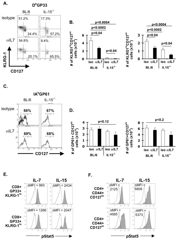
After acute LCMV infection, most effector CD8+ T cells were comprised of two major subpopulations (3), a population of KLRG1hiCD127lo cells and another population that is KLRG1loCD127hi (Fig. 2A). Given that KLRG1loCD127hi CD8+ T cells expressed both CD127 and CD122, it was logical that IL-7 and IL-15 might be redundant for maintaining this effector subpopulation. To determine the relative redundancy of IL-7 and IL-15 on effector CD4+ and CD8+ T cell survival, we infected groups of either BL/6 or IL-15−/− mice and treated them with either isotype control antibody or anti-IL-7 neutralizing antibody between days 10 and 20 after infection.
Figure 2. IL-7 and IL-15 contribute redundantly for effector CD8+, but not CD4+ T cell survival.
Groups of BL/6 or IL-15−/− mice (N = 5/group) were infected with LCMV and treated i.p. with either isotype control (MPC11) or α-IL-7 (M25) antibody (3 mg/mouse on days 11, 13, 15, 17, 19) and sacrificed on day 20. Spleen cells were stained with Dbgp33-tetramers and with antibodies against CD8, KLRG1, and CD127 or with IAbgp61-tetramers and with antibodies against CD4, CD16/32, and CD127. Dot plots show (A) KLRG1 by CD127 staining in Dbgp33-sp. CD8+ T cells from either BL/6 (left panels) or IL-15−/− mice (right panels) treated with either isotype control (top panels) or α-IL-7 antibody (bottom panels). (B) Graphs show the total numbers of KLRG1hiCD127lo versus KLRG1loCD127hi Dbgp33-sp. CD8+ T cells. p values shown are from a Student’s t-test analysis. (C) Histograms show CD127 staining of I-Abgp61-sp. CD4+ T cells from BL/6 or IL-15−/− mice treated with either isotype control or M25 antibody. Gray histogram shows staining of I-Abgp61-sp. T cells with an isotype control antibody for CD127. (D) Graphs show total numbers of CD127hi versus CD127lo I-Abgp61-sp CD4+ T cells from BL/6 or IL-15−/− mice treated with either MPC11 or M25 antibody mice. Data are representative of 3 independent experiments. (E, F) Groups of BL/6 mice (n=3) were infected with LCMV and sacrificed on 8 days later. pStat5 levels were determined in splenocytes following stimulation with cytokines (10ng/ml for 20 minutes) in either (E) Dbgp33-specific CD8+ T cells or (F) CD4+ CD44hi cells. Histograms show pSTAT5 levels in cells treated with either media alone (open histograms) or with IL-7 or IL-15 (gray histograms). Data are representative of 2 independent experiments.
In BL/6 mice, we found that IL-7 was critical for survival of some KLRG1hiCD127lo and some KLRG1loCD127hi CD8+ T cells (Fig. 2B). In contrast, IL-15 was critical for survival of most KLRG1hiCD127lo and some KLRG1loCD127hi effector CD8+ T cells (Fig. 2B). Neutralization of IL-7 in IL-15−/− mice further decreased the numbers of both subsets of CD8+ T cells (Fig. 2B). Further, Bcl-2 levels correlated with the cells loss as the combined effects of IL-7 and IL-15 were required to maintain high levels of Bcl-2 in both subsets of CD8+ T cells (Fig. S1A,B). At no time was KLRG1 expression observed on LCMV-sp. CD4+ T cells, although CD127 expression was increased on most of these cells by day 20 after infection (Fig. 2C). Thus, although KLRG1 marked subpopulations of LCMV-sp. effector CD8+ T cells, the lack of KLRG1 on effector CD4+ T cells, left CD127 as a potential marker to identify effector CD4+ subpopulations. Neither the individual or combined loss of IL-7 and IL-15 had a significant effect on the total numbers of CD4+ gp61-sp. T cells, irrespective of their CD127 expression (Fig. 2C and 2D). To ensure the effectiveness of IL-7 neutralization, we assessed pre-B cells in the bone marrow as described (6). In both BL/6 and IL-15−/− mice, administration of M25 caused a ~20-fold loss of IgM+B220int BM pre-B cells (Fig. S1C). Thus, dynamic regulation of cytokine receptors controls cytokine availability and thereby contributes to effector CD8+, but not CD4+, T cell survival.
STAT5 is critical to maintain effector CD8+ T cells during LCMV infection
STAT5 is known to be a common downstream signaling molecule for both IL-7 and IL-15 (24). To determine if IL-7 or IL-15 could activate STAT5, we cultured effector T cells with the cytokines and assessed STAT5 phosphorylation (pSTAT5) using a phospho-STAT5-specific monoclonal antibody and intracellular flow cytometry. IL-7 drove STAT5 phosphorylation in KLRG1hi CD8+ T cells, and higher pSTAT5 in KLRG1lo LCMV-sp. CD8+ T cells (Fig. 2E), consistent with the increased expression of CD127 on the latter population. Although the pSTAT5 stain worked in conjunction with the Dbgp33-tetramer stain, it was technically infeasible with the IAbgp61-tetramer stain. Instead, we co-stained with CD44, a marker of activation. Recent work has shown that multiple LCMV epitopes are recognized by CD4+ T cells, and suggest that early after infection, most activated CD44hi T cells are LCMV-specific (25). Similar to the results in effector CD8+ T cells, IL-7 drove modest pSTAT5 in CD127lo CD4+ T cells, but stronger pSTAT5 in CD127hi CD4+ T cells (Fig. 2F). In contrast, IL-15 drove pSTAT5 similarly in all effector CD4+ and CD8+ T cells (Fig. 2E and F).
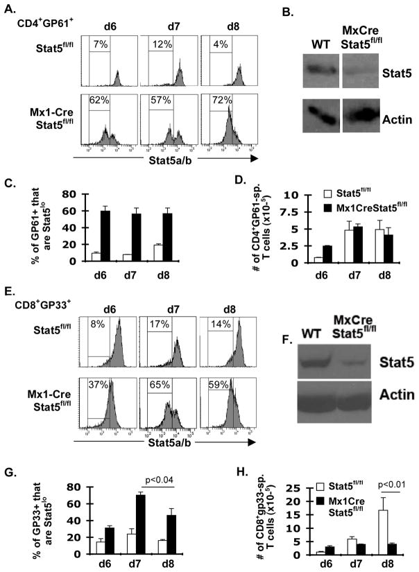
As both IL-7 and IL-15 activated STAT5, we next tested the requirement of STAT5 for survival of effector T cells using conditional STAT5-deficient mice (26). Because cell-lineage specific ablation of STAT5 affects both T cell development and peripheral naïve T cell survival (11), we used an inducible method of deletion. As LCMV infection drives very high systemic levels of type I interferon (Fig. S2A), we used a transgenic system in which Cre expression is controlled by the αIFN-inducible Mx1 promoter, to inducibly delete STAT5 during the course of the response to LCMV. Using a STAT5-specific antibody and intracellular flow cytometry, we found that by day 6 after LCMV infection, most CD4+ IAbgp61-sp. T cells in Mx1Cre-STAT5fl/fl mice had significantly decreased expression of STAT5 (Fig. 3A). Similar decreases in STAT5 levels were observed using SDS-PAGE and Western blotting of purified CD4+ T cell lysates from LCMV-infected Mx1Cre-STAT5fl/fl versus STAT5fl/fl mice (Fig. 3B). From day 6 to day 8 the frequency of CD4+ IAbgp61-sp T cells that were STAT5lo remained constant (Fig. 3C). Further, the total numbers of CD4+ IAbgp61+ T cells were slightly increased in Mx1Cre-STAT5fl/fl mice on day 6 after infection, but were unchanged on days 7 and 8 after LCMV infection (Figure 3D). The slight increase in LCMV-sp. CD4+ T cells in Mx1Cre-STAT5fl/fl mice on day 6 was not reproducible (data not shown).
Figure 3. STAT5 is critical for maintaining effector CD8+, but not CD4+, T cells.
Groups of STAT5fl/fl or Mx1Cre-STAT5fl/fl mice (N = 3–5/group) were infected with LCMV and sacrificed 6–8 days later. Spleen cells were surface stained with either IAbgp61 or with Dbgp33 tetramer along with antibodies against CD8 or CD4 and intracellularly with antibody against total STAT5. (A, C, E, G) Frequencies of MHC tetramer+ cells that were STAT5lo (C,G) and total overall numbers of tetramer+ T cells (D, H) are shown. Data from day 8 were pooled between 2 independent experiments. Shown are p values from a Student’s t-test analysis. (B, F) Groups of mice of either STAT5fl/fl or Mx1Cre-STAT5fl/fl mice (N = 3–5/group) were infected with LCMV and sacrificed 8 days later. Total CD4+ (B) or CD8+ (F) were purified using a panT cell isolation kit (Miltenyi Biotech) and 1×10⁶ cell equivalents were subjected to SDS-PAGE and Western blotting for either STAT5 or for actin. Data in figures 3B and 3F were done in separate experiments. The bands displayed in figures 3B for actin and STAT5 were from separate lanes run on the same gel. Data are representative of 2 independent experiments.
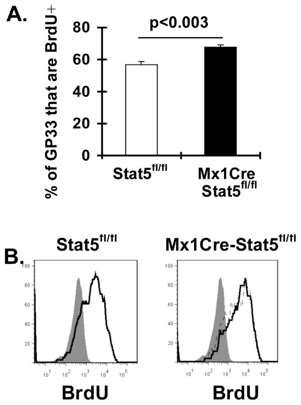
In contrast, the frequency of LCMV-sp. CD8+ T cells that were STAT5lo in Mx1Cre-STAT5fl/fl mice was increased on day 6, increased further on day 7, and then decreased by day 8 after infection (Fig. 3E and G). SDS-PAGE and Western blotting again confirmed the loss of STAT5 in purified CD8+ T cells (Fig. 3F). While the total numbers of LCMV-sp. CD8+ T cells were not different between Mx1Cre-STAT5fl/fl versus STAT5fl/fl mice on day 7 after LCMV infection, they were significantly lower in Mx1Cre-STAT5fl/fl mice by day 8 after infection. The total number of CD8+gp33-sp. T cells on day 8 after infection was also not different between BL/6 and Mx1Cre Tg mice, arguing against a non-specific effect of Cre on the T cell response (data not shown). As previous work showed that STAT5 was critical for perforin expression (27, 28), it was possible that the loss of STAT5 might result in persistent viral infection that could influence CD8+ T cell responses. However, no significant differences in viral loads were observed from either the livers or spleens of STAT5fl/fl nor Mx1Cre-STAT5fl/fl mice on days 6 or 7 after infection (Fig. S2B). It was also possible that the diminution of T cell numbers in infected Mx1Cre-STAT5fl/fl mice reflected a STAT5 contribution to T cell proliferation. However, using in vivo BrdU labeling, the frequency of CD8+gp33-sp. T cells that were BrdU+ was actually increased in Mx1Cre-STAT5fl/fl compared to STAT5fl/fl mice on day 8 after infection (Fig. 4A). In addition, there was no difference in BrdU uptake in cells that were STAT5lo compared to those that were STAT5hi at this timepoint (Fig. 4B). Together, these data show that STAT5 is critical to maintain CD8+, but not CD4+, effector T cells.
Figure 4. In vivo proliferation of CD8+gp33-sp. T cells in Mx1Cre-Stat5fl/fl mice on day 8 after infection.
Groups of either Mx1CreStat5fl/fl versus Stat5fl/fl (N=5 mice/group) mice were infected i.p. with LCMV. On day 7 after infection, mice were injected i.p. with 0.8mg of BrdU in the morning and evening and sacrificed on day 8 after infection. One control mouse did not receive BrdU. Spleen cells were stained with antibodies against CD8, KLRG1,CD127 and intracellularly against STAT5 and BrdU. (A) Results show the frequency of CD8+ GP33-sp. T cells were BrdU+ from either Stat5fl/fl versus Mx1CreStat5fl/fl mice. (B) Representative histograms show BrdU staining intensity in CD8+gp33-sp. T cells from an LCMV-infected STAT5fl/fl mouse (left histogram, 60% BrdU+) versus a Mx1CreStat5fl/fl mouse after gating on STAT5hi (dark line, 67% BrdU+) versus STAT5lo (dashed line, 68% BrdU+) cells. The shaded histogram shows BrdU staining intensity in CD8+gp33+ T cells from the control mouse not injected with BrdU.
STAT5 is critical to maintain both effector CD8+ T cell subpopulations
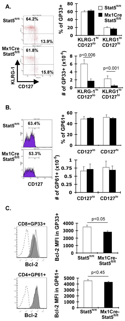
Because IL-15 was critical for KLRG1hiCD127lo cells, but IL-7 and IL-15 acted in concert to maintain KLRG1loCD127hi cells, we next tested the requirement for STAT5 on these two sub-populations. Importantly, deletion of Stat5 was similar in both CD8+ T cell subpopulations (Fig. S3A). The frequency of KLRG1hiCD127lo and KLRG1loCD127hi CD8+ T cells was similar in both STAT5fl/fl and Mx1Cre-STAT5fl/fl mice (Fig. 5A). However, both subsets of effector CD8+ T cells were significantly reduced in Mx1Cre-STAT5fl/fl compared to STAT5fl/fl mice (Fig. 5A). As expected, the frequencies of CD127hi and total numbers of LCMV-sp. CD4+ T cells were not different between STAT5fl/fl and Mx1Cre-STAT5fl/fl mice (Fig. 5B).
Figure 5. STAT5 is critical for maintaining Bcl-2 levels in effector CD8+, but not CD4+, T cells.
Groups of either STAT5fl/fl or Mx1Cre-STAT5fl/fl mice (N = 5/group) were infected with LCMV and sacrificed 15 days later. (A) Representative dot plots show KLRG1 versus CD127 staining in gated CD8+Dbgp33-sp. T cells from either STAT5fl/fl (top plot) or Mx1Cre-STAT5fl/fl (bottom plot). Graphs show the frequency (top) and total numbers (bottom) of effector CD8+ subpopulations. Numbers of KLRG1hiCD127lo and KLRG1loCD127hi cells were significantly decreased in Mx1Cre-STAT5fl/fl mice (Students t-test). (B) Representative histograms show CD127 staining in gated CD4+ IAbgp61-sp. T cells from either STAT5fl/fl (top histogram) or Mx1Cre-STAT5fl/fl (bottom histogram). Graphs show the frequency (top) and total numbers (bottom) of effector CD4+ subpopulations. (C) Representative histograms show Bcl-2 staining in gated CD8+ gp33-sp. T cells (top histogram) or IAbgp61-sp. T cells (bottom histogram) from either STAT5fl/fl (open histogram) or Mx1Cre-STAT5fl/fl (shaded histogram). Isotype control staining is shown by the dashed line. Graphs show the mean fluorescence intensity (MFI) of the Bcl-2 stain in CD8+ Dbgp33-sp. (top graph) or in CD4+ IAbgp61-sp. cells (bottom graph) from either STAT5fl/fl or Mx1Cre-STAT5fl/fl mice. Bcl-2 levels were significantly decreased in CD8+ Dbgp33-sp. T cells from Mx1Cre-STAT5fl/fl mice (Students t-test). Data are representative of 2 independent experiments.
Stat5 is critical for IL-7 and IL-15-driven upregulation of Bcl-2
As IL-15 was critical for Bcl-2 expression (Fig. 1B) and STAT5 was critical for maintenance of effector CD8+ T cells (Fig. 3G, 3H) we next determined the role of STAT5 in promoting Bcl-2 expression. Even though fewer CD8+gp33-sp. cells were STAT5lo by day 15 after infection compared to day 8 (31.3% +/− 4.8 versus 46.1% +/− 8.1 respectively, Fig. S3B, C)), Bcl-2 levels in LCMV-sp. CD8+ T cells from Mx1Cre-STAT5fl/fl mice were significantly reduced compared to cells from STAT5fl/fl mice (Fig. 5C). Further, levels of Bcl-2 were significantly lower in STAT5lo (Bcl-2 MFI = 2116 ± 101) compared to STAT5hi (Bcl-2 MFI = 3356 ± 158) CD8+gp33-sp. T cells from Mx1Cre-STAT5fl/fl mice (p<0.0001; Students t-test). As expected, Bcl-2 levels in CD4+ gp61-sp. T cells were not affected by STAT5 deletion (Fig. 5C). Moreover, the frequencies of CD4+ gp61-sp. T cells that were STAT5lo were similar between days 8 and 15 (56.5%+/− 6.55 vs. 50.6% +/− 0.98, respectively, Fig. S3B).
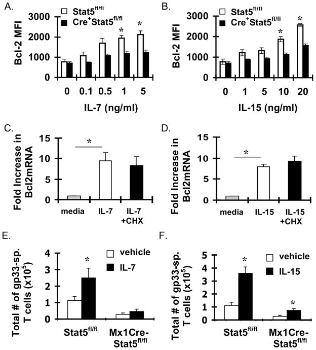
To assess the requirement for STAT5 in upregulation of Bcl-2 by IL-7 and IL-15, we cultured spleen cells from d7 LCMV-infected STAT5fl/fl or Mx1Cre-STAT5fl/fl mice with IL-7 or IL-15 and assessed levels of Bcl-2 within gp33-sp. CD8+ T cells the next day. Notably, both IL-7 and IL-15 increased expression of Bcl-2 in a dose dependent fashion in T cells from STAT5fl/fl mice, while upregulation of Bcl-2 was significantly impaired in T cells from Mx1Cre-STAT5fl/fl mice (Fig. 6A and 6B). The slight induction that was observed in response to IL-7 and IL-15 in Mx1Cre-STAT5fl/fl T cells was likely due to incomplete deletion of STAT5 within these cells, as only cells expressing high levels of STAT5 increased expression of Bcl-2 in response to IL-7 or IL-15 (Fig. S4). Combined, these data demonstrate that both IL-7 and IL-15 require STAT5 to induce Bcl-2 within effector CD8+ T cells.
Figure 6. IL-7 and IL-15 require Stat5 to drive Bcl-2 expression in vitro and to maintain effector T cell responses in vivo.
Groups of STAT5fl/fl versus Mx1Cre-STAT5fl/fl mice (N = 3 mice/group) were infected with LCMV and sacrificed 7 days after infection. Spleen cells were cultured with the indicated concentrations of (A) IL-7 or (B) IL-15 overnight. Cells were then stained with Db-gp33 Tetramers, antibodies against CD8, KLRG1, and intracellularly with an α-Bcl-2antibody. Results show the MFI of the Bcl-2 stain within CD8+gp33-sp. T cells +/− SEM. (C, D) BL/6 mice were infected with LCMV, sacrificed on day 7 and purified splenic CD8+ T cells were cultured in triplicate with IL-7 or IL-15 with or without 2 μM cycloheximide (CHX) for 3 hours. Cells were harvested, RNA was isolated, and cDNA generated and subjected to real-time RT-PCR for Bcl-2. Results show the fold change in Bcl-2 mRNA expression in response to (C) IL-7 or (D) IL-15 +/− S.D. (E, F) Groups of STAT5fl/fl versus Mx1Cre-STAT5fl/fl mice (N = 5 mice/group) were infected with LCMV and treated with either PBS, with (E) IL-7/α-IL-7 immune complexes or with (F) IL-15/IL-15Rα complexes on days 10, 12, 14 and sacrificed on day 15 after infection. Spleen cells were stained with Db-gp33 tetramers, antibodies against CD8, KLRG1, and IL-7Rα. Results show the total number of Db-gp33-sp. T cells +/− SEM. * = statistically significant difference between cytokine treated vs untreated control (Student’s t-test). Data are representative of 2 independent experiments.
The mechanism by which STAT5 controls Bcl-2 expression is controversial. One report suggested that the effects of STAT5 are indirect (i.e. STAT5 inducing expression of another factor that induces Bcl-2 transcription) (29). Other reports have suggested direct effects of STAT5 on Bcl-2 transcription (30, 31). To determine which of these mechanism(s) contributes to cytokine-driven Bcl-2 expression in T cells, we cultured purified CD8+ T cells from LCMV-infected BL/6 mice with IL-7 or IL-15 with or without cycloheximide for 3 hours and then measured Bcl-2 mRNA levels by real-time RT-PCR. Interestingly, while both IL-7 and IL-15 both drove significant induction of Bcl-2 within 3 hours, the presence of cycloheximide did not reduce Bcl-2 mRNA levels in response to these cytokines (Fig. 6C and 6D) providing evidence that the effect of STAT5 on Bcl-2 expression is direct (i.e. it did not require new protein synthesis).
IL-7 and IL-15 depend on Stat5 to maintain effector T cell responses
As both IL-7 and IL-15 required STAT5 to induce Bcl-2 expression in vitro, We next determined if IL-7 or IL-15 required for STAT5 to maintain effector T cell responses in vivo. To do this, we infected mice with LCMV, and starting on day 10 after infection treated mice with long-acting forms of either IL-7 or IL-15 (6, 32) every other day until day 15 after infection. On day 15 we found that, both IL-7 and IL-15 significantly increased numbers of CD8+ gp33-sp. T cells in Stat5fl/fl mice (Fig. 6E and 6F). In contrast, IL-7 was unable to significantly increase gp33-sp. CD8+ T cells in Mx1Cre-STAT5fl/fl mice (Fig. 6E). While IL-15 did significantly increase gp33-sp. T cells in Mx1Cre-STAT5fl/fl mice; the increase was less than in STAT5fl/fl mice (Fig. 6F) and the levels of STAT5 were enhanced in Mx1Cre-STAT5fl/fl compared to STAT5fl/fl controls (data not shown). Thus, IL-15 likely enhanced selection of STAT5hi T cells in vivo. Combined, these data show that IL-7 and IL-15 largely require STAT5 for maintaining effector CD8+ T cells in vivo.
Discussion
Here we demonstrate that STAT5 is critical to maintain effector CD8+T cells. In this model, Stat5 is deleted in multiple tissues as the Mx1Cre transgene is expressed ubiquitously. It is possible that some of the effects we observe could be due to deletion of Stat5 in non-T cells. For instance, STAT5 signaling in dendritic cells (DCs) may influence survival of effector T cells as it has recently been shown that IL-7 can act on DCs to regulate the size of the CD4 T cell pool under lymphopenic conditions (33). However, whether this axis operates under normal physiologic conditions or during viral infection remains unclear. If there were a non-T cell effect of Stat5 that was dominant, we would have expected to see similar effects on CD4+ and CD8+ T cell responses as expansion of both cells require DCs, and this was clearly not the case. Although we cannot completely rule out non-T cell effects of Stat5, our data clearly show that despite similar deletion of Stat5, CD8+ effector T cells rely on Stat5 considerably more than do effector CD4+ T cells. We were somewhat surprised to find that IL-15 was not required for maintaining effector CD4+ T cell responses, as a recent report showed that IL-15 contributes to survival of memory CD4+ T cells (34). It is possible that IL-15 becomes a more prominent survival factor for memory CD4+ T cells than for effector CD4+ T cells. Indeed, we found that expression of CD122 was quite transient on effector CD4+ T cells (data not shown), and previous work showed that increased expression of CD122 on memory CD4+ T cells (34), may facilitate their dependence on IL-15. We note that our data do not imply that IL-15 is not involved in effector CD4+ T cell survival, just that it is not strictly required. Thus, mechanisms maintaining effector CD4+ T cells remain unclear. Previous work has shown that Bcl-3, a NF-κB p50 family member, can promote survival of activated CD4+ T cells (35). Further, activated CD4+ T cells have dramatically increased expression of A1 (1, 2), and A1 expression can be regulated by NF-κB signaling (36–38). A1 can also antagonize Bim, making A1 a logical candidate for promoting effector CD4+ T cell survival. Future experiments will evaluate the requirement for A1 in the survival of effector CD4+ T cells.
Our data in effector CD8+ T cells are consistent with previous reports showing that in cell lines, IL-7 and IL-15 control expression of Bcl-2 via STAT5 (29, 31, 39). However, other reports have suggested that STAT5 is not critical for Bcl-2 induction (40, 41). In one of these reports, mice were developed in which tyrosine 449 in the IL-7Rα gene was mutated to a phenylalanine (41). The Y449F mutation incapacitates both STAT5 and PI-3K activation in response to IL-7 in lymphocytes and thymocytes (39, 41). Using T cells from these Y449F mice, the authors showed that although IL-7 was unable to induce detectable pSTAT5, it was able to increase expression of Bcl-2 (41). From these data it was concluded that Bcl-2 upregulation was independent of STAT5 activation. However, at baseline, Bcl-2 levels were significantly lower in peripheral CD4+ and CD8+ T cells directly isolated from Y449F mice (41), suggesting that, physiologically, IL-7RαY449 signaling controls expression of Bcl-2 in vivo. Consistent with this, another group showed, in a thymocyte cell line, that Bcl-2 upregulation and STAT5 activation in response to IL-7 signaling required Y449F of IL-7Rα (39). Furthermore, mice deficient in JAK3, which is required for IL-7 and IL-15 activation of STAT5, exhibited a profound loss of peripheral CD8+ T cells but had nearly normal levels of CD4+ T cells (42–44). Moreover, JAK3-deficiency dramatically impaired Bcl-2 expression in CD8+CD4-, but not CD4+CD8- thymocytes (45). Thus, the simplest explanation is that, under physiologic conditions, IL-7 and IL-15 require STAT5 to maintain Bcl-2 expression within CD8+ T cells.
Our data also suggest that STAT5 maintains Bcl-2 directly as cycloheximide failed to block Bcl-2 induction by IL-7 and IL-15. A previous paper showed in a pro-B cell line that cycloheximide blocked induction of Bcl-2 by IL-2, leading the authors to conclude that the effects of STAT5 on Bcl-2 induction by IL-2 were largely indirect (29). However, in that study the effect of cycloheximide was only partial and these results were not reported to be significant. Our results in primary T cells suggest that the effects of IL-7 on Bcl-2 induction are direct and do not require new protein synthesis. These data are supported by a recent report showing that, in mast cells, STAT5 can bind to a site in intron 2 of the Bcl-2 gene (30). Further work will be required to determine if cytokines promote STAT5 binding to this site in intron-2 in activated CD8+ T cells.
These data have implications for the development of T cell memory that emerges from the effector pool. We and others have previously shown that the pro-apoptotic molecule, Bim, is critical for limiting the numbers of effector T cells that can become memory T cells (20, 46, 47). Here our data suggest that IL-7 and/or IL-15 signaling through Stat5 is likely critical to maintain levels of Bcl-2 sufficient to combat Bim and develop into memory T cells. Thus, cytokines may have use as potential vaccine adjuvants. Optimal vaccines would promote robust CD4+ and CD8+ T cell responses that would be long-lived. As we and others have shown, common gamma chain cytokines can significantly enhance CD4 and CD8 T cell responses (6, 7, 48). Although these cytokines enhanced short-term T cell responses, their effects were transient once the cytokines were withdrawn. This makes sense given that survival of nearly all populations of T cells is achieved, at least in part, via competition for limiting amounts of cytokines. However, we note that, in general these studies were done in viral infections in which the T cell responses are extraordinarily robust. It remains unclear if cytokine adjuvants, given short-term, may enhance long-term memory responses under conditions in which less robust T cell responses are generated. This approach could be beneficial for enhancing sub-optimal vaccine responses which currently require several rounds of boosting.
Supplementary Material
Acknowledgments
This work was supported by a Public Health Service Grant AI057753 (D.A.H.).
The authors thank Drs. Edith Janssen, Brian Ladle, Chris Karp, and Ms. Jana Raynor for critical review of the manuscript. The authors thank Dr. Allan Zajac for gift of Db-gp33-monomers, Ms. Alicia Browning for help with viral plaque assays, and Dr. Lothar Hennighausen for the Stat5fl/fl mice.
References
- 1.D'Cruz LM, Rubinstein MP, Goldrath AW. Surviving the crash: transitioning from effector to memory CD8+ T cell. Semin Immunol. 2009;21:92–98. doi: 10.1016/j.smim.2009.02.002. [DOI] [PMC free article] [PubMed] [Google Scholar]
- 2.Hildeman D, Jorgensen T, Kappler J, Marrack P. Apoptosis and the homeostatic control of immune responses. Current opinion in immunology. 2007;19:516–521. doi: 10.1016/j.coi.2007.05.005. [DOI] [PMC free article] [PubMed] [Google Scholar]
- 3.Joshi NS, Cui W, Chandele A, Lee HK, Urso DR, Hagman J, Gapin L, Kaech SM. Inflammation directs memory precursor and short-lived effector CD8(+) T cell fates via the graded expression of T-bet transcription factor. Immunity. 2007;27:281–295. doi: 10.1016/j.immuni.2007.07.010. [DOI] [PMC free article] [PubMed] [Google Scholar]
- 4.Hand TW, Morre M, Kaech SM. Expression of IL-7 receptor alpha is necessary but not sufficient for the formation of memory CD8 T cells during viral infection. Proc Natl Acad Sci U S A. 2007;104:11730–11735. doi: 10.1073/pnas.0705007104. [DOI] [PMC free article] [PubMed] [Google Scholar]
- 5.Haring JS, Jing X, Bollenbacher-Reilley J, Xue HH, Leonard WJ, Harty JT. Constitutive expression of IL-7 receptor alpha does not support increased expansion or prevent contraction of antigen-specific CD4 or CD8 T cells following Listeria monocytogenes infection. J Immunol. 2008;180:2855–2862. doi: 10.4049/jimmunol.180.5.2855. [DOI] [PubMed] [Google Scholar]
- 6.Tripathi P, Mitchell TC, Finkelman F, Hildeman DA. Cutting Edge: Limiting Amounts of IL-7 Do Not Control Contraction of CD4+ T Cell Responses. J Immunol. 2007;178:4027–4031. doi: 10.4049/jimmunol.178.7.4027. [DOI] [PMC free article] [PubMed] [Google Scholar]
- 7.Rubinstein MP, Lind NA, Purton JF, Filippou P, Best JA, McGhee PA, Surh CD, Goldrath AW. IL-7 and IL-15 differentially regulate CD8+ T-cell subsets during contraction of the immune response. Blood. 2008;112:3704–3712. doi: 10.1182/blood-2008-06-160945. [DOI] [PMC free article] [PubMed] [Google Scholar]
- 8.Yajima T, Yoshihara K, Nakazato K, Kumabe S, Koyasu S, Sad S, Shen H, Kuwano H, Yoshikai Y. IL-15 regulates CD8+ T cell contraction during primary infection. J Immunol. 2006;176:507–515. doi: 10.4049/jimmunol.176.1.507. [DOI] [PubMed] [Google Scholar]
- 9.Sasaki T, Irie-Sasaki J, Jones RG, Oliveira-dos-Santos AJ, Stanford WL, Bolon B, Wakeham A, Itie A, Bouchard D, Kozieradzki I, Joza N, Mak TW, Ohashi PS, Suzuki A, Penninger JM. Function of PI3Kgamma in thymocyte development, T cell activation, and neutrophil migration. Science. 2000;287:1040–1046. doi: 10.1126/science.287.5455.1040. [DOI] [PubMed] [Google Scholar]
- 10.Yao Z, Cui Y, Watford WT, Bream JH, Yamaoka K, Hissong BD, Li D, Durum SK, Jiang Q, Bhandoola A, Hennighausen L, O'Shea JJ. Stat5a/b are essential for normal lymphoid development and differentiation. Proc Natl Acad Sci U S A. 2006;103:1000–1005. doi: 10.1073/pnas.0507350103. [DOI] [PMC free article] [PubMed] [Google Scholar]
- 11.Seki Y, Yang J, Okamoto M, Tanaka S, Goitsuka R, Farrar MA, Kubo M. IL-7/STAT5 cytokine signaling pathway is essential but insufficient for maintenance of naive CD4 T cell survival in peripheral lymphoid organs. J Immunol. 2007;178:262–270. doi: 10.4049/jimmunol.178.1.262. [DOI] [PubMed] [Google Scholar]
- 12.Swainson L, Kinet S, Mongellaz C, Sourisseau M, Henriques T, Taylor N. IL-7-induced proliferation of recent thymic emigrants requires activation of the PI3K pathway. Blood. 2007;109:1034–1042. doi: 10.1182/blood-2006-06-027912. [DOI] [PubMed] [Google Scholar]
- 13.Mazzucchelli R, Durum SK. Interleukin-7 receptor expression: intelligent design. Nat Rev Immunol. 2007;7:144–154. doi: 10.1038/nri2023. [DOI] [PubMed] [Google Scholar]
- 14.Okkenhaug K, Bilancio A, Farjot G, Priddle H, Sancho S, Peskett E, Pearce W, Meek SE, Salpekar A, Waterfield MD, Smith AJ, Vanhaesebroeck B. Impaired B and T cell antigen receptor signaling in p110delta PI 3-kinase mutant mice. Science. 2002;297:1031–1034. doi: 10.1126/science.1073560. [DOI] [PubMed] [Google Scholar]
- 15.Imada K, Bloom ET, Nakajima H, Horvath-Arcidiacono JA, Udy GB, Davey HW, Leonard WJ. Stat5b is essential for natural killer cell-mediated proliferation and cytolytic activity. J Exp Med. 1998;188:2067–2074. doi: 10.1084/jem.188.11.2067. [DOI] [PMC free article] [PubMed] [Google Scholar]
- 16.Nakajima H, Liu XW, Wynshaw-Boris A, Rosenthal LA, Imada K, Finbloom DS, Hennighausen L, Leonard WJ. An indirect effect of Stat5a in IL-2-induced proliferation: a critical role for Stat5a in IL-2-mediated IL-2 receptor alpha chain induction. Immunity. 1997;7:691–701. doi: 10.1016/s1074-7613(00)80389-1. [DOI] [PubMed] [Google Scholar]
- 17.Moriggl R, Topham DJ, Teglund S, Sexl V, McKay C, Wang D, Hoffmeyer A, van Deursen J, Sangster MY, Bunting KD, Grosveld GC, Ihle JN. Stat5 is required for IL-2-induced cell cycle progression of peripheral T cells. Immunity. 1999;10:249–259. doi: 10.1016/s1074-7613(00)80025-4. [DOI] [PubMed] [Google Scholar]
- 18.Hildeman D, Yanez D, Pederson K, Havighurst T, Muller D. Vaccination against persistent viral infection exacerbates CD4+ T-cell-mediated immunopathological disease. J Virol. 1997;71:9672–9678. doi: 10.1128/jvi.71.12.9672-9678.1997. [DOI] [PMC free article] [PubMed] [Google Scholar]
- 19.Wojciechowski S, Tripathi P, Bourdeau T, Acero L, Grimes HL, Katz JD, Finkelman FD, Hildeman DA. Bim/Bcl-2 balance is critical for maintaining naive and memory T cell homeostasis. J Exp Med. 2007;204:1665–1675. doi: 10.1084/jem.20070618. [DOI] [PMC free article] [PubMed] [Google Scholar]
- 20.Wojciechowski S, Jordan MB, Zhu Y, White J, Zajac AJ, Hildeman DA. Bim mediates apoptosis of CD127(lo) effector T cells and limits T cell memory. Eur J Immunol. 2006;36:1694–1706. doi: 10.1002/eji.200635897. [DOI] [PMC free article] [PubMed] [Google Scholar]
- 21.Jiang Z, Georgel P, Du X, Shamel L, Sovath S, Mudd S, Huber M, Kalis C, Keck S, Galanos C, Freudenberg M, Beutler B. CD14 is required for MyD88-independent LPS signaling. Nat Immunol. 2005;6:565–570. doi: 10.1038/ni1207. [DOI] [PubMed] [Google Scholar]
- 22.Kaech SM, Tan JT, Wherry EJ, Konieczny BT, Surh CD, Ahmed R. Selective expression of the interleukin 7 receptor identifies effector CD8 T cells that give rise to long-lived memory cells. Nat Immunol. 2003;4:1191–1198. doi: 10.1038/ni1009. [DOI] [PubMed] [Google Scholar]
- 23.Khanolkar A, Fuller MJ, Zajac AJ. CD4 T cell-dependent CD8 T cell maturation. J Immunol. 2004;172:2834–2844. doi: 10.4049/jimmunol.172.5.2834. [DOI] [PubMed] [Google Scholar]
- 24.Lin JX, Migone TS, Tsang M, Friedmann M, Weatherbee JA, Zhou L, Yamauchi A, Bloom ET, Mietz J, John S, et al. The role of shared receptor motifs and common Stat proteins in the generation of cytokine pleiotropy and redundancy by IL-2, IL-4, IL-7, IL-13, and IL-15. Immunity. 1995;2:331–339. doi: 10.1016/1074-7613(95)90141-8. [DOI] [PubMed] [Google Scholar]
- 25.Dow C, Oseroff C, Peters B, Nance-Sotelo C, Sidney J, Buchmeier M, Sette A, Mothe BR. Lymphocytic choriomeningitis virus infection yields overlapping CD4+ and CD8+ T-cell responses. J Virol. 2008;82:11734–11741. doi: 10.1128/JVI.00435-08. [DOI] [PMC free article] [PubMed] [Google Scholar]
- 26.Cui Y, Riedlinger G, Miyoshi K, Tang W, Li C, Deng CX, Robinson GW, Hennighausen L. Inactivation of Stat5 in mouse mammary epithelium during pregnancy reveals distinct functions in cell proliferation, survival, and differentiation. Mol Cell Biol. 2004;24:8037–8047. doi: 10.1128/MCB.24.18.8037-8047.2004. [DOI] [PMC free article] [PubMed] [Google Scholar]
- 27.Yu CR, Ortaldo JR, Curiel RE, Young HA, Anderson SK, Gosselin P. Role of a STAT binding site in the regulation of the human perforin promoter. J Immunol. 1999;162:2785–2790. [PubMed] [Google Scholar]
- 28.Zhang J, Scordi I, Smyth MJ, Lichtenheld MG. Interleukin 2 receptor signaling regulates the perforin gene through signal transducer and activator of transcription (Stat)5 activation of two enhancers. J Exp Med. 1999;190:1297–1308. doi: 10.1084/jem.190.9.1297. [DOI] [PMC free article] [PubMed] [Google Scholar]
- 29.Lord JD, McIntosh BC, Greenberg PD, Nelson BH. The IL-2 receptor promotes lymphocyte proliferation and induction of the c-myc, bcl-2, and bcl-x genes through the trans-activation domain of Stat5. J Immunol. 2000;164:2533–2541. doi: 10.4049/jimmunol.164.5.2533. [DOI] [PubMed] [Google Scholar]
- 30.Li G, Miskimen KL, Wang Z, Xie XY, Brenzovich J, Ryan JJ, Tse W, Moriggl R, Bunting KD. STAT5 requires the N-domain for suppression of miR15/16, induction of bcl-2, and survival signaling in myeloproliferative disease. Blood. 2010;115:1416–1424. doi: 10.1182/blood-2009-07-234963. [DOI] [PMC free article] [PubMed] [Google Scholar]
- 31.Qin JZ, Zhang CL, Kamarashev J, Dummer R, Burg G, Dobbeling U. Interleukin-7 and interleukin-15 regulate the expression of the bcl-2 and c-myb genes in cutaneous T-cell lymphoma cells. Blood. 2001;98:2778–2783. doi: 10.1182/blood.v98.9.2778. [DOI] [PubMed] [Google Scholar]
- 32.Rubinstein MP, Kovar M, Purton JF, Cho JH, Boyman O, Surh CD, Sprent J. Converting IL-15 to a superagonist by binding to soluble IL-15R{alpha} Proc Natl Acad Sci U S A. 2006;103:9166–9171. doi: 10.1073/pnas.0600240103. [DOI] [PMC free article] [PubMed] [Google Scholar]
- 33.Guimond M, Veenstra RG, Grindler DJ, Zhang H, Cui Y, Murphy RD, Kim SY, Na R, Hennighausen L, Kurtulus S, Erman B, Matzinger P, Merchant MS, Mackall CL. Interleukin 7 signaling in dendritic cells regulates the homeostatic proliferation and niche size of CD4+ T cells. Nat Immunol. 2009;10:149–157. doi: 10.1038/ni.1695. [DOI] [PMC free article] [PubMed] [Google Scholar]
- 34.Purton JF, Tan JT, Rubinstein MP, Kim DM, Sprent J, Surh CD. Antiviral CD4+ memory T cells are IL-15 dependent. J Exp Med. 2007;204:951–961. doi: 10.1084/jem.20061805. [DOI] [PMC free article] [PubMed] [Google Scholar]
- 35.Mitchell TC, Hildeman D, Kedl RM, Teague TK, Schaefer BC, White J, Zhu Y, Kappler J, Marrack P. Immunological adjuvants promote activated T cell survival via induction of Bcl-3. Nat Immunol. 2001;2:397–402. doi: 10.1038/87692. [DOI] [PubMed] [Google Scholar]
- 36.Grumont RJ, I, Rourke J, Gerondakis S. Rel-dependent induction of A1 transcription is required to protect B cells from antigen receptor ligation-induced apoptosis. Genes Dev. 1999;13:400–411. doi: 10.1101/gad.13.4.400. [DOI] [PMC free article] [PubMed] [Google Scholar]
- 37.Lee HH, Dadgostar H, Cheng Q, Shu J, Cheng G. NF-kappaB-mediated up-regulation of Bcl-x and Bfl-1/A1 is required for CD40 survival signaling in B lymphocytes. Proc Natl Acad Sci U S A. 1999;96:9136–9141. doi: 10.1073/pnas.96.16.9136. [DOI] [PMC free article] [PubMed] [Google Scholar]
- 38.Zong WX, Edelstein LC, Chen C, Bash J, Gelinas C. The prosurvival Bcl-2 homolog Bfl-1/A1 is a direct transcriptional target of NF-kappaB that blocks TNFalpha-induced apoptosis. Genes Dev. 1999;13:382–387. doi: 10.1101/gad.13.4.382. [DOI] [PMC free article] [PubMed] [Google Scholar]
- 39.Jiang Q, Li WQ, Hofmeister RR, Young HA, Hodge DR, Keller JR, Khaled AR, Durum SK. Distinct regions of the interleukin-7 receptor regulate different Bcl2 family members. Mol Cell Biol. 2004;24:6501–6513. doi: 10.1128/MCB.24.14.6501-6513.2004. [DOI] [PMC free article] [PubMed] [Google Scholar]
- 40.Lindemann MJ, Benczik M, Gaffen SL. Anti-apoptotic signaling by the interleukin-2 receptor reveals a function for cytoplasmic tyrosine residues within the common gamma (gamma c) receptor subunit. J Biol Chem. 2003;278:10239–10249. doi: 10.1074/jbc.M209471200. [DOI] [PubMed] [Google Scholar]
- 41.Osborne LC, Dhanji S, Snow JW, Priatel JJ, Ma MC, Miners MJ, Teh HS, Goldsmith MA, Abraham N. Impaired CD8 T cell memory and CD4 T cell primary responses in IL-7R alpha mutant mice. J Exp Med. 2007;204:619–631. doi: 10.1084/jem.20061871. [DOI] [PMC free article] [PubMed] [Google Scholar]
- 42.Nosaka T, van Deursen JM, Tripp RA, Thierfelder WE, Witthuhn BA, McMickle AP, Doherty PC, Grosveld GC, Ihle JN. Defective lymphoid development in mice lacking Jak3. Science. 1995;270:800–802. doi: 10.1126/science.270.5237.800. [DOI] [PubMed] [Google Scholar]
- 43.Park SY, Saijo K, Takahashi T, Osawa M, Arase H, Hirayama N, Miyake K, Nakauchi H, Shirasawa T, Saito T. Developmental defects of lymphoid cells in Jak3 kinase-deficient mice. Immunity. 1995;3:771–782. doi: 10.1016/1074-7613(95)90066-7. [DOI] [PubMed] [Google Scholar]
- 44.Thomis DC, Gurniak CB, Tivol E, Sharpe AH, Berg LJ. Defects in B lymphocyte maturation and T lymphocyte activation in mice lacking Jak3. Science. 1995;270:794–797. doi: 10.1126/science.270.5237.794. [DOI] [PubMed] [Google Scholar]
- 45.Wen R, Wang D, McKay C, Bunting KD, Marine JC, Vanin EF, Zambetti GP, Korsmeyer SJ, Ihle JN, Cleveland JL. Jak3 selectively regulates Bax and Bcl-2 expression to promote T-cell development. Mol Cell Biol. 2001;21:678–689. doi: 10.1128/MCB.21.2.678-689.2001. [DOI] [PMC free article] [PubMed] [Google Scholar]
- 46.Prlic M, Bevan MJ. Exploring regulatory mechanisms of CD8+ T cell contraction. Proc Natl Acad Sci U S A. 2008;105:16689–16694. doi: 10.1073/pnas.0808997105. [DOI] [PMC free article] [PubMed] [Google Scholar]
- 47.Reckling S, Divanovic S, Karp CL, Wojciechowski S, Belkaid Y, Hildeman D. Proapoptotic Bcl-2 family member Bim promotes persistent infection and limits protective immunity. Infect Immun. 2008;76:1179–1185. doi: 10.1128/IAI.01093-06. [DOI] [PMC free article] [PubMed] [Google Scholar]
- 48.Nanjappa SG, Walent JH, Morre M, Suresh M. Effects of IL-7 on memory CD8 T cell homeostasis are influenced by the timing of therapy in mice. J Clin Invest. 2008;118:1027–1039. doi: 10.1172/JCI32020. [DOI] [PMC free article] [PubMed] [Google Scholar]
Associated Data
This section collects any data citations, data availability statements, or supplementary materials included in this article.


