Abstract
Th17 cells promote a variety of autoimmune diseases, including psoriasis, multiple sclerosis, rheumatoid arthritis, and inflammatory bowel disease. TGF-β is required for conversion of naive T cells to Th17 cells, but the mechanisms regulating this process are unknown. Integrin αvβ8 on DCs can activate TGF-β, and this process contributes to the development of induced Tregs. Here, we have now shown that integrin αvβ8 expression on DCs plays a critical role in the differentiation of Th17 cells. Th17 cells were nearly absent in the colons of mice lacking αvβ8 expression on DCs. In addition, these mice and the DCs harvested from them had an impaired ability to convert naive T cells into Th17 cells in vivo and in vitro, respectively. Importantly, mice lacking αvβ8 on DCs showed near-complete protection from experimental autoimmune encephalomyelitis. Our results therefore suggest that the integrin αvβ8 pathway is biologically important and that αvβ8 expression on DCs could be a therapeutic target for the treatment of Th17-driven autoimmune disease.
Introduction
For the adaptive immune system to elicit an effective response to different types of pathogens, naive T cells must undergo a series of epigenetic changes that specialize them for specific responses. Th17 cells develop from naive T cells and aid in the destruction of certain bacteria and fungi. The cytokines IL-17A, IL-17F, and IL-22, produced by these cells, support the recruitment and activation of neutrophils and drive the production of antimicrobial peptides by epithelial cells (1). Because of the prominent role these cells play in host defense and autoimmune disease, the mechanisms that regulate Th17 development are a topic of intensive investigation.
IL-6 and TGF-β are sufficient to differentiate Th17 cells from naive CD4+ T cells (2–4). Other cytokines, including IL-21 and IL-23, augment Th17 conversion and expand the population (5, 6). TGF-β directs the transcriptional program of Th17 cells by inducing expression of the Th17 lineage-specific transcription factor RORγt (7). TGF-β also stimulates expression of Foxp3, a transcription factor that is required for Treg function, and IL-6 is required to mediate the stabilization of RORγt and repression of Foxp3 (8). TGF-β is also necessary for expression of the IL-23 receptor on T cells, which allows the Th17 population to expand in response to IL-23 (4). While TGF-β is known to be a critical factor for Th17 differentiation and function, the mechanisms that regulate TGF-β activity in Th17 development have not been described.
TGF-β is produced in a latent form and must be activated to exert a biological effect. Each of the 3 TGF-β mammalian genes encode a translation product consisting of an N-terminal propeptide (termed “latency-associated peptide” [LAP]) and bioactive TGF-β. This product is cleaved intracellularly by furin, and LAP associates with TGF-β in a noncovalent fashion to form the small latent complex. In most cells, LAP is covalently attached to 1 of 3 latent TGF-β binding proteins prior to secretion to form a large latent complex (9). TGF-β is generally inactive when it is incorporated into a small latent complex or large latent complex and is unable to bind TGF-β receptors. Latent TGF-β has been reported to be activated by proteases, thrombospondin-1, reactive oxygen species, and, at least for TGF-β1 and -β3, the integrins αvβ6 and αvβ8 (10, 11). A prominent role for integrin-mediated TGF-β activation is suggested by recent evidence that mice expressing a mutant form of TGF-β1 that cannot be activated by integrins phenocopy mice completely lacking TGF-β1 (12), and mice lacking αvβ8 treated at birth with antibodies blocking αvβ6 express all of the phenotypic features of mice lacking TGF-β1 and TGF-β3 (13).
We have recently shown that mice lacking αvβ8 on DCs develop colitis and autoimmunity and have a quantitative defect in development of CD4+ Foxp3+ Tregs in the colon (14), demonstrating a critical role for this pathway in maintenance of immune homeostasis. However, the importance of this pathway in the induction of effector Th17-mediated immune responses is completely unknown. Because of the prominent role of TGF-β in Th17 cell induction and the absence of any data describing how TGF-β gets activated to induce these cells, we sought to determine what role integrin αvβ8 on DCs plays in the development of Th17 cells.
Results
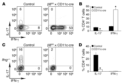
We examined the colons of mice lacking αvβ8 on DCs for the presence of IL-17–producing CD4+ T cells. Intracellular flow cytometry analysis of cells isolated from the colonic lamina propria of β8fl/fl × CD11c-cre mice showed a near absence of IL-17–expressing CD4+ T cells (0.6% ± 0.2%), whereas 8.5% ± 1.3% of colonic CD4+ T cells in control mice expressed IL-17 (Figure 1, A and B). In contrast, in β8fl/fl × CD11c-cre mice a majority (52% ± 4.4%) of CD4+ T cells expressed IFN-γ (Figure 1B). Since IFN-γ impedes development of Th17 cells in vitro, we examined whether increased production of IFN-γ was responsible for impaired Th17 development in β8fl/fl × CD11c-cre mice by crossing them to mice deficient in IFN-γ. The colons of Ifng–/– β8fl/fl × CD11c-cre mice also showed a dramatic reduction in the number of CD4+ IL-17+ cells, with only 3.1% ± 0.5% of the CD4+ cells expressing IL-17 compared with 26% ± 4.0% in control Ifng–/– mice (Figure 1, C and D). Interestingly, Ifng–/– β8fl/fl × CD11c-cre mice exhibited colitis and enlarged mesenteric lymph nodes that closely resembled β8fl/fl × CD11c-cre mice, suggesting that the colitis seen in the absence of αvβ8 on DCs occurs independently of IFN-γ (data not shown). We previously reported that β8fl/fl × CD11c-cre mice have a decreased frequency of Tregs in the colon but a normal proportion in the spleen (14). Since Tregs are significant producers of TGF-β, we questioned whether a decreased frequency of Tregs in the colon could decrease the amount of total TGF-β available for Th17 development in β8fl/fl × CD11c-cre mice. We found no significant difference in the amount of TGF-β in the colons from control and β8fl/fl × CD11c-cre mice (417 ± 29 and 384 ± 13 pg/ml; P = 0.16). These data indicate that excess production of IFN-γ and total TGF-β concentrations were not responsible for the absence of Th17 cells in the colons of β8fl/fl × CD11c-cre mice.
Figure 1. Impaired Th17 development in β8fl/fl × CD11c-cre mice.
(A and C) Flow cytometry plots of control and β8fl/fl × CD11c-cre (A) or Ifng–/– control and Ifng–/– β8fl/fl × CD11c-cre (C) colon lamina propria CD4+ T cells stained for IL-17 and IFN-γ after activation with PMA/ionomycin. Numbers in quadrants indicate the percentage of positive cells. (B and D) Percentage of IL-17– and IFN-γ–producing CD4+ T cells isolated from the colon lamina propria of control and β8fl/fl × CD11c-cre (B) or Ifng–/– control and Ifng–/– β8fl/fl × CD11c-cre (D) mice after activation with PMA/ionomycin. Data represent the mean ± SEM of 3 independent experiments and more than 6 mice per group. *P < 0.005.
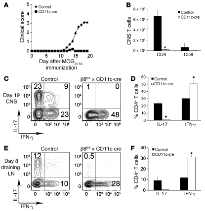
To determine the potential clinical relevance of impaired IL-17 production by T cells stimulated with αvβ8-deficient DCs, we employed the EAE disease model. The EAE model has been extensively studied and depends on the formation of pathogenic IL-17–producing T cells. We immunized control and β8fl/fl × CD11c-cre mice for EAE and scored them based on paralysis severity. β8fl/fl × CD11c-cre mice were almost completely protected from paralysis (3.1 ± 0.2 mean peak EAE clinical score in control mice vs. 0.2 ± 0.1 in β8fl/fl × CD11c-cre mice; Figure 2A). We then examined the CNS of mice at peak disease for the presence of Th17 cells. As expected, induction of EAE caused a marked accumulation of CD4+ and CD8+ T cells in the CNS of control mice, but the numbers of T cells were dramatically reduced in the CNS of β8fl/fl × CD11c-cre mice (Figure 2B). Of the CD4+ T cells that were present in the CNS of in β8fl/fl × CD11c-cre mice, 1.4% ± 0.4% expressed IL-17 (Figure 2, C and D). In contrast, 23% ± 2% of CD4+ T cells produced IL-17 in the CNS of control mice (Figure 2, C and D).
Figure 2. β8fl/fl × CD11c-cre mice are protected from EAE.
(A) Clinical scores of control and β8fl/fl × CD11c-cre mice after EAE immunization. (B) Total number of CD4 and CD8 T cells isolated from the CNS of control or β8fl/fl × CD11c-cre mice on day 19 after EAE immunization. (C) Flow cytometry plots of CD4+ T cells from the CNS of control or β8fl/fl × CD11c-cre mice on day 19 after EAE immunization. Cells were activated with PMA/ionomycin and then stained for IL-17 and IFN-γ. (D) Percentage of IL-17– and IFN-γ–producing CD4+ T cells from the CNS of control and β8fl/fl × CD11c-cre mice. (E) Flow cytometry plots of CD4+ T cells from the draining lymph nodes of control or β8fl/fl × CD11c-cre mice on day 8 after EAE immunization. Cells were activated with PMA/ionomycin and then stained for IL-17 and IFN-γ. (F) Percentage of IL-17– and IFN-γ–producing CD4+ T cells from the draining lymph nodes of control and β8fl/fl × CD11c-cre mice. Data represent the means ± SEM of 3 independent experiments and at least 10 mice in each group. *P < 0.01.
Since Th17 cells were nearly absent in the CNS of β8fl/fl × CD11c-cre mice and these mice also exhibited a marked reduction in the total number of CNS T lymphocytes, we examined the periphery of these mice for Th17 cells before onset of EAE. At day 8 after immunization for EAE, we observed 9.2% ± 3% of CD4+ T cells producing IL-17 in the draining lymph nodes of control mice, but only 0.6% ± 0.1% in the draining lymph nodes of β8fl/fl × CD11c-cre mice (Figure 2, E and F). To determine whether mice lacking integrin αvβ8 on DCs could develop EAE if the T cell priming phase was bypassed, we transferred activated 2D2 TCR transgenic T cells (transgenic for a T cell receptor to the MOG35–55 peptide derived from myelin oligodendrocyte protein) into control and β8fl/fl × CD11c-cre mice. Control and β8fl/fl × CD11c-cre mice developed similar EAE after passive immunization with activated 2D2 T cells (Supplemental Figure 1; supplemental material available online with this article; doi: 10.1172/JCI43786DS1). We also checked whether β8 expression on microglia, an important antigen-presenting cell in the CNS, was affected in β8fl/fl × CD11c-cre mice. We found no difference in microglia β8 mRNA expression from control and β8fl/fl × CD11c-cre mice (1.3 × 10⁻³ ± 4.2 × 10⁻⁴ and 1.1 × 10⁻³ ± 2.5 × 10⁻⁴; P = 0.35). These data support a critical role for induction of Th17 cells by αvβ8 on DCs in periphery during the development of EAE.
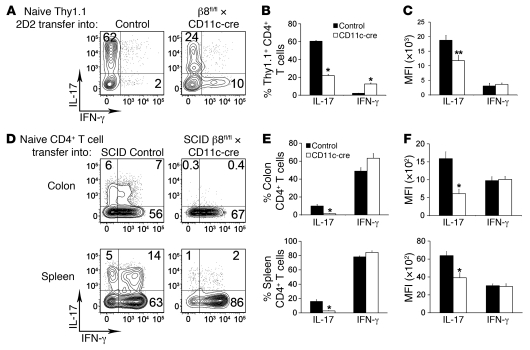
To assess whether integrin αvβ8 on DCs participates in the conversion of naive CD4+ T cells to Th17 cells, we transferred Thy1.1-labeled naive CD4+ 2D2 TCR transgenic T cells into control and β8fl/fl × CD11c-cre mice immunized with an emulsion of MOG35–55 and CFA. The Thy1.1 label allowed us to identify the transferred cells among endogenous CD4+ T cells upon re-isolation from the draining lymph nodes and spleen. When transferred Thy1.1+ 2D2 T cells were re-stimulated and analyzed 8 days after immunization, 60% ± 1.1% of the cells produced IL-17 (Figure 3, A and B). However, only 22% ± 1.4% Thy1.1+ 2D2 T cells produced IL-17 after transfer into β8fl/fl × CD11c-cre mice (Figure 3, A and B). Additionally, IL-17–expressing 2D2 T cells recovered from β8fl/fl × CD11c-cre mice produced significantly less IL-17 as measured by MFI of IL-17 staining (Figure 3C). We also determined the role of integrin αvβ8 on DCs in the conversion of naive CD4+ T cells to Th17 cells by transferring polyclonal naive CD4+ T cells into β8fl/fl × CD11c-cre mice crossed onto the SCID background. SCID mice lack functional T and B cells and therefore allowed us to study the role of integrin αvβ8 on DCs in Th17 differentiation in the absence of endogenous lymphocytes. When naive CD4+ T cells were transferred into SCID control mice and recovered from the colonic lamina propria and spleen 12 days later, 10% ± 1.2% and 16.1% ± 2.4%, respectively, produced IL-17 upon restimulation (Figure 3, D and E). However, only 1.2% ± 0.3% and 2.8% ± 0.6% of CD4+ T cells recovered from the colonic lamina propria and spleen, respectively, of SCID β8fl/fl × CD11c-cre mice produced IL-17. Furthermore, the few IL-17–expressing CD4+ T cells isolated from SCID β8fl/fl × CD11c-cre mice produced significantly less IL-17 as measured by MFI of IL-17 staining (Figure 3F). These data indicate that integrin αvβ8 on DCs is critical for the development of Th17 cells in response to antigen challenge in vivo.
Figure 3. Blunted Th17 conversion of naive CD4+ T cells adoptively transferred into β8fl/fl × CD11c-cre mice.
(A) Flow cytometry plots of Thy1.1 2D2 TCR transgenic T cells isolated from the spleen and draining lymph nodes 8 days after transfer into control or β8fl/fl × CD11c-cre mice immunized with MOG plus CFA. Isolated cells were activated with PMA/ionomycin and stained for IL-17 and IFN-γ. Numbers in quadrants indicate the percentage of positive cells. (B) Percentage of IL-17– and IFN-γ–producing Thy1.1+ CD4+ T cells isolated from control and β8fl/fl × CD11c-cre mice. (C) MFI of IL-17 and IFN-γ staining in Thy1.1+CD4+ T cells isolated from control and β8fl/fl × CD11c-cre mice. (D) Flow cytometry plots of polyclonal CD4+ T cells isolated from the colonic lamina propria (top) or spleen (bottom) 12 days after transfer into SCID control or SCID β8fl/fl × CD11c-cre mice. Isolated cells were activated with PMA/ionomycin and stained for IL-17 and IFN-γ. Numbers in quadrants indicate the percentage of positive cells. (E) Percentage of IL-17– and IFN-γ–producing CD4+ T cells isolated from control and β8fl/fl × CD11c-cre mice. (F) MFI of IL-17 and IFN-γ staining in CD4+ T cells isolated from control and β8fl/fl × CD11c-cre mice. Data represent the mean ± SEM of 2 independent experiments and 3 to 5 mice per group. *P < 0.005; **P < 0.01.
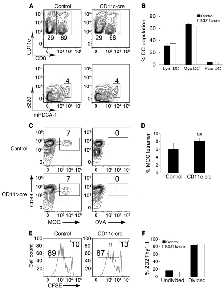
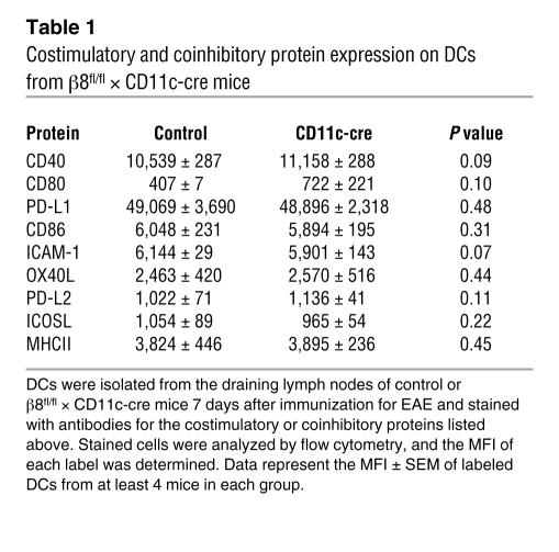
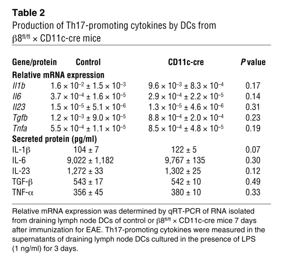
To examine the mechanism by which loss of αvβ8 on DCs protected mice from EAE and impaired Th17 differentiation, we characterized DCs from the draining lymph nodes of β8fl/fl × CD11c-cre mice. DCs have been categorized into the lymphoid, myeloid, and plasmacytoid groups based on their developmental origin and function. The presence of these DC populations in the draining lymph nodes of control or β8fl/fl × CD11c-cre mice was determined 7 days after immunization for EAE. We observed 31.4% ± 1.7% lymphoid, 66.8% ± 1.7% myeloid, and 3.9% ± 0.6% plasmacytoid DCs in control mice and 35.1% ± 3.4% lymphoid, 62.6% ± 3.4% myeloid, and 4.5% ± 0.6% plasmacytoid DCs in β8fl/fl × CD11c-cre mice (Figure 4, A and B). Next, we checked the ability of DCs from β8fl/fl × CD11c-cre mice to stimulate the proliferation of naive T cells after immunization for EAE. At day 7 after immunization, there were 6.0% ± 1.5% myelin oligodendrocyte glycoprotein–specific (MOG-specific) T cells after enrichment for tetramer-labeled cells in control mice and 8.1% ± 0.8% in β8fl/fl × CD11c-cre mice (Figure 4, C and D). Furthermore, we saw that MOG-specific TCR transgenic T cells (clone 2D2) adoptively transferred into control and β8fl/fl × CD11c-cre mice underwent a similar number of cell divisions following EAE immunization (Figure 4, E and F). We then analyzed the expression of costimulatory and coinhibitory molecules on DCs from β8fl/fl × CD11c-cre mice. As shown in Table 1, no significant differences were found in the expression of CD40, CD80, CD86, PD-L1, PD-L2, ICAM-1, OX40L, ICOSL, or MHCII. Finally, we examined the ability of DCs from β8fl/fl × CD11c-cre mice to produce cytokines related to the development of Th17 cells. As shown in Table 2, we found no significant differences in the levels of Il1b, Il6, Il23, Tgfb, or Tnfa mRNA or secreted protein. We also measured the effects of CFA immunization on DC β8 integrin expression in control mice and found that CFA slightly reduced β8 mRNA, from 5.0 × 10⁻⁵ ± 7.3 × 10⁻⁶ to 2.4 × 10⁻⁵ ± 3.2 × 10⁻⁶ (P = 0.037). Collectively, these data suggest that DCs from β8fl/fl × CD11c-cre mice are similar to control mice in their composition, ability to stimulate naive CD4 T cell proliferation, and capacity to make cytokines that promote Th17 differentiation.
Figure 4. Characterization of draining lymph node DCs in β8fl/fl × CD11c-cre mice.
(A) Flow cytometry plots of MHCII+CD11c+ DCs isolated from the draining lymph nodes of control or β8fl/fl × CD11c-cre mice 7 days after in vivo stimulation with CFA. DCs within plots are gated on CD8+ (lymphoid) and CD8– (myeloid) DCs (top) or B220+mPDCA-1+ (plasmacytoid; bottom). Numbers next to the gates indicate the percentage of CD8+ or CD8– MHCII+CD11c+ cells (top) or B220+mPDCA-1+MHCII+CD11c+ cells (bottom). (B) Percentage of lymphoid (Lym), myeloid (Mye), and plasmacytoid (Plas) DCs isolated from control β8fl/fl × CD11c-cre mice. (C) Flow cytometry plots of CD4+ T cells isolated from control or β8fl/fl × CD11c-cre mice 7 days after immunization with MOG plus CFA and stained with MHCII tetramers loaded with MOG or OVA peptide. Numbers above the gates indicate the percentage of cells labeled with tetramer after enrichment for tetramer-positive cells. (D) Percentage of CD4+ T cells labeled with MOG tetramer after enrichment for tetramer-positive cells isolated from control or β8fl/fl × CD11c-cre mice. (E) Flow cytometry histograms of CFSE-stained Thy1.1+2D2+ TCR transgenic T cells isolated from control or β8fl/fl × CD11c-cre mice 7 days after immunization with MOG plus CFA. Numbers above gates indicate the percentage of divided (left gate) and undivided (right gate) cells. (F) Percentage of undivided and divided 2D2+ Thy1.1+ cells isolated from control or β8fl/fl × CD11c-cre mice.
Table 1 .
Costimulatory and coinhibitory protein expression on DCs from β8fl/fl × CD11c-cre mice
Table 2 .
Production of Th17-promoting cytokines by DCs from β8fl/fl × CD11c-cre mice
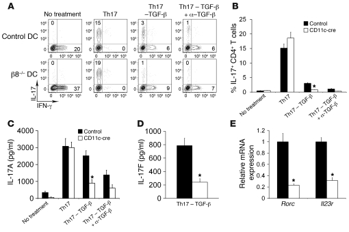
Integrin αvβ8 controls the activation of TGF-β1 and -β3 through interaction with an Arg-Gly-Asp sequence on the LAP of the latent TGF-β complex (11). To determine whether integrin αvβ8 on DCs regulates Th17 development through activation of latent TGF-β, we used an in vitro assay to monitor Th17 development in the presence of control or αvβ8-deficient DCs. As shown in Figure 5, αvβ8-deficient DCs cultured with naive CD4+ 2D2 T cells under conditions that drive Th17 development (i.e., IL-1β and IL-6), but in the absence of exogenous TGF-β, showed a reduced ability to promote IL-17 production by T cells. However, IL-17 production after culture with αvβ8-deficient DCs was restored by the addition of exogenous TGF-β (Figure 5, A–C). Furthermore, the addition of a TGF-β–neutralizing antibody significantly reduced IL-17 production by T cells cultured with control DCs but had a minimal effect on αvβ8-deficient DCs (Figure 5, A–C). To confirm that IL-17 produced by T cells in this assay was from bona fide Th17 cells, we measured IL-17F, another canonical Th17 cytokine. Indeed, IL-17F was detected in the supernatant of cultures grown without the addition of exogenous TGF-β and was significantly reduced in cultures with αvβ8-deficient DCs (Figure 5D). Furthermore, we measured mRNA expression of 2 genes that have been shown previously to be regulated by TGF-β — Rorc, which encodes the Th17-specific transcription factor RORγt, and Il23r, which encodes the IL-23 receptor, a receptor required for clonal expansion of Th17 cells. We detected transcripts of these genes in T cells from cultures with control DCs but found a dramatic reduction in expression of these genes in T cells from cultures with αvβ8-deficient DCs (Figure 5E).
Figure 5. DCs from β8fl/fl × CD11c-cre mice fail to drive Th17 development through a defect in TGF-β activation.
(A) Flow cytometry plots of naive CD4+ 2D2 TCR transgenic T cells after culture with DCs from control or β8fl/fl × CD11c-cre mice for 5 days. Cells were activated with PMA/ionomycin for 4 hours and then stained for IL-17 and IFN-γ. Cultures were grown under the following conditions: no treatment, Th17 (see Methods), Th17 without TGF-β, and Th17 with TGF-β–neutralizing antibody. (B) Percentage of IL-17–producing CD4+ 2D2 T cells after cultured as described in A. (C) Measurement of IL-17A in the supernatants of the cultures described in A using ELISA. (D) Measurement of IL-17F in the supernatant of the Th17 without the TGF-β condition described in A. (E) qRT-PCR analysis of the Rorc and Il23r genes from T cells after culture under the Th17 without the TGF-β condition described in A. Data represent the means ± SEM of at least 3 independent experiments with 8 mice in each group. *P < 0.01.
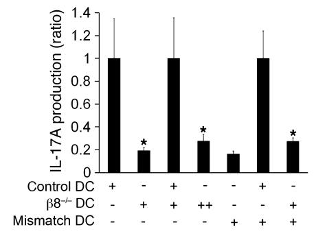
The mechanism by which integrin αvβ8 on DCs presents active TGF-β to induce IL-17 expression in T cells is unknown. One possibility is that latent TGF-β is constitutively activated by αvβ8 on DCs to create a surrounding milieu of active TGF-β (i.e., a field effect). Alternatively, αvβ8 might only induce IL-17 expression in T cells if it activates TGF-β in the context of antigen presentation. This would be a teleologically attractive mechanism to tightly regulate, in space and time, this important immunologic decision. To distinguish between these mechanisms, we cultured naive CD4+ 2D2 T cells under Th17-polarizing conditions (in the absence of exogenous TGF-β) with mixtures of αvβ8-deficient DCs and αvβ8-containing DCs that express either matched or mismatched MHCII. Thus, MHCII-mismatched DCs would be unable to present MOG peptides to 2D2 T cells. We first established that a 1:1 mixture of control and αvβ8-deficient DCs was able to completely rescue the defect in IL-17 induction seen with αvβ8-deficient DCs alone (Figure 6). We then cultured αvβ8-expressing, MHCII-mismatched (H2q MHC haplotype from FVB mice) DCs at a 4:1 mixture with αvβ8-deficient DCs (H2b MHC haplotype from C57BL/6 mice). MHCII-mismatched DCs were unable to rescue the impaired IL-17 production seen with αvβ8-deficient DCs (Figure 6). These data suggest that Th17 cell induction requires simultaneous presentation of antigen (by MHCII) and activation of TGF-β by integrin αvβ8 on the same DCs.
Figure 6. Th17 development through integrin αvβ8 on DCs is dependent on MHCII-TCR engagement.
DCs were isolated from control, β8fl/fl × CD11c-cre, and MHCII-mismatched (H2q MHC haplotype from FVB strain) mice and cultured in different combinations with naive CD4+ 2D2 TCR transgenic T cells under Th17-driving conditions without exogenous TGF-β. Supernatants were collected and analyzed by ELISA for the presence of IL-17. Data are means ± SEM of the ratio of IL-17 produced between control and β8fl/fl × CD11c-cre wells under each condition. Twice as many β8fl/fl × CD11c-cre DCs were present in 1 condition (indicated by ++) to account for a doubling in the number of DCs in the control plus β8–/– group. MHCII-mismatched DCs were present in cultures at a 4:1 ratio, compared with control or β8–/– DCs. *P < 0.05.
Discussion
How TGF-β is regulated in the extracellular space and presented to naive T cells during Th17 development has been unknown. Our results strongly suggest that the integrin αvβ8 on DCs is required for maximal Th17 development. This effect appears to be due to TGF-β activation by this integrin, since the defect in Th17 induction by DCs lacking αvβ8 can be completely rescued by the addition of exogenous TGF-β. Furthermore, these data suggest that activation of TGF-β by integrin αvβ8 on DCs may be a novel target in the development of therapeutics for autoimmune diseases.
We tested the in vivo relevance of TGF-β activation by integrin αvβ8 on DCs in the EAE disease model. The EAE model is widely used to study the formation of pathogenic T cells to self-molecules and has been utilized as a model of multiple sclerosis. Our result that mice lacking αvβ8 on DCs are almost completely protected from EAE suggests that TGF-β activation by αvβ8 on DCs is a required event in the formation of pathogenic self-reactive T cells. The nearly complete absence of CD4+ T cells in the CNS of EAE-immunized mice lacking αvβ8 on DCs suggests that these mice may be protected from EAE in part due to impaired formation of MOG-reactive Th17 cells. This concept is also supported by our observation that at day 8 following EAE immunization, a time point before the onset of symptomatic EAE, Th17 cells failed to develop in the draining lymph nodes of mice lacking in αvβ8 on DCs. These data agree with a previous report that mice engineered to express a dominant-negative TGF-β receptor-II in T cells are protected from EAE through a defect in Th17 cell induction (15). Interestingly, transgenic mice that overexpress TGF-β within T cells develop more severe EAE than wild-type mice and also exhibit greater numbers of Th17 cells in the CNS (2). While Th17 cells are regarded as essential for EAE, several recent studies have called into question which type or types of CD4+ T cells are encephalitogenic in this model (16, 17). In the present study, the scarcity of Th17 cells found in the CNS and periphery of EAE-primed mice lacking αvβ8 on DCs, and their dramatic protection from EAE, indicates that TGF-β activated via αvβ8 on DCs is an important step in the generation of encephalitogenic CD4+ T cells.
An absolute requirement for TGF-β in Th17 development has been controversial. A combination of TGF-β and IL-6 was first reported to be sufficient to induce Th17 differentiation in mice (2–4). Subsequently, 2 groups reported that TGF-β was not required for the conversion of naive T cells to Th17 cells in humans (18, 19). However, the human naive T cells used in those studies were isolated using protocols that may have introduced TGF-β contamination and were cultured with serum containing TGF-β. Subsequently, 3 groups used rigorous methods to exclude TGF-β contamination and showed that TGF-β is required for Th17 differentiation by human T cells (20–22). However, a recent study that used mice lacking the transcription factors required to drive Th1 and Th2 differentiation suggested that TGF-β may function indirectly in Th17 conversion by suppressing Th1 and Th2 factors, which in turn suppress RORγt (23). Nonetheless, TGF-β appears to be crucial for Th17 induction in vivo and at the site of T cell priming, as administration of a TGF-β–blocking antibody delivered with antigen and CFA abrogates Th17 induction and EAE (15). The data presented in this study are also in line with previous findings on a requirement for TGF-β in the generation of intestinal lamina propria Th17 cells. Lamina propria Th17 cells are absent in Tgfb1C33S/C33S knockin mice, which have impaired TGF-β activation through inhibition of covalent TGF-β attachment to latent TGF-β binding protein 1 (24). Furthermore, Atarashi et al. demonstrated that an αvβ8-expressing CD11c+ DC lamina propria subset preferentially induces Th17 generation from naive T cells in vitro (25).
We were concerned that the increased production of IFN-γ by T cells from mice lacking αvβ8 on DCs might contribute to the markedly impaired Th17 cell induction in these animals. Our finding that mice deficient in both IFN-γ and αvβ8 on DCs still have a dramatic defect in Th17 cell induction excludes that explanation. We also saw that some MOG-specific TCR transgenic T cells adoptively transferred into mice lacking αvβ8 on DCs were able to express IL-17, albeit at lower levels than those transferred into control mice. This finding suggests that the presence αvβ8 on DCs may not be an absolute requirement for the generation of Th17 cells, or alternatively, that a small number of memory cells were present in the naive transferred cells.
The present study did not examine which cell type or types are responsible for producing the latent TGF-β required for Th17 development. A previous report from Li et al. showed that deletion of the Tgfb gene in T cells protects mice from EAE and Th17 development (26). In context with the findings presented here, these data suggest a model whereby T cells provide their own latent TGF-β, which then gets activated by αvβ8 on DCs. However, DCs also produce significant amounts of TGF-β, and it has been reported that a subset of DCs in the CNS that preferentially induce Th17 cells also produce elevated levels of TGF-β (27). These data suggest that TGF-β signaling within the immune system may be regulated by both expression and activation of the latent form.
The spatial and temporal regulation of TGF-β in the extracellular space during Th17 development has been unknown. In the current study, we provide a partial answer to this question by showing that αvβ8 integrin-mediated TGF-β activation by DCs can only induce antigen-specific T cells to become Th17 cells if the same cell that is expressing the integrin is also actively presenting antigen in the context of MHCII. Since TGF-β signaling in T cells has the potential to direct the formation of a pro-inflammatory (i.e., Th17) or immunosuppressive (i.e., Treg) response to antigen, it makes teleologic sense for this process to be tightly regulated in space and time. Our results suggest that one important factor is precise spatial regulation of delivery of the active TGF-β, potentially allowing the same DCs to provide active TGF-β and other polarizing cytokines (for example, IL-6 in the context of Th17 cell induction). Presentation of active TGF-β when T cells are in close proximity to DCs would allow for precise control of T cell fate at a time when T cells are receiving signals from costimulatory and coinhibitory molecules as well as secreted cytokines.
Collectively, the data presented here indicate that integrin αvβ8 on DCs facilitates the development of Th17 cells, and consequently contributes to IL-17–mediated CNS inflammation, through activation of TGF-β. This finding suggests that the αvβ8 integrin, or the steps upstream and downstream of αvβ8-mediated TGF-β activation, could be novel targets for treatment of multiple sclerosis and other autoimmune diseases.
Methods
Mice.
C57BL/6 β8fl/fl × CD11c-cre (14) and C57BL/6 2D2 TCR transgenic (28) mice have been previously described. C57BL/6 SCID mice (strain B6.CB17-Prkdcscid/SzJ) were purchased from The Jackson Laboratory and crossed to C57BL/6 β8fl/fl × CD11c-cre. FVB/NJ mice were purchased from The Jackson Laboratory. Mice used for all experiments were 8–12 weeks old and were housed under specific pathogen–free conditions in the Animal Barrier Facility of the University of California, San Francisco. All experiments were approved by the Institutional Animal Care and Use Committee of the University of California, San Francisco.
Flow cytometry.
For intracellular cytokine analysis, cells were incubated for 4 hours at 37°C in complete RPMI containing 50 ng/ml PMA (Sigma-Aldrich), 1 μM ionomycin (Sigma-Aldrich), and 2 μM monensin (eBioscience). Cells were then washed with ice-cold PBS and blocked for 20 minutes at 4°C with PBS containing 2 μg/ml α-CD16/32 (UCSF hybridoma core) and 10 % heat-inactivated normal rat serum (Sigma-Aldrich). After washing with PBS, cells were incubated for 30 minutes in PBS with cell-surface antibodies and Aqua Live/Dead Fixable Dead cell stain (Invitrogen). Cells were then fixed and permeabilized with eBioscience Fix/Perm solution according to the manufacturer’s instructions. Intracellular cytokines were labeled with antibodies for 45 minutes at 4°C in permeabilization buffer (eBioscience). Cells were analyzed on an LSRII (BD Biosciences), and post-analysis of flow cytometry data was performed with FlowJo software (Tree Star Inc.). The following antibodies were used in experiments and were purchased from BD Biosciences — Pharmingen or eBioscience: anti-CD4 (clone RM4-5), anti-CD8 (clone 53-6.7), anti-CD11c (clone N418), anti-MHCII (clone M5/114.15.2), anti-B220 (clone RA3-6B2), anti-mPDCA1 (clone eBio927), anti-CD44 (clone IM7), anti-Thy1.1 (clone HIS51), anti–IL-17 (clone eBio17B7), anti–IFN-γ (clone XMG1.2), anti–PD-L1 (clone MIH5), anti–PD-L2 (clone 122), anti-ICOSL (clone HK5.3), anti-CD40 (clone 1C10), anti-CD80 (clone 16-10A1), anti-CD86 (clone GL1), anti–ICAM-1 (clone YN1/1.7.4), and anti-OX40L (clone RM134L).
MOG tetramer staining.
CD4 T cells expressing a MOG-specific TCR were stained with PE-labeled I-A(b) GWYRSPFSRVVH tetramer (NIH Tetramer Core Facility at Emory University) as previously described with modifications (29). Briefly, cells (1 × 10⁸/ml) from the draining lymph nodes and spleen of mice at day 7 after EAE immunization were incubated with a 1:100 dilution of tetramer in HEPES-buffered DMEM plus 2% FBS for 1 hour at room temperature. Cells were then washed and incubated with anti-PE microbeads (Miltenyi Biotec) in HEPES-buffered DMEM plus 2% FBS for 30 minutes at 4°C. After washing, tetramer-labeled cells were enriched by passing over a magnetic column (LS column; Miltenyi Biotec) according to the manufacturer’s instructions. Eluted cells were stained for cell surface markers and analyzed on an LSRII as described above.
Lymphocyte isolation from colonic lamina propria.
Lymphocytes were isolated from the colonic lamina propria as previously described with slight modification (30). Briefly, colons were removed, washed with ice-cold PBS, and cut into 2- to 3-cm-long pieces. Pieces were predigested in 5 ml of HBSS containing 5 mM EDTA and 1 mM DTT and rotated (180 rotations/min) for 20 minutes at 37°C to remove debris and intraepithelial lymphocytes. Pieces were then washed with PBS and predigested a second time. Pieces were next cut to 0.5 cm and digested in serum-free RPMI containing 0.1 U/ml liberase blendzyme 3 (Roche) and 10 U/ml DNase I (Roche) at 37°C for 20 minutes with rotation. After vortexing, the supernatants were collected and placed on ice while the pieces were digested 2 more times. Supernatants from all 3 digestions were combined and washed with complete RPMI. Cells were then separated on a 40%/80% Percoll (Sigma-Aldrich) gradient and analyzed by flow cytometry.
EAE induction.
Mice were given s.c. injections into the flanks and nape with 100 μl of an emulsion containing 200 μg MOG35–55 peptide (MEVGWYRSPFSRVVHLYRNGK; Genemed Synthesis Inc.) and 200 μg Mycobacterium tuberculosis H37 RA (Difco) in CFA. Mice also received 200 ng pertussis toxin (List Biologicals) i.p. on the day of immunization and 2 days later. Mice were scored for clinical disease in a blinded fashion as follows: 1, tail paralysis; 2, hindlimb weakness; 3, partial hindlimb paralysis; 4, complete hindlimb paralysis; 5, moribund.
Lymphocyte isolation from the CNS.
Lymphocytes were isolated from the CNS of mice as previously described (27). Briefly, mice were perfused with PBS and spinal cords removed. Spinal cords were cut into 2- to 3-cm-long pieces and digested in 0.2 U/ml liberase blendzyme 3 (Roche) and 50 U/ml DNase I (Roche) at 37°C for 30 minutes. Lymphocytes were then enriched by separation on a 30%/70% percoll gradient and analyzed by flow cytometry.
Adoptive transfer of naive CD4 T cells.
Naive Thy1.1+ 2D2 T cells were isolated as described below and 7.5 × 10⁴ were transferred i.v. into control or β8fl/fl × CD11c-cre mice. In some cases cells were labeled with CFSE (Invitrogen) before transfer by incubation with 5 μM CFSE for 5 minutes at room temperature. Mice were then immunized for EAE as described above. Lymph nodes and spleens were removed 8 days after transfer, and Thy1.1+ 2D2 cells were analyzed by intracellular flow cytometry. Polyclonal naive CD4 T cells were isolated from the spleen and lymph nodes of a wild-type sex-matched C57BL/6 mouse using the CD4+CD62L+ T cell isolation kit (Miltenyi Biotec), and 2.0 × 10⁶ cells were transferred i.v. into SCID control or SCID β8fl/fl × CD11c-cre mice.
Activation of 2D2 T cells for EAE passive immunization.
CD4 T cells from C57BL/6 2D2 TCR transgenic mice were activated for EAE passive immunization as previously described (31). Briefly, 3 × 10⁷ naive 2D2 T cells and 3 × 10⁶ DCs were cultured on 10-cm dishes in the presence of MOG (10 μg/ml), IL-12 (20 ng/ml), IL-18 (25 ng/ml), and IL-2 (100 U/ml) for 4 days. Cells were then activated on plates coated with anti-CD3 (1 μg/ml)/anti-CD28 (1 μg/ml) in the presence of IL-12 (2 ng/ml), IL-18 (2.5 ng/ml), and IL-2 (10 U/ml) for 24 hours. Cells were then washed 3 times with PBS and transferred i.v. into mice.
DC isolation.
DCs were isolated from the draining lymph nodes of β8fl/fl × CD11c-cre or sex-matched littermate control mice 7–8 days after receiving s.c. injections into the flanks and nape with 100 μl of an emulsion containing equal parts PBS and 200 μg M. tuberculosis H37 RA (Difco) in CFA. Lymph nodes were digested at 37°C for 30 minutes in serum-free RPMI containing 0.14 U/ml liberase (Roche) and 50 U/ml DNase I (Roche). CD11c+ cells were selected with anti-CD11c magnetic beads (Miltenyi Biotec) according to the manufacturer’s instructions. CD11c+ DCs used were typically 90%–95% pure.
In vitro Th17 differentiation assay and cytokine measurement.
DCs were isolated as described above and plated at 20,000 cells/well in complete Iscove modified Dulbecco medium (IMDM; Sigma-Aldrich). Naive 2D2 T cells were isolated from the lymph nodes and spleen of sex-matched 2D2 TCR transgenic mice using the CD4+ CD62L+ T cell isolation kit (Miltenyi Biotec), and 50,000 cells were added to each well along with MOG35–55 (10 μg/ml), IL-6 (50 ng/ml; Peprotech), IL-1β (10 ng/ml; Peprotech), FICZ (6-formylindolo [3,2-b] carbazole; 300 nM; Biomol), and TGF-β (1 ng/ml; R&D Systems) or TGF-β–neutralizing antibody (clone 1d11; 50 μg/ml). 2D2 T cells were harvested 5 days later and analyzed by flow cytometry or quantitative RT-PCR (qRT-PCR). Cytokines in the culture supernatants were measured with IL-17A (eBioscience) and IL-17F (eBioscience) ELISA reagent sets according to the manufacturer’s instructions. Measurement of TGF-β in colon tissue and IL-1β, IL-6, IL-23, TGF-β, and TNF-α production by DCs was performed using the Mouse FlowCytomix Kit (eBioscience) according to the manufacturer’s instructions.
qRT-PCR.
RNA was isolated from T cells and DC using an RNeasy Mini Kit (Qiagen). cDNA transcription and qPCR were performed using a SYBR-Greener Two-Step qRT-PCR kit (Invitrogen). Samples were amplified on an ABI 7900HT thermocycler and normalized to β-actin expression. Primers used were as follows: B-actin forward: TGTTACCAACTGGGACGACA, reverse: GGGGTGTTGAAGGTCTCAAA; Rorc forward: GACAGGGAGCCAAGTTCTCA, reverse: GGTGATAACCCCGTAGTGGA; Il23r forward: TTCAGATGGGCATGAATGTTTCT, reverse: CCAAATCCGAGCTGTTGTTCTAT; Il1b forward: GCAACTGTTCCTGAACTCAACT, reverse: ATCTTTTGGGGTCCGTCAACT; Il6 forward: TAGTCCTTCCTACCCCAATTTCC, reverse: TTGGTCCTTAGCCACTCCTTC; Il23 forward: ATGCTGGATTGCAGAGCAGTA, reverse: ACGGGGCACATTATTTTTAGTCT; Tgfb forward: CTCCCGTGGCTTCTAGTGC, reverse: GCCTTAGTTTGGACAGGATCTG; Tnf forward: CCCTCACACTCAGATCATCTTCT, reverse: GCTACGACGTGGGCTACAG; Itgb8 forward: CTGAAGAAATACCCCGTGGA, reverse: ATGGGGAGGCATACAGTCT.
Statistics.
The statistical significance of differences between groups was calculated with a 2-tailed Student’s t test. Differences with a P value of less than 0.05 were considered statistically significant.
Supplementary Material
Acknowledgments
This work was supported by NIH grants HL64353, HL53949, HL083950, AI024674, and U19 AI077439 (to D. Sheppard); NIH grant U19 AI056388 (to J. Bluestone); ADA Mentor-Based Award (to S. Bailey-Bucktrout); NIH Ruth L. Kirschstein National Research Service Award HL095314 (to A. Melton); and American Lung Association of California Research Training Fellowship Award (to A. Melton).
Footnotes
Conflict of interest: The authors have declared that no conflict of interest exists.
Citation for this article: J Clin Invest. 2010;120(12):4436–4444. doi:10.1172/JCI43786.
See the related Commentary beginning on page 4185.
References
- 1.Korn T, Bettelli E, Oukka M, Kuchroo VK. IL-17 and Th17 cells. Annu Rev Immunol. 2009;27:485–517. doi: 10.1146/annurev.immunol.021908.132710. [DOI] [PubMed] [Google Scholar]
- 2.Bettelli E, et al. Reciprocal developmental pathways for the generation of pathogenic effector TH17 and regulatory T cells. Nature. 2006;441(7090):235–238. doi: 10.1038/nature04753. [DOI] [PubMed] [Google Scholar]
- 3.Veldhoen M, Hocking RJ, Atkins CJ, Locksley RM, Stockinger B. TGFbeta in the context of an inflammatory cytokine milieu supports de novo differentiation of IL-17-producing T cells. Immunity. 2006;24(2):179–189. doi: 10.1016/j.immuni.2006.01.001. [DOI] [PubMed] [Google Scholar]
- 4.Mangan PR, et al. Transforming growth factor-beta induces development of the T(H)17 lineage. Nature. 2006;441(7090):231–234. doi: 10.1038/nature04754. [DOI] [PubMed] [Google Scholar]
- 5.Korn T, et al. IL-21 initiates an alternative pathway to induce proinflammatory T(H)17 cells. Nature. 2007;448(7152):484–487. doi: 10.1038/nature05970. [DOI] [PMC free article] [PubMed] [Google Scholar]
- 6.Zhou L, et al. IL-6 programs T(H)-17 cell differentiation by promoting sequential engagement of the IL-21 and IL-23 pathways. Nat Immunol. 2007;8(9):967–974. doi: 10.1038/ni1488. [DOI] [PubMed] [Google Scholar]
- 7.Ivanov II, et al. The orphan nuclear receptor RORgammat directs the differentiation program of proinflammatory IL-17+ T helper cells. Cell. 2006;126(6):1121–1133. doi: 10.1016/j.cell.2006.07.035. [DOI] [PubMed] [Google Scholar]
- 8.Zhou L, et al. TGF-beta-induced Foxp3 inhibits T(H)17 cell differentiation by antagonizing RORgammat function. Nature. 2008;453(7192):236–240. doi: 10.1038/nature06878. [DOI] [PMC free article] [PubMed] [Google Scholar]
- 9.Rifkin DB. Latent transforming growth factor-beta (TGF-beta) binding proteins: orchestrators of TGF-beta availability. J Biol Chem. 2005;280(9):7409–7412. doi: 10.1074/jbc.R400029200. [DOI] [PubMed] [Google Scholar]
- 10.Annes JP, Munger JS, Rifkin DB. Making sense of latent TGFbeta activation. J Cell Sci. 2003;116(pt 2):217–224. doi: 10.1242/jcs.00229. [DOI] [PubMed] [Google Scholar]
- 11.Mu D, et al. The integrin alpha(v)beta8 mediates epithelial homeostasis through MT1-MMP-dependent activation of TGF-beta1. J Cell Biol. 2002;157(3):493–507. doi: 10.1083/jcb.200109100. [DOI] [PMC free article] [PubMed] [Google Scholar]
- 12.Yang Z, et al. Absence of integrin-mediated TGFbeta1 activation in vivo recapitulates the phenotype of TGFbeta1-null mice. J Cell Biol. 2007;176(6):787–793. doi: 10.1083/jcb.200611044. [DOI] [PMC free article] [PubMed] [Google Scholar]
- 13.Aluwihare P, et al. Mice that lack activity of alphavbeta6- and alphavbeta8-integrins reproduce the abnormalities of Tgfb1- and Tgfb3-null mice. J Cell Sci. 2009;122(pt 2):227–232. doi: 10.1242/jcs.035246. [DOI] [PMC free article] [PubMed] [Google Scholar]
- 14.Travis MA, et al. Loss of integrin alpha(v)beta8 on dendritic cells causes autoimmunity and colitis in mice. Nature. 2007;449(7160):361–365. doi: 10.1038/nature06110. [DOI] [PMC free article] [PubMed] [Google Scholar]
- 15.Veldhoen M, Hocking RJ, Flavell RA, Stockinger B. Signals mediated by transforming growth factor-beta initiate autoimmune encephalomyelitis, but chronic inflammation is needed to sustain disease. Nat Immunol. 2006;7(11):1151–1156. doi: 10.1038/ni1391. [DOI] [PubMed] [Google Scholar]
- 16.Yang Y, et al. T-bet is essential for encephalitogenicity of both Th1 and Th17 cells. J Exp Med. 2009;206(7):1549–1564. doi: 10.1084/jem.20082584. [DOI] [PMC free article] [PubMed] [Google Scholar]
- 17.McGeachy MJ, et al. TGF-beta and IL-6 drive the production of IL-17 and IL-10 by T cells and restrain T(H)-17 cell-mediated pathology. Nat Immunol. 2007;8(12):1390–1397. doi: 10.1038/ni1539. [DOI] [PubMed] [Google Scholar]
- 18.Acosta-Rodriguez EV, Napolitani G, Lanzavecchia A, Sallusto F. Interleukins 1beta and 6 but not transforming growth factor-beta are essential for the differentiation of interleukin 17-producing human T helper cells. Nat Immunol. 2007;8(9):942–949. doi: 10.1038/ni1496. [DOI] [PubMed] [Google Scholar]
- 19.Wilson NJ, et al. Development, cytokine profile and function of human interleukin 17-producing helper T cells. Nat Immunol. 2007;8(9):950–957. doi: 10.1038/ni1497. [DOI] [PubMed] [Google Scholar]
- 20.Yang L, et al. IL-21 and TGF-beta are required for differentiation of human T(H)17 cells. Nature. 2008;454(7202):350–352. doi: 10.1038/nature07021. [DOI] [PMC free article] [PubMed] [Google Scholar]
- 21.Manel N, Unutmaz D, Littman DR. The differentiation of human T(H)-17 cells requires transforming growth factor-beta and induction of the nuclear receptor RORgammat. Nat Immunol. 2008;9(6):641–649. doi: 10.1038/ni.1610. [DOI] [PMC free article] [PubMed] [Google Scholar]
- 22.Volpe E, et al. A critical function for transforming growth factor-beta, interleukin 23 and proinflammatory cytokines in driving and modulating human T(H)-17 responses. Nat Immunol. 2008;9(6):650–657. doi: 10.1038/ni.1613. [DOI] [PubMed] [Google Scholar]
- 23.Das J, et al. Transforming growth factor beta is dispensable for the molecular orchestration of Th17 cell differentiation. J Exp Med. 2009;206(11):2407–2416. doi: 10.1084/jem.20082286. [DOI] [PMC free article] [PubMed] [Google Scholar]
- 24.Ivanov II, et al. Specific microbiota direct the differentiation of IL-17-producing T-helper cells in the mucosa of the small intestine. Cell Host Microbe. 2008;4(4):337–349. doi: 10.1016/j.chom.2008.09.009. [DOI] [PMC free article] [PubMed] [Google Scholar]
- 25.Atarashi K, et al. ATP drives lamina propria T(H)17 cell differentiation. Nature. 2008;455(7214):808–812. doi: 10.1038/nature07240. [DOI] [PubMed] [Google Scholar]
- 26.Li MO, Wan YY, Flavell RA. T cell-produced transforming growth factor-beta1 controls T cell tolerance and regulates Th1- and Th17-cell differentiation. Immunity. 2007;26(5):579–591. doi: 10.1016/j.immuni.2007.03.014. [DOI] [PubMed] [Google Scholar]
- 27.Bailey SL, Schreiner B, McMahon EJ, Miller SD. CNS myeloid DCs presenting endogenous myelin peptides ‘preferentially’ polarize CD4+ T(H)-17 cells in relapsing EAE. Nat Immunol. 2007;8(2):172–180. doi: 10.1038/ni1430. [DOI] [PubMed] [Google Scholar]
- 28.Bettelli E, Pagany M, Weiner HL, Linington C, Sobel RA, Kuchroo VK. Myelin oligodendrocyte glycoprotein-specific T cell receptor transgenic mice develop spontaneous autoimmune optic neuritis. J Exp Med. 2003;197(9):1073–1081. doi: 10.1084/jem.20021603. [DOI] [PMC free article] [PubMed] [Google Scholar]
- 29.Moon JJ, et al. Naive CD4(+) T cell frequency varies for different epitopes and predicts repertoire diversity and response magnitude. Immunity. 2007;27(2):203–213. doi: 10.1016/j.immuni.2007.07.007. [DOI] [PMC free article] [PubMed] [Google Scholar]
- 30.Weigmann B, Tubbe I, Seidel D, Nicolaev A, Becker C, Neurath MF. Isolation and subsequent analysis of murine lamina propria mononuclear cells from colonic tissue. Nat Protoc. 2007;2(10):2307–2311. doi: 10.1038/nprot.2007.315. [DOI] [PubMed] [Google Scholar]
- 31.Ito A, et al. Transfer of severe experimental autoimmune encephalomyelitis by IL-12- and IL-18-potentiated T cells is estrogen sensitive. J Immunol. 2003;170(9):4802–4809. doi: 10.4049/jimmunol.170.9.4802. [DOI] [PubMed] [Google Scholar]
Associated Data
This section collects any data citations, data availability statements, or supplementary materials included in this article.


