Abstract
Background
Pancreatic β-cells release insulin via an electrogenic response triggered by an increase in plasma glucose concentrations. The critical plasma glucose concentration has been determined to be ~3 mM, at which time both insulin and GABA are released from pancreatic β-cells. Taurine, a β-sulfonic acid, may be transported into cells to balance osmotic pressure. The taurine transporter (TauT) has been described in pancreatic tissue, but the function of taurine in insulin release has not been established. Uptake of taurine by pancreatic β-cells may alter membrane potential and have an effect on ion currents. If taurine uptake does alter β-cell current, it might have an effect on exocytosis of cytoplasmic vesicle. We wished to test the effect of taurine on regulating release of insulin from the pancreatic β-cell.
Methods
Pancreatic β-cell lines Hit-TI5 (Syrian hamster) and Rin-m (rat insulinoma) were used in these studies. Cells were grown to an 80% confluence on uncoated cover glass in RPMI media containing 10% fetal horse serum. The cells were then adapted to a serum-free, glucose free environment for 24 hours. At that time, the cells were treated with either 1 mM glucose, 1 mM taurine, 1 mM glucose + 1 mM taurine, 3 mM glucose, or 3 mM glucose + 1 mM taurine. The cells were examined by confocal microscopy for cytoplasmic levels of insulin.
Results
In both cell lines, 1 mM glucose had no effect on insulin levels and served as a control. Cells starved of glucose had a significant reduction (p<0.001) in the level of insulin, but this level was significantly higher than all other treatments. As expected, the 3 mM glucose treatment resulted in a statistically lower (p<0.001) insulin level than control cells. Interestingly, 1 mM taurine also resulted in a statistically lower level of insulin (p<0.001) compared to controls when either no glucose or 1 mM glucose was present. Cells treated with 1 mM taurine plus 3 mM glucose showed a level of insulin similar to that of 3 mM glucose alone.
Conclusions
Taurine administration can alter the electrogenic response in β-cell lines, leading to a change in calcium homeostasis and a subsequent decrease in intracellular insulin levels. The consequence of these actions could represent a method of increasing plasma insulin levels leading to a decrease in plasma glucose levels.
Background
The endocrine pancreas is regulated by neurotransmitters, including the use of glutamate and GABA to regulate insulin and glucagon release as well as somatostatin to regulate both α- and β-cell activities. For insulin release, a rise in extracellular glucose to ~3 mM initiates co-release of insulin and GABA [1]. The elevated glucose likely stimulates the release of insulin and GABA from the synapse-like microvesicles (SLMV) through changes in ion currents in β-cells [2,3]. While the insulin is free to move to the circulatory system through the interstitial fluid, GABA binds to GABAA receptors on αcells, causing the hyperpolarization of the α-cells, and inhibiting release of glucagon. Glutamate released from α-cells binds to GluR4 receptors on δ-cells, increasing the release of SST [4]. SST binds either to the SSTR2 receptor on the α-cell or the SSTR1 and/or SSTR5 receptor on the β-cell. SSTR2 activation maintains the GABA-initiated inhibition of glucagon from α-cells, while the SSTR1/5 receptors are responsible for inhibiting insulin release [5].
Taurine (2-aminoethanesulfonic acid) is a sulfur-containing amino acid and is developmentally high in neonates, especially in the brain, and the levels decline to reach stable adult concentrations that are second to those of glutamate in the brain. In the adult, taurine is responsible for maintaining intracellular osmotic balance in a variety of cells examined [6-8]. In the non-obese diabetic mouse model, taurine alters islet development [9]. Previous work done in our laboratory demonstrate that taurine administration during early development in the mouse causes an increase in the number and size of pancreatic islets, without affecting the exocrine portion of the organ [10]. Further, resting plasma glucose levels in these mice were significantly lower than in age-matched controls (manuscript in preparation). When we examined the relative immunoreactivity of insulin, glucagon, and somatostatin in the pancreas of these mice, we found a significant increase in the levels of all three islet markers.
To determine the level at which taurine may interact with the pancreatic β-cells, we set out to test our hypothesis that taurine influences insulin release through its electrogenic transport into the cell. We used immunohistochemistry to address relative intracellular levels of insulin in Hit-T15 (pancreatic β-cell line) and Rin-m (insulinoma) cells.
Methods
Cell Culture. Pancreatic β-cell lines Hit-T15 from Syrian hamster (CRL-1777; ATCC, Manassas VA) or Rin-m (CRL-2057; ATCC) were used for these experiments. Our rationale to use two cell lines arises from the biochemical differences between the two lines. The Hit cells exhibit characteristics of normal β-cells in that insulin production decreases with age and they express receptors for glucagon and somatostatin. The Rin-m cells are pancreatic islet tumor cells and overproduce insulin. All cells were from early passages to prevent passage-dependent decreases of insulin synthesis by the Hit cells. Cells were grown in RPMI medium containing 2 mM L-glutamine, 1.5 g/L sodium bicarbonate, 10% horse serum, 2.5% fetal bovine serum, 50 U penicillin and 50 μg streptomycin (Invitrogen, Carlsbad CA) at 37oC, 5% CO2. For treatment, cells were plated in complete medium on sterile cover glass at 5,000 cells/cm2 until ~80% confluent. The media were removed and replaced with glucose-free RPMI medium that was supplemented with either 1 mM glucose, 1 mM taurine, 3 mM glucose, 1 mM glucose + 1 mM taurine, or 3 mM glucose + 1 mM taurine. Immunohistochemistry. Following treatment with the supplemented media, cultures were rinsed in 37oC phosphate-buffered saline (PBS) then fixed in 4% paraformaldehyde in PBS. The fixed cells were rinsed in PBS and nonspecific binding blocked with 4% nonfat dry milk, 4% normal goat serum and 0.05% Triton X-100 in PBS. Following blocking, the cultures were incubated with a mouse anti-insulin antibody (Zymed) diluted 1:400 in antibody dilution buffer (1% nonfat dry milk, 2% normal goat serum in PBS). Antibodies were incubated for overnight at 4oC with constant agitation. Excess unbound antibody was removed by rinsing in antibody dilution buffer. Following these rinses, the primary antibodies were detected using a goat anti-mouse IgG conjugated with Alexa Fluor 488 (Invitrogen Molecular Probes, Carlsbad CA) or goat anti-mouse IgG conjugated with Alexa Fluor 633 (Invitrogen Molecular Probes, Carlsbad CA). The secondary antibodies were diluted 1: 500 in antibody dilution buffer and cultures incubated for 1 hour at room temperature with constant agitation. Excess unbound secondary antibody was removed by rinses in PBS. The cover glass were placed on a drop of antifade (Slow Fade Gold Plus with DAPI; Invitrogen, Carlsbad CA) and sealed to the microscope slide with nail polish and imaged by confocal microscopy (Leica SP2 AOBS). Laser lines utilized were 405 nm for DAPI, 488 nm for Alexa Fluor, and 633 nm for Cy5. Images were collected using a sequential scan mode to prevent overlap of the DAPI and Alexa Fluor signals. Post-acquisition analyses of relative fluorescence activities were performed by both the Leica software and Imaris X64 (Bitplane Scientific Software, Saint Paul MN).
Statistical Analyses. Data from each set of experiments were analyzed by InStat Software (GraphPad Software, La Jolla CA). A one-way ANOVA indicated a significant difference amongst the means (p< 0.001) The Bonferroni multiple comparisons test was then used to compare treatments.
Results
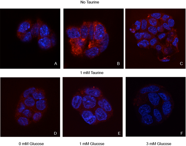
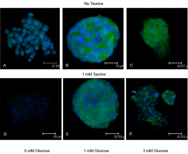
Pancreatic β-cells respond to glucose when interstitial fluid levels reach ~3 mM. When we incubate either β-cell line in 1 mM glucose, we find that there are substantial amounts of insulin present in the cytoplasm (Figs. 1b, 2b, 3). Because of the physiological relatedness of this condition, we use the 1 mM glucose as a control for these experiments. When the cells are glucose-starved, there is a significant reduction in the amount of cytoplasmic insulin (Figs. 1a, 2a, 3), likely due to ATP-dependent changes in the KATP-channels. When the cells are treated with 3 mM glucose to approximate in vivo conditions, we see a substantial decrease (p<0.001) in insulin immunoreactivity in the cells (Figs. 1c, 2c, 3).
Figure 1.

Insulin immunoreactivity in Hit pancreatic β-cell line. To test the efficacy of taurine in releasing insulin from the pancreatic β-cell line cultures were treated with no taurine (A-C) or 1 mM taurine (D-F) in increasing glucose concentrations. Cultures received either 0 mM glucose (A, D), the subthreshold concentration 1 mM glucose (B, E), or a concentration shown to initiate glucose release (3 mM; C, F). For controls, we chose the 1 mM glucose without taurine treatment, a condition that does not promote insulin release from β-cells. Cultures were immunostained for insulin (red). Blue structures are nuclei counterstained with DAPI. Insulin levels were significantly decreased in all cultures compared to the control cultures (see Figure 3).
Figure 2.

Insulin immunoreactivity in Rin pancreatic insulinoma cell line. To test the efficacy of taurine in releasing insulin from the insulinoma cell line cultures were treated with no taurine (A-C) or 1 mM taurine (D-F) in increasing glucose concentrations. Cultures received either 0 mM glucose (A, D), the subthreshold concentration 1 mM glucose (B, E), or a concentration shown to initiate glucose release (3 mM; C, F). Cultures were immunostained for insulin (green). Blue structures are nuclei counterstained with DAPI.
Figure 3.
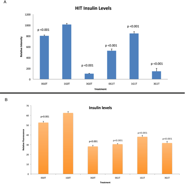
Comparison of insulin levels in Hit and Rin cells. Relative fluorescence intensity, a marker for amounts of protein present in the cell, from cells represented in Figures 1 and 2 For statistical analyses, all p values are related to control cultures treated with 1 mM glucose only. A: Relative fluorescence intensity for insulin in the β-cell line Hit-T15. B: Relative fluorescence intensity for insulin in the β-cell line Rin-m. Using 1 mM glucose in the absence of taurine (1G0T) as a control, all treatments resulted in significantly reduced insulin immunoreactivity. Legend: 0G0T (no glucose or taurine), 3G0T (3 mM glucose, no taurine), 0G1T (no glucose, 1 mM taurine), 1G1T (1 mM each glucose plus taurine), 3G1T (3 mM glucose, 1 mM taurine).
Interestingly, when cells are treated with 1 mM taurine, there is also a significant decrease (p<0.001) in cytoplasmic insulin levels in the cells (Figs., 1d, 2d, 3). In the Hit cells, this decrease is similar to that seen in the glucose-deprived condition (0G0T), with a p value > 0.05 comparing the two treatments. Comparing all other groups, each observed value was statistically different than all others with the exception of the cells treated with 3 mM glucose and 3 mM glucose plus taurine. This suggests that the 3 mM glucose is the overriding stimulus for insulin release.
For the insulinoma cells, again all treatments resulted in a significant decrease in insulin immunoreactivity (p<0.001) compared to controls. As with the Hit cells, insulin immunoreactivity in the presence of 3 mM glucose, while significantly lower than controls, were not significantly different from each other (p>0.05). Of interest, we found that there were no significant differences between Rin cells treated with 3 mM glucose and those treated with 1 mM taurine in the absence of glucose (p>0.05; Fig. 3). Likewise, when taurine is in the presence of glucose, the concentration of glucose does not affect the amount of insulin released as both treatments resulted in no significantly different values for insulin levels (p>0.05). This would seem to indicate that in the insulinoma cell line, either 3 mM glucose or 1 mM taurine could elicit insulin release as effectively as the other.
The Hit cells treated with 1 mM taurine contained more insulin than those treated with 3 mM glucose (Fig. 3), suggesting that different mechanisms exist in normal β-cells than insulinoma cells with regard to sensitivity to taurine-regulated insulin release. When cells were treated with 1 mM glucose (subthreshold) plus 1 mM taurine, both cell lines responded with a significant reduction (p<0.001) in the amount insulin retained compared to controls (Figs. 1e, 2e, 3). In both cell lines, this treatment resulted in significantly higher insulin levels compared to cells treated with 3 mM glucose (p<0.001 for Hit cells; p<0.01 for Rin cells). For cells treated with 3 mM glucose plus 1 mM taurine, the insulin levels were significantly reduced compared to controls (p<0.001; Figs. 1f, 2f, 3). The insulin levels for the synergistic treatment were not significantly higher than the treatment with 3 mM glucose alone (p>0.05).
Discussion
We demonstrate here that taurine transport into pancreatic β-cells may initiate the calcium-dependent release of insulin and GABA, independent of extracellular glucose concentrations. Experimentally, glucose concentrations are considered low if < 2.8 mM but appropriate for insulin release at concentrations above 11 mM, although lower concentrations have elicited physiological responses. Glucose concentrations at 3 mM can regulate glucagon release from pancreatic α-cells, presumably through release of GABA from β-cells [1]. Since insulin and GABA may be co-released, we chose to use 3 mM glucose for our studies. In both cell lines used, this glucose concentration was sufficient to bring about a reduction in intracellular insulin levels compared to concentrations less that 2.8 mM.
Insulin exocytosis begins with the uptake of glucose by the β-cells [2,3] and subsequent breakdown via glycolysis. Subsequent generation of ATP closes ATP-dependent K+ channels [11]. Glucose deprivation appears to increase KATP channel activity through cAMP-activated protein kinase-dependent recruitment of these channel proteins [12]. We demonstrated here that glucose deprivation suppresses insulin release, presumably by cAMP-activated protein kinase-dependent increase in KATP channel expression at the cell surface [12]. In the paper by Lim et al., the use of 3 mM glucose did not significantly alter KATP-dependent currents; however, in that study the islets were pre-treated with glucose for 2 hr at room temperature. It may be that in our study, the environmental parameters may also contribute to the observed changes in insulin levels. In our study, treatment was for 24 h and at 37oC and may more appropriately describe differences seen between our data at 0 mM, 1 mM and 3 mM.
With the significant reduction in insulin using 3 mM glucose compared to 1 mM, we used these two conditions as physiologically relevant. When these cell lines were treated with 1 mM taurine in the absence of glucose, taurine alone was capable of significantly reducing the cytoplasmic insulin levels (Figs 1-3). These data may explain our previous observations that chronic taurine exposure may result in a chronic hypoglycemia. We have found that mice chronically fed taurine in their drinking water are hypoglycemic compared to age-matched controls (data not shown). We further found that when taurine-fed mice are challenged with glucose following a 12 hour fast, there is a delay of 30 min in peak plasma glucose levels, suggesting that these animals are better prepared to handle a sudden rise in glucose. If taurine transport into β-cells increases insulin release, one would expect to find that chronic release of insulin might facilitate glucose uptake, resulting in a chronic depression in plasma glucose concentration.
Additionally, taurine is a known agonist of the GABAA receptor [13-20]. In pancreatic islets, the GABAA receptor is expressed on α-cells where glucagon is synthesized and released. Taurine binding to the GABAA receptor results in a hyperpolarization of α-cells, thus inhibiting glucagon release. For our in vivo observations, the hypoglycemia observed in chronic taurine administration may be attributed to activation of insulin release via transport into the β-cells, inhibition of glucagon release via interaction of taurine with the GABAA receptors on the α-cells, or a combination of these events.
Conclusions
The transport of taurine into pancreatic β-cells may be sufficient to cause the release of insulin. We believe that the mechanism by which this can occur in β-cells involves the interaction of taurine with the ATP-sensitive K-channels. In these cells in vivo, a rise in intracellular glucose levels results in a concomitant increase in cytoplasmic ATP, NADH and NADPH, all required for insulin exocytosis [21]. In part, the mechanism of this action requires an increase in the ATP:ADP ratio, which closes ATP-dependent K+ channels [21]. Closure of these K+-channels depolarizes the β-cells and activates voltage-gated calcium channels. The resulting increase in cytosolic calcium is responsible in part for the exocytosis of the insulin-containing large dense-core vesicles (LDVC) [21]. The resulting exocytosis of the LDVC also releases GABA, which is found in both the LDVC and synapse-like microvesicles (SLMV) [22]. Therefore, release of insulin and GABA from the pancreatic β-cells requires regulation of both K+ and Ca2+ currents.
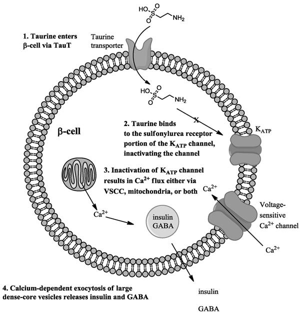
Taurine has been shown to inhibit the KATP-channels in skeletal muscle fibers [23]. In these fibers, taurine binds either to the sulfonylurea receptor (SUR) portion of channel, or to some associated protein that in turn binds to the SUR [23]. We propose the following mechanism by which taurine might independently affect insulin release from the pancreatic β-cells: First, taurine is taken up by the taurine transporter (TauT) on the β-cells (Fig 4, step 1). Once in the cytoplasm, taurine binds to the SUR portion of the channel (or associated protein which in turn binds to the SUR) and inactivates the ATP-sensitive K+-channel (Fig 4, step 2). The inhibition of the ATP-sensitive K+-channel leads to the opening of voltage-gated calcium channels (Fig 4, step 3) or alternately the efflux of calcium from intracellular stores. In neurons, taurine-modulated calcium flux has been reported [24]. Ultimately, the increase in cytoplasmic calcium concentration results in the exocytotic release of the LDCV containing insulin and GABA.
Figure 4.
Proposed scheme by which taurine may affect insulin and GABA release from pancreatic β-cells. In this scheme, 1: taurine is taken up into the β-cells via the taurine transporter (TauT). Once in the cytoplasm, 2: taurine inactivates the ATP-sensitive K+-channels either directly via interaction with the sulfonylurea receptor on the channel, or indirectly via an associated protein that binds to the SUR. The inactivation of ATP-sensitive K+-channels leads to 3: an increase in intracellular Ca++ either via voltage-sensitive calcium channels or mitochondrial buffering. Once there is a critical Ca++ concentration, 4: exocytosis of the large dense-core vesicles containing insulin and GABA occurs. In the in vivo pancreas, GABA would bind to GABAA receptors on α-cells and inactivate glucagon release.
If taurine is administered chronically, the metabolic relevance could manifest as chronic hypoglycemia. The significance of this in the treatment of type II diabetes could mean that in certain cases, higher plasma insulin levels could alleviate increases in plasma glucose concentrations.
Competing interests
The authors have no competing interests.
Authors' contributions
WJL conceived of the study, designed the study, performed the statistical analysis and drafted the manuscript. CC performed the immunohistochemical analyses of the Hit cells; AS performed the immunohistochemical analyses of the Rin cells; JB and JT were responsible for the cell cultures and confocal analyses. AEI participated in the study design and coordination as well as edited the manuscript. All authors read and approved the final manuscript.
Contributor Information
William J L’Amoreaux, Email: William.Lamoreaux@csi.cuny.edu.
Christina Cuttitta, Email: cmariecuttitta@yahoo.com.
Allison Santora, Email: allison.santora@gmail.com.
Jonathan F Blaize, Email: Jonathan.Blaize@gmail.com.
Janto Tachjadi, Email: jtachjadi@yahoo.com.
Abdeslem El Idrissi, Email: Abdeslem.Elidrissi@csi.cuny.edu.
Acknowledgements
The authors wish to acknowledge funding from the Professional Staff Congress of CUNY (WJL and AEI), FRAXA (AEI), and the National Science Foundation for funds for the purchase of the confocal microscope (DBI0421046).
This article has been published as part as part of Journal of Biomedical Science Volume 17 Supplement 1, 2010: Proceedings of the 17th International Meeting of Taurine. The full contents of the supplement are available online at http://www.jbiomedsci.com/supplements/17/S1.
References
- Gilon P, Bertrand G, Loubatieres-Mariani MM, Remacle C, Henquin JC. The influence of gamma-aminobutyric acid on hormone release by the mouse and rat endocrine pancreas. Endocrinology. 1991;129:2521–2529. doi: 10.1210/endo-129-5-2521. [DOI] [PubMed] [Google Scholar]
- Newgard CB, McGarry JD. Metabolic coupling factors in pancreatic beta-cell signal transduction. Annu Rev Biochem. 1995;64:689–719. doi: 10.1146/annurev.bi.64.070195.003353. [DOI] [PubMed] [Google Scholar]
- Tal M, Wu YJ, Leiser M, Surana M, Lodish H, Fleischer N, Weir G, Efrat S. [Val12] HRAS downregulates GLUT2 in beta cells of transgenic mice without affecting glucose homeostasis. Proc Natl Acad Sci U S A. 1992;89:5744–5748. doi: 10.1073/pnas.89.13.5744. [DOI] [PMC free article] [PubMed] [Google Scholar]
- Muroyama A, Uehara S, Yatsushiro S, Echigo N, Morimoto R, Morita M, Hayashi M, Yamamoto A, Koh DS, Moriyama Y. A novel variant of ionotropic glutamate receptor regulates somatostatin secretion from delta-cells of islets of Langerhans. Diabetes. 2004;53:1743–1753. doi: 10.2337/diabetes.53.7.1743. [DOI] [PubMed] [Google Scholar]
- Wang XP, Norman MA, Yang J, Cheung A, Moldovan S, Demayo FJ, Brunicardi FC. Double-gene ablation of SSTR1 and SSTR5 results in hyperinsulinemia and improved glucose tolerance in mice. Surgery. 2004;136:585–592. doi: 10.1016/j.surg.2004.05.042. [DOI] [PubMed] [Google Scholar]
- Lambert IH. Regulation of the cellular content of the organic osmolyte taurine in mammalian cells. Neurochem Res. 2004;29:27–63. doi: 10.1023/B:NERE.0000010433.08577.96. [DOI] [PubMed] [Google Scholar]
- Kreisman NR, Olson JE. Taurine enhances volume regulation in hippocampal slices swollen osmotically. Neuroscience. 2003;120:635–642. doi: 10.1016/S0306-4522(03)00359-2. [DOI] [PubMed] [Google Scholar]
- Haskew-Layton RE, Rudkouskaya A, Jin Y, Feustel PJ, Kimelberg HK, Mongin AA. Two distinct modes of hypoosmotic medium-induced release of excitatory amino acids and taurine in the rat brain in vivo. PLoS ONE. 2008;3:e3543. doi: 10.1371/journal.pone.0003543. [DOI] [PMC free article] [PubMed] [Google Scholar]
- Arany E, Strutt B, Romanus P, Remacle C, Reusens B, Hill DJ. Taurine supplement in early life altered islet morphology, decreased insulitis and delayed the onset of diabetes in non-obese diabetic mice. Diabetologia. 2004;47:1831–1837. doi: 10.1007/s00125-004-1535-z. [DOI] [PubMed] [Google Scholar]
- El Idrissi A, Boukarrou L, L'Amoreaux W. Taurine supplementation and pancreatic remodeling. Adv Exp Med Biol. 2009;643:353–358. full_text. [PubMed] [Google Scholar]
- Maechler P. Mitochondria as the conductor of metabolic signals for insulin exocytosis in pancreatic beta-cells. Cell Mol Life Sci. 2002;59:1803–1818. doi: 10.1007/PL00012507. [DOI] [PMC free article] [PubMed] [Google Scholar]
- Lim A, Park SH, Sohn JW, Jeon JH, Park JH, Song DK, Lee SH, Ho WK. Glucose deprivation regulates KATP channel trafficking via AMP-activated protein kinase in pancreatic beta-cells. Diabetes. 2009;58:2813–2819. doi: 10.2337/db09-0600. [DOI] [PMC free article] [PubMed] [Google Scholar]
- del Olmo N, Bustamante J, del Rio RM, Solis JM. Taurine activates GABA(A) but not GABA(B) receptors in rat hippocampal CA1 area. Brain Res. 2000;864:298–307. doi: 10.1016/S0006-8993(00)02211-3. [DOI] [PubMed] [Google Scholar]
- El Idrissi A. Taurine and brain excitability. Adv Exp Med Biol. 2006;583:315–322. doi: 10.1007/978-0-387-33504-9_35. full_text. [DOI] [PubMed] [Google Scholar]
- El Idrissi A. Taurine improves learning and retention in aged mice. Neurosci Lett. 2008;436:19–22. doi: 10.1016/j.neulet.2008.02.070. [DOI] [PubMed] [Google Scholar]
- El Idrissi A, Boukarrou L, Heany W, Malliaros G, Sangdee C, Neuwirth L. Effects of taurine on anxiety-like and locomotor behavior of mice. Adv Exp Med Biol. 2009;643:207–215. doi: 10.1007/978-0-387-75681-3_21. full_text. [DOI] [PubMed] [Google Scholar]
- El Idrissi A, L'Amoreaux WJ. Selective resistance of taurine-fed mice to isoniazide-potentiated seizures: in vivo functional test for the activity of glutamic acid decarboxylase. Neuroscience. 2008;156:693–699. doi: 10.1016/j.neuroscience.2008.07.055. [DOI] [PubMed] [Google Scholar]
- El Idrissi A, Messing J, Scalia J, Trenkner E. Prevention of epileptic seizures by taurine. Adv Exp Med Biol. 2003;526:515–525. doi: 10.1007/978-1-4615-0077-3_62. [DOI] [PubMed] [Google Scholar]
- El Idrissi A, Trenkner E. Taurine as a modulator of excitatory and inhibitory neurotransmission. Neurochem Res. 2004;29:189–197. doi: 10.1023/B:NERE.0000010448.17740.6e. [DOI] [PubMed] [Google Scholar]
- Sergeeva OA, Fleischer W, Chepkova AN, Warskulat U, Haussinger D, Siebler M, Haas HL. GABAA-receptor modification in taurine transporter knockout mice causes striatal disinhibition. J Physiol. 2007;585:539–548. doi: 10.1113/jphysiol.2007.141432. [DOI] [PMC free article] [PubMed] [Google Scholar]
- Maechler P. Novel regulation of insulin secretion: the role of mitochondria. Curr Opin Investig Drugs. 2003;4:1166–1172. [PubMed] [Google Scholar]
- Braun M, Wendt A, Karanauskaite J, Galvanovskis J, Clark A, MacDonald PE, Rorsman P. Corelease and differential exit via the fusion pore of GABA, serotonin, and ATP from LDCV in rat pancreatic beta cells. J Gen Physiol. 2007;129(z):221–231. doi: 10.1085/jgp.200609658. [DOI] [PMC free article] [PubMed] [Google Scholar]
- Tricarico D, Barbieri M, Camerino DC. Taurine blocks ATP-sensitive potassium channels of rat skeletal muscle fibres interfering with the sulphonylurea receptor. Br J Pharmacol. 2000;130:827–834. doi: 10.1038/sj.bjp.0703385. [DOI] [PMC free article] [PubMed] [Google Scholar]
- El Idrissi A. Taurine increases mitochondrial buffering of calcium: role in neuroprotection. Amino Acids. 2008;34:321–328. doi: 10.1007/s00726-006-0396-9. [DOI] [PubMed] [Google Scholar]


