Abstract
Retroperitoneal benign lipomas are extremely rare and represent about 2.9% of all primary retroperitoneal tumors. About 80% of the tumors in the retroperitoneal cavities are malignant neoplasms. We experienced a case of a retroperitoneal lipoma simulating an ovarian mature cystic teratoma. A diagnosis was correctly made by magnetic resonance imaging (MRI) prior to surgery, and a total tumorectomy was performed. The retroperitoneal lipoma was recognized to have arisen from the urinary bladder. Histological sections revealed a tumor consisting of typical adipose cells without atypia. These types of lipomas should be carefully followed-up because they often recur and undergo malignant transformations.
Key words: lipoma, retroperitoneum, ovarian mature cystic teratoma, urinary bladder, magnetic resonance imaging.
Introduction
Primary retroperitoneal tumors represent about 0.2% of all neoplasms. Out of these about 80% of the tumors are malignant neoplasms.1 Retroperitoneal benign lipomas are extremely rare, and represent about 2.9% of all primary retroperitoneal tumors.2 Herein we present a case of a large retroperitoneal lipoma prior to resection. The tumor was identified as having arisen from the urinary bladder at the time of surgery.
Case Report
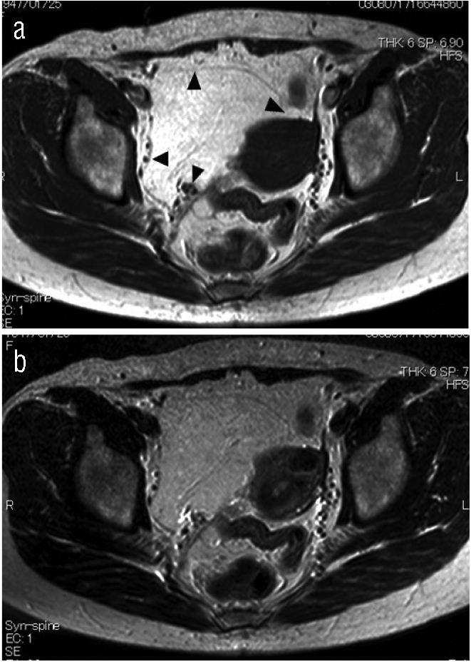
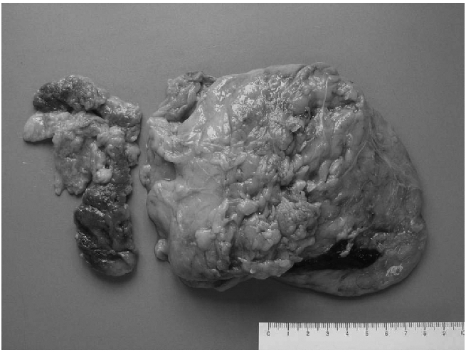
A 61-year old Japanese woman, gravida 3, complained of gluteal pain. She did not have a medical history of surgery or concomitant medications. Upon a pelvic examination, an ill-defined and soft mass was detected in the right pelvic cavity. She had fullness in the right adnexal region. Transvaginal ultrasound sonography showed an echogenic mass without any solid portions in the right adnexal region. These observations suggested a mature cystic tumor of the ovary containing fat. An MRI revealed a mass measuring 15 cm in the right retropelvic cavity, which showed high intensities on T1 and T2-weighted images that were the same intensity as fatty tissues (Figure 1). Another MRI showed that the tumor was in the retropelvic cavity, because the right round ligament and broad uterine ligament were detected over this tumor and the right iliac artery and vein touched the tumor. The patient underwent surgery with a diagnosis of a retroperitoneal lipoma. Macroscopically, an encapsulated, yellowish and soft mass lay between the urinary bladder and the right iliac vessels in the right retropelvic space (Figure 2). The tumor penetrated the right lateral umbilical ligament, and had feeding vessels from that ligament. The tumor showed strong adherence to the urinary bladder but did not infiltrate the surrounding organs. The tumor was thought to be a large lipoma arising from the urinary bladder, therefore, a total tumorectomy was performed.
Figure 1.

Retropelvic mass showed very high intensities on T1-(a) and T2-(b) weighted images that were the same intensity as fatty tissues. This mass was diagnosed as a retropelvic lipoma by observing the position of the uterine broad ligament (arrow), the round ligament (arrow), the uterine artery (arrow) and the lateral umbilical ligament (arrow).
Figure 2.

Retropelvic lipoma after its removal.
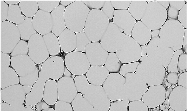
Histological sections revealed a tumor consisting of typical adipose cells without atypia (Figure 3). Neither lipoblasts nor malignant cells were observed in the sections, and the histo-pathological diagnosis was that of a lipoma. The patient had no post-operative complications, experienced less gluteal pain, and was discharged on post-operative day 8. She had an uneventful recovery during the post-operative follow-up period.
Figure 3.

Histological examination reveals an adipose tissue neoplasm without cytologic atypia.
Discussion
Most lipomas are superficial, and retroperitoneal lipomas are very rare. When lipomas occur in retroperitoneal locations, they are mostly renal in origin.3 A few reported cases have a pancreatic origin,4 and so on. Pelvic lipomas are exceedingly rare, but can arise from the iliac vessels and paravaginal tissues.5 One reported case with pelvic origins demonstrated a urinary bladder outlet obstruction secondary to a pelvic lipoma.6 To our knowledge, the present case arising from the urinary bladder is the first such report in the world. The lipoma presented herein was characterized by the diffuse overgrowth of fatty tissues in the perivesical spaces, but lacked any urinary obstructions. The patient experienced only slight gluteal pain.
MRIs present greater diagnostic precision than ultrasonographies (USGs) and computerized tomographies (CTs). In the present case, the transvaginal ultrasound sonograph showed an echogenic mass without any solid portion in the right adnexal region, which suggested a mature cystic teratoma of the ovary containing fat. On the MRIs, the lipomatous components showed very high signal intensities on the T1-weighted and T2-weighted images. Furthermore, the diagnosis of retropelvic tumors is aided by observing the position of the uterine broad ligament, the round ligament, the uterine artery and the lateral umbilical ligament. Thus, we were able to diagnose this tumor as a retropelvic lipoma prior to surgery. Therefore, MRIs are effective for diagnosing retroperitoneal lipomas.
Benign subcutaneous lipomas and malignant liposarcomas are well-documented and are characterized by chromosome 12 aberrations. Recently, some retroperitoneal lipomas were shown to have no detectable rearrangements of chromosome 12 or its amplification.7 On the other hand, retroperitoneal lipomas have shown rearrangements in chromosomes 1 and 8. Therefore, there are different chromosomal rearrangements between retroperitoneal lipomas and malignant liposarcomas. However, there are no significant optical differences in CTs or MRIs observed prior to surgery between lipomas and well-differentiated liposarcomas.8 Thus, one should consider the possibility of a liposarcoma when trying to resect a retroperitoneal lipoma. Furthermore, because these types of lipomas are known to recur and undergo malignant transformation, subjects with resected retroperitoneal lipomas must have careful follow-up.
References
- 1.Armstrong JR, Cohen I. Primary malignant retroperitoneal tumors. Am J Surg. 1965;110:937–43. doi: 10.1016/0002-9610(65)90181-9. [DOI] [PubMed] [Google Scholar]
- 2.Pai MR, Naik R, Raughuveer CV. Primary retroperitoneal tumors: a 25 year study. Indian J Med Sci. 1995;49:139–41. [PubMed] [Google Scholar]
- 3.Cavazza A, Giunta A, Pedrazzoli C, et al. Extrarenal retroperitoneal angiomyolipoma: description of a case and review of the literature. Pathologica. 2001;91:44–9. [PubMed] [Google Scholar]
- 4.Moriki T, Ohtsuki Y, Takahashi T, et al. Lipoma-like tumor mass probably arising in the retroperitoneal heterotopic pancreas: A previously undescribed lesion. Pathol Int. 2004;54:527–31. doi: 10.1111/j.1440-1827.2004.01661.x. [DOI] [PubMed] [Google Scholar]
- 5.Hull WB, Blumenfeld ML, Jacques D. Large paravaginal pelvic lipoma: A case report. J Reprod Med. 1999;44:636–8. [PubMed] [Google Scholar]
- 6.Berens BM, Azavan A. Bladder outlet obstruction due to pelvic lipoma: computerized tomography, magnetic resonance imaging and radiographic evaluation. J Urol. 1991;145:138–9. doi: 10.1016/s0022-5347(17)38270-8. [DOI] [PubMed] [Google Scholar]
- 7.Foa C, Mainguene C, Dupre F, et al. Rearrangement involving chromosomes 1 and 8 in a retroperitoneal lipoma. Cancer Genet Cytogen. 2002;133:156–9. doi: 10.1016/s0165-4608(01)00573-8. [DOI] [PubMed] [Google Scholar]
- 8.Kimura N, Morita T, Murata A, et al. A case of giant retroperitoneal lipoma. Nihon Rinsyo Gekagakkaishi. 2002;63:2030–4. [Google Scholar]


