Summary
Chronic pain remains one of the most widespread and under treated medical conditions. Opioid drugs, such as morphine, are some of the most effective analgesics available. However, their utility for the treatment of chronic pain are limited by side effects including tolerance and dependence. Morphine induces its biological actions primarily through the G-protein coupled mu-opioid peptide receptor (MOP-R) [1], which is also the target of the endogenous opioids. However, unlike endogenous MOP-R ligands, morphine fails to promote substantial endocytosis of the MOP-R both in vitro [2–5] and in vivo [6–11]. Endocytosis of the MOP-R serves at least two important functions in regulating signal transduction. First, desensitization and endocytosis act as an “OFF” switch by uncoupling receptors from their cognate G protein. Second, endocytosis and receptor recycling serve as an “ON” switch, resensitizing receptors by recycling them to the plasma membrane where agonist can regain access to the receptors. Thus, due to poor endocytosis, both the OFF and ON function of the MOP-R are altered in response to morphine compared to endogenous ligands. To examine whether poor endocytosis contributes to morphine tolerance and dependence, we generated a knock-in mouse that expresses a mutant MOP-R that undergoes rapid endocytosis in response to morphine. Morphine remains an excellent antinociceptive agent in these mice. In addition, the mutant mice display substantially reduced antinociceptive tolerance and attenuated physical dependence. These data suggest that opioid drugs with pharmacological profile of morphine and the ability to promote endocytosis of the receptor could provide analgesia while having a reduced liability for the side effects of tolerance and dependence.
Results
Generation of a novel MOP-R knock-in mouse with altered trafficking properties
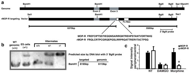
To directly examine whether endocytosis of the MOP-R in response to morphine alters the development of tolerance and dependence in vivo, we generated a knock-in mouse expressing the rMOP-R mutant receptor that internalizes in response to morphine [12]. In this rMOP-R, a portion of the cytoplasmic tail of the MOP-R, encoded entirely within exon 3, has been replaced with sequence from the delta opioid peptide receptor (see Fig. 1a). Mice expressing the rMOP-R were identified by Southern (DNA) blot analysis (Fig. 1a and b). The specific mutation introduced to the MOP-R gene [12] was contained entirely within Exon 3, which is common to all splice variants that have been described. Endocytic trafficking of the wild-type MOP-R and the rMOP-R mutant receptor examined in striatal neurons cultured from wild-type and mutant mice demonstrated that morphine promote rMOP-R but not MOP-R endocytosis in response to morphine (Fig. 1c and Supplemental Fig. 1).
Figure 1. Generation of rMOP-R knock-in mice.
a. Schematic of targeting strategy A Sal1-Sac1 genomic fragment containing the MOP-R sequence, including Exons 2 and 3 was modified to contain the rMOP-R sequence (inset). A cassette containing resistance to G418 (Fx-Neo) and flanked by Lox P sites was inserted in the intron downstream of exon 3 for selection of ES clones. b. Detection of homologous recombinants. Genomic DNA was digested with BamHI and subjected to DNA hybridization with a ~1.1kb BglII fragment (see a). Targeted loci were confirmed by the presence of a band at ~8kb. The intact locus gave a band at ~6kb. c. Quantification of endocytosis. MetaMorph software was used to quantify the intensity of receptor signal at the plasma membrane versus the cytosol for each treatment condition and each genotype (MOP-R in black, rMOP-R in white). Data is plotted as the ratio of signal located within 0.3 μm of the surface (peripheral) versus the amount in the cytosol (central). See supplemental Fig. 1 for representative neurons and schematic.
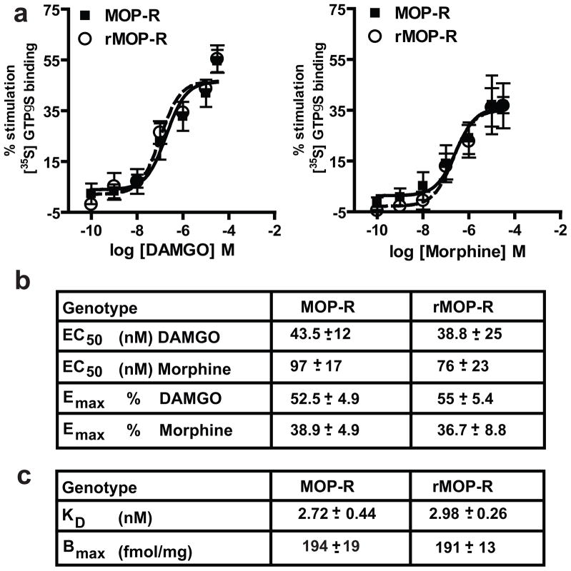
The mutant mice were viable, had no gross phenotypic abnormalities and showed normal baseline pain responses (hot-plate latency, 56°C: wild-type, 4.88 ± 0.33 seconds; mutant, 4.55 ± 0.32 seconds and see Fig. 4 saline treatments). Consistent with their equivalent baseline pain responses, there were no genotypic differences in MOP-R distribution in the spinal cord or multiple brain regions important for the antinociceptive and reinforcing properties of opiates (Supplemental Fig. 2 and data not shown). In addition, ligand affinity, receptor number and receptor G-protein coupling were unaltered in the rMOP-R mice (Fig. 2a-c).
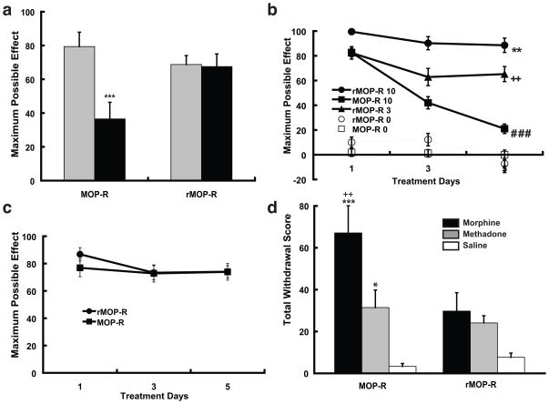
Figure 4. Opioid tolerance and dependence in MOP-R wild-type and rMOP-R knock-in mice. a. Acute morphine tolerance.
Mice (n=9) were initially treated with either saline (gray bars) or a high dose of morphine (100 mg/kg, sc, black bars). 24 h later, mice were challenged with an acute equi-antinociceptive dose of morphine (3 mg/kg for rMOP-R and 10 mg/kg for MOP-R, see Fig. 4B). Data are presented as mean ± SEM of MPE. Wild-type MOP-R mice exhibited significant acute tolerance, showing a 43% reduction in MPE when pretreated with 100 mg/kg of morphine compared to saline 24 h before (MOP-R comparing saline vs. morphine pretreatment, student’s t-test, ***p<.001). In contrast, rMOP-R knock-in mice showed no evidence of tolerance and maintained the same level of morphine-induced antinociception whether they were pretreated with 100 mg/kg of morphine or saline 24 h before. b. Chronic morphine tolerance. Mice were treated twice daily with morphine (10 mg/kg, sc) for 5 days and antinociception was assessed following the first injection of morphine each day. Mean ± SEM of MPE across days are presented. A two-way ANOVA revealed that mice treated chronically with morphine (n=17) behaved differently corresponding to genotype as indicated by a significant group effect [F(2,42)=27.95, p<.001] and group X treatment days effect [F(4,84)=12.09, p<.001]. Post-hoc comparisons (Tukey’s) revealed the source of the interaction. rMOP-R knock-in mice treated with 10 mg/kg of morphine had significantly longer response latencies across days compared to wild-type mice treated with the same dose of morphine (10 mg/kg) and rMOP-R knock-in mice chronically treated with a lower (equi-antinociceptive) dose of morphine (3 mg/kg), [rMOP-R 10 significantly different from MOP-R 10 and rMOP-R 3, **p<.01]. Additionally, rMOP-R knock-in mice chronically treated with an equi-antinociceptive dose of morphine (3 mg/kg) showed significantly greater antinociception than did wild-type mice chronically treated with a higher dose of morphine (10 mg/kg) across the tolerance development days (rMOP-R 3 vs. MOP-R 10, ++p<.01). Only wild-type mice chronically treated with morphine (10 mg/kg) showed a significant decrease in antinociception from Day 1 to Day 5 (MOP-R 10 Day 1 vs. Day 5, ###p<.001). Thus, the development of antinociceptive tolerance to morphine was evident in wild-type mice but attenuated in the knock in mice, whether they were chronically treated with the same (10 mg/kg) or equi-antinociceptive (3 mg/kg) dose of morphine. c. Methadone tolerance. Mice were similarly treated to the protocol in experiment in Fig. 6b but injected twice daily with methadone (4 mg/kg, sc) for 5 days. For both genotypes, there was no evidence of tolerance development with both groups expressing comparable levels of methadone-antinociception following the first and last injection. Methadone antinociception was equivalent in both genotypes across all days. d. Naloxone precipitated withdrawal. Groups of mice were chronically treated with 10 mg/kg of morphine (black bars, n=10-12), 4 mg/kg of methadone (grey bars, n=9) or saline (white bars, n=6) at the same intervals described for Fig 4b and c. Mice were challenged with naloxone (2 mg/kg, sc) 30 min following the final treatment injection. Notably, the chronic dose of morphine used (10 mg/kg) corresponded to a functionally higher dose in the rMOP-R knock-in mice relative to wild-type mice (see Fig 3a, b). Standard withdrawal behaviors including jumping, wet-dog shakes, paw licks and paw tremors were scored by an observer blind to genotype. Total withdrawal scores (the sum of all individual withdrawal behaviors) ± SEM are presented and group differences were analyzed with the LSD test. Compared to saline-treated mice, MOP-R wild-type mice displayed a significantly higher incidence of withdrawal when chronically treated with morphine or methadone (MOP-R MOR vs. MOP-R SAL, ***p<.001; MOP-R METH vs. MOP-R SAL, *p<.05), with a higher degree of withdrawal associated with chronic morphine treatment (MOP-R MOR vs. MOP-R METH, ++p<.01). In contrast, rMOP-R knock-in mice displayed similar levels of withdrawal regardless of pretreatment drug. For rMOP-R knock-in mice, both morphine and methadone pretreatment resulted in similar levels of moderate withdrawal, comparable to methadone pretreatment in MOP-R wild-type mice.
Figure 2. Pharmacological characterization of wild-type MOP-R and rMOP-R knock-in mice.
a, b. Receptor-G protein coupling. Agonist mediated GTPγS binding was measured in brain membranes of wild-type MOP-R (squares) and rMOP-R knock-in mice (circles) with increasing concentrations of DAMGO or morphine. Data were analyzed by nonlinear regression using GraphPad Prism software and are presented as means ± SEM of at least three experiments performed in triplicate. There were no significant genotypic differences in either EC50 or Emax. c. Ligand affinity and receptor number. [3H] Naloxone binding in whole brain membranes from MOP-R and rMOP-R mice. Saturation binding assays were performed on membranes (50-100 μg per well) with increasing concentrations of [3H] naloxone (0 to 15 nM, 55.9Ci/mmol). Nonspecific binding was measured in the presence of 10 μM naloxone. Binding parameters were determined by Scatchard analysis of specific binding. Data are means ± SEM of three experiments performed in duplicate. There were no statistically significant differences between the genotypes (One way ANOVA with Tukey’s post-hoc test). Bmax, maximum binding capacity; KD, dissociation constant.
Antinociception in rMOP-R knock-in mice versus wild-type MOP-R mice
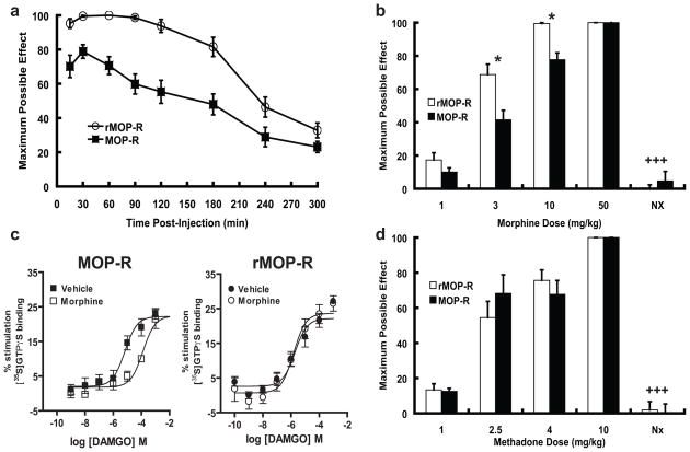
Morphine-induced antinociception was evaluated by measuring response latencies in the hot-plate test. We tested a dose of morphine (10 mg/kg) known to induce robust antinociception in mice. The acute antinociceptive effect of this dose of morphine was significantly enhanced and prolonged in knock-in mice relative to their wild-type littermates (Fig. 3a). A dose of 3 mg/kg in the mutant mouse was equi-antinociceptive to 10 mg/kg in the wild-type mouse (Fig. 3b). Both genotypes reached a ceiling effect at the highest dose tested, 50 mg/kg. The opioid antagonist naloxone completely reversed the antinociceptive effects of morphine in both wild-type and mutant mice (Fig. 3b).
Figure 3. Antinociception in wild-type MOP-R and rMOP-R knock-in mice.
a. Enhanced and prolonged morphine-induced antinociception in rMOP-R knock-in mice. Antinociceptive responses were measured with the hot-plate response latency test (56°C) after morphine treatment (10 mg/kg, sc). A response endpoint was defined as latency to either lick the fore- or hindpaws or flick the hindpaws. To avoid tissue damage, mice were exposed to the hot-plate for a maximum of 20s. Data are reported as the mean ± SEM of percent maximum possible effect (MPE) using the following formula: 100% × [(drug response time – basal response time)/(20 s – basal response time)]. A two-way analysis of variance revealed that the MPE curve for rMOP-R mice (n=17) mice was significantly greater and prolonged relative to the MOP-R mice (n=17) as indicated by a significant genotype [p<.001, F(1,7)=28.05] and genotype X time interaction effect [p<.001, F(1,7)=4.97]. b. Dose-dependent morphine-antinociception. Antinociceptive responses were determined with the hot-plate test and data are reported as mean ± SEM of MPE (see a). Separate groups of mice for both genotypes (n=7–9) were injected with the doses of morphine indicated and assessed for antinociception 30 min later. To test whether the antinociceptive responses were mediated by opioid receptors, a final grouping was injected with morphine (10 mg/kg) followed by naloxone (2 mg/kg). rMOP-R knock-in mice showed enhanced antinociception at 3 and 10 mg/kg doses (rMOP-R vs. MOP-R scores for MPE at respective morphine doses, student’s t-test, *p<.03) with the latter dose inducing the maximum possible response (100%) in the mutant mice. At the highest dose tested (50 mg/kg) both genotypes exhibited the maximum possible response (100%). For both genotypes, antinociception induced by 10 mg/kg of morphine was reversed by treatment with 2 mg/kg of the opioid antagonist naloxone (morphine 10 mg/kg with and without naloxone 2 mg/kg treatment for each genotype respectively, student’s t-test +++p<.001). c. MOP-R desensitization in the brainstem following acute morphine treatment. Agonist-mediated [35S]GTPγS binding was measured in brainstem membranes of MOP-R and rMOP-R mice with increasing concentrations of DAMGO. Left Panel. Binding in MOP-R mice was significantly reduced (p<0.01) following acute morphine-treatment (10 mg/kg s.c. 30 min; EC50 = 428 ± 141 μM; open squares ) compared to vehicle-treated mice (EC50 = 2.35 ± 0.9 μM; closed squares). Right Panel. Binding in rMOP-R mice was not significantly changed (p>0.05) following acute morphine-treatment (EC50 = 3.29 ± 1.4 μM; open circles) compared to vehicle-treated mice (EC50 = 1.16 ± 0.6 μM; closed circles). Data were analyzed by nonlinear regression using GraphPad Prism software and are presented as means ± SEM of at least three experiments performed in triplicate. d. Enhanced antinociception in rMOP-R knock-in mice is morphine-specific. Separate groups of mice for both genotypes (n=8-10) were injected with the doses of methadone indicated (1-10 mg/kg,) and assessed for antinociception. Methadone induced a dose-dependent increase in antinociceptive response with no genotypic differences. For both genotypes, antinociception induced by 4 mg/kg of methadone was reversed by treatment with 2 mg/kg of the opioid antagonist naloxone (methadone 4 mg/kg with and without naloxone 2 mg/kg treatment for each genotype respectively, student’s t-test +++p<.001). Thus, enhanced opioid-induced antinociception observed in the rMOP-R knock-in mice is agonist-specific, and naloxone-reversible.
We propose that the enhanced antinociception in the mutant mice reflects the restoration of the ON function provided by receptor endocytosis and recycling. Specifically, we propose that morphine-occupied MOP receptors become partially desensitized in wild-type mice and fail to resensitize due to poor endocytosis; whereas in the rMOP-R knock-in mice, receptors are also desensitized but are rapidly resensitized by endocytosis and recycling. Consistent with this hypothesis, MOP receptors in wild-type mice given a single 10 mg/kg dose of morphine showed significant receptor-G protein uncoupling (Fig. 3c, left panel). Clearly not all MOP receptors in these mice were desensitized, since morphine is still an excellent acute antinociceptive agent in wild-type mice. Nevertheless, MOP-Rs in the brainstem of wild-type mice treated with morphine showed a 200-fold shift in the EC50 of DAMGO (Fig. 3c, left panel) compared to wild-type mice treated with vehicle. In contrast, receptors in rMOP-R mice given the same dose of morphine, showed no desensitization (Fig. 3c, right panel). These data suggest that the reduced morphine antinociception in the wild-type compared to the rMOP-R mice reflects partial desensitization of MOP-Rs that is not reversed by endocytosis and recycling.
If this were the case, we would expect mice of both genotypes to show equivalent antinociception to an agonist that promotes endocytosis of the receptor in both genotypes. Indeed, there were no significant genotypic differences in antinociception induced by methadone (1–10 mg/kg; Fig. 3d), a MOP-R agonist that promotes rapid internalization of both the wild-type MOP-R and mutant rMOP-R. Thus, the enhanced opioid antinociception observed in the rMOP-R knock-in mice is specific to morphine. Together with our immunohistochemical and pharmacological data (Supplemental Fig. 2 and Fig. 2), these data suggest that the enhanced morphine antinociception in the rMOP-R knock-in mice cannot be accounted for by differences in MOP-R distribution, ligand affinity, receptor number or receptor G-protein coupling. Rather, these data suggest that facilitating MOP-R endocytosis enhances morphine antinociception by reversing rapid desensitization.
Acute antinociceptive tolerance in rMOP-R knock-in mice versus wild-type MOP-R mice
It has been hypothesized that MOP-R desensitization contributes to acute morphine tolerance. If this were the case, rMOP-R mice would be expected to develop reduced acute tolerance compared to wild-type mice. To examine this, we evaluated the acute antinociceptive effect of equi-antinociceptive doses of morphine (3 mg/kg in rMOP-R and 10 mg/kg in MOP-R, see Fig 3b) 24 hours following pretreatment with a high dose of morphine (100 mg/kg) or saline. The day following pretreatment, baseline response latencies between genotypes were similar (rMOP-R, 5.76 ± 0.47 secs; MOP-R, 5.86, ± 0.51 secs). Indicative of the acute tolerance that is typically observed in this paradigm [13], wild-type MOP-R mice that had been pretreated with 100 mg/kg of morphine showed a 43% reduction in antinociception compared to mice that had received saline the day before (Fig. 4a). In contrast, the rMOP-R knock-in mice maintained similar levels of morphine antinociception regardless of whether they had received morphine or saline pretreatment the day before (Fig. 4a). Thus, the rMOP-R knock-in mice did not develop acute antinociceptive tolerance to morphine.
Chronic antinociceptive tolerance in rMOP-R knock-in mice versus wild-type MOP-R mice
While acute tolerance to high doses of opioids is most relevant to acute pain, during the treatment of chronic pain, analgesic tolerance typically develops over the course of repeated administrations of moderate levels of drug. Thus, we evaluated the development of tolerance following twice daily administrations of morphine (10 mg/kg) over 5 days. Wild-type mice in this paradigm developed antinociceptive tolerance (Fig. 4b, squares). In contrast, their rMOP-R littermates, treated with the same dose of morphine (10 mg/kg) at the same intervals, showed no evidence of tolerance, exhibiting as much antinociception on the last day of drug treatment as they did on the first day (Fig. 4b, circles).
To rule out the possibility that the lack of tolerance in the mutant mice was an artifact of enhanced morphine antinociception (Fig. 3a,b), a separate group of knock-in mice were treated chronically with an equi-antinociceptive dose of morphine (3 mg/kg, see Fig. 3b) given at the same intervals. These rMOP-R knock-in mice still showed reduced tolerance, maintaining similar levels of antinociception over the course of treatment (Fig 4b, triangles). Thus, reduced morphine tolerance in the knock-in relative to wild-type mice cannot be attributed to enhanced morphine antinociception.
These results suggest that endocytosis of the receptor reduces the development of antinociceptive tolerance. If this were the case, one would expect that opiate agonists, such as methadone, that promote endocytosis of the MOP-R would have reduced liability for promoting tolerance in wild-type mice. In addition, wild-type and rMOP-R mice should show equivalent responsiveness to chronic methadone. To examine this hypothesis, we evaluated the development of tolerance to methadone in MOP-R and rMOP-R mice. In order to directly compare tolerance to morphine versus methadone, a dose of methadone was chosen (4 mg/kg, see Fig. 3d) that was equi-antinociceptive to the morphine dose administered in Fig. 4b. At this dose, neither genotype showed evidence of tolerance across treatment days (Fig. 4c). In addition, responsiveness to methadone during all treatment days was equivalent in MOP-R (Fig. 4c, squares) and rMOP-R mice (Fig. 4c circles). Thus, reduced chronic opioid tolerance in rMOP-R mice relative to MOP-R mice is specific to morphine.
As was the case for reduced acute tolerance, reduced chronic tolerance to morphine in the mutant mice may reflect, at least in part, that MOP-Rs in the wild-type mice are desensitized (Fig. 3c) but are unable to resensitize due to poor endocytosis of the receptor. Facilitating receptor internalization and recycling (i.e., restoring the ON function of endocytosis) may protect against the development of both acute (Fig. 4a) and chronic tolerance (Fig. 4b).
However, receptor desensitization alone cannot explain antinociceptive tolerance to morphine. Specifically, if all MOP-Rs were desensitized in morphine tolerant mice, then displacement of morphine from these non-signaling receptors with antagonist should have no behavioral effect. However, morphine tolerant animals show substantial naloxone-precipitated withdrawal signs (see for example [14]), indicating that receptors continue to signal actively in morphine tolerant animals despite the lack of antinociception. Hence, mechanisms other than receptor desensitization are likely contributing to tolerance.
Morphine withdrawal in rMOP-R knock-in mice versus wild-type MOP-R mice
We next examined whether facilitating endocytosis in the rMOP-R mice affected the development of morphine dependence. Following chronic treatment with morphine, mice were challenged with the opioid antagonist, naloxone (2 mg/kg), 30 min following the final morphine injection. Global withdrawal responses were scored by an observer who was blind to genotype (Fig 4d). Wild-type mice expressed robust withdrawal responses compared to mutant mice, which were chronically treated with the same amount of morphine (10 mg/kg) but at a functionally higher dose (see Fig. 3a, b). Consistent with the hypothesis that enhanced receptor endocytosis decreases withdrawal, chronic methadone treatment (4 mg/kg given at the same intervals as morphine), promoted substantially less withdrawal than did morphine in wild-type mice (Fig. 4d). In fact, the moderate level of methadone withdrawal in wild-type mice was equivalent to that produced by either morphine or methadone in the rMOP-R mice (Fig. 4d). Hence, we have generated a mouse line that retains morphine antinociceptive potency with markedly reduced morphine tolerance and dependence.
Discussion
In summary, here we report that mice expressing a mutant rMOP-R with altered receptor trafficking properties in response to morphine show enhanced morphine-induced antinociception, reduced morphine tolerance, and reduced naloxone-precipitated withdrawal compared to their wild-type littermates. These knock-in mice otherwise show normal ligand affinity, receptor number, receptor G-protein coupling and receptor distribution, consistent with the fact that both their basal pain responses as well as methadone antinociception are equivalent to that of their wild-type littermates.
These data are consistent with the hypothesis that enhanced endocytosis of the MOP-R in response to morphine can reduce antinociceptive tolerance and dependence while retaining the antinociceptive efficacy of morphine. It is important to note that endocytosis is only one step in a cascade of highly conserved events that occurs following G-protein coupled receptor activation. When receptors are activated by endogenous ligand, they are rapidly desensitized by phosphorylation and interaction with arrestin and then endocytosed. Following endocytosis, MOP-Rs are functionally resensitized by recycling to the plasma membrane. Many groups have demonstrated in vitro that morphine-activated receptors elude this natural cycle of receptor desensitization, endocytosis and resensitization that is induced by endogenous MOP-R ligands [15-18]. Similarly, morphine has been found to be a poor inducer of receptor endocytosis in vivo [6–10]. However, in vivo, subtleties also emerge, because in some cases, desensitization has not been detected [19], whereas in other cases, desensitization of the morphine-activated receptor by GRK/arrestin and/or PKC does appear to occur [20–27]. Thus, receptor desensitization may be either brain region specific, incomplete, or both.
In the context of regionally-specific or incomplete receptor desensitization, the failure to endocytose the morphine-bound receptor has the potential to affect signal transduction in at least two ways. First, in cells or brain regions where morphine does not cause substantial receptor desensitization, prolonged receptor activation may trigger downstream adaptive responses that contribute to morphine tolerance and dependence. In rMOP-R mice, this prolonged receptor activation is replaced by pulsatile receptor activation due to restoration of the OFF/ON switch of endocytosis. Second, in cells or brain regions where receptors do become desensitized after morphine activation, failure to endocytose would prevent functional resensitization of the receptor. In rMOP-R mice, resensitization would be restored. Notably, even in regions where desensitization appears to occur (Fig. 3c), a significant number of receptors remain coupled. These remaining receptors would exhibit prolonged activation in wild-type mice and pulsatile activation in knock-in mice.
Disruption of arrestin appears to enhance morphine antinociception [21] and delay tolerance [21], presumably by decreasing the degree of morphine-induced desensitization. However, arrestin knock-out mice show levels of withdrawal equivalent to their wild-type littermates [28], indicating that there are still a substantial number of functionally coupled MOP-Rs even in animals with intact arrestin. Promoting morphine-induced endocytosis would be expected to both facilitate receptor resensitization and alleviate the compensatory adaptive changes associated with dependence. Thus, while both preventing receptor desensitization and facilitating receptor endocytosis/resensitization are effective strategies to enhance morphine antinociception and prevent tolerance, the latter has the added benefits of 1) reducing morphine dependence and 2) specificity to the MOP-R.
All opioids, when given at high enough concentration for a long enough period of time, including methadone, can induce tolerance and dependence. However, when given at equi-antinociceptive doses, opioids induce different degrees of tolerance and dependence [29–31] and see Fig. 4c, and some ligands even appear to cause these effects by different mechanisms [32, 33]. Hence, given the complex pharmacology of the various opioid ligands, it has been difficult to isolate the effect of endocytosis on tolerance and dependence.
The present results provide a genetic “proof-of-concept” that endocytosis, is an important mechanism that can delay tolerance and dependence. Notably, the use of the rMOP-R knock-in mice allowed the same opioid drug to be compared in mice that appear to differ only in their MOP-R trafficking properties. Importantly, even if mechanisms other than endocytosis are contributing to the behavioral differences in the MOP-R and rMOP-R mice, these mice will provide a powerful tool for delineating which of the adaptive changes that have been observed in wild-type animals following chronic morphine treatment are relevant to behavioral tolerance and dependence.
Supplementary Material
Acknowledgments
The authors thank Mark von Zastrow for support in the development of this project and Brigitte Kieffer for supplying the pBK2 plasmid containing the genomic fragment used to generate the targeting vector. We thank Randall Armstrong for assistance with the DNA blot, Yuichiro Inoue, Ling Wang, and Marian Logrip for assistance with primary culture and Brigitte Kieffer, Mark Von Zastrow, Howard L. Fields, Dorit Ron and Randy Hampton for critical reading of the manuscript. This work was supported by the National Institute on Drug Abuse (NIDA) grant DA015232 and funds provided by the state of California for medical research on alcohol and substance abuse through the University of California San Francisco (UCSF), both to J.L.W.
Footnotes
Publisher's Disclaimer: This is a PDF file of an unedited manuscript that has been accepted for publication. As a service to our customers we are providing this early version of the manuscript. The manuscript will undergo copyediting, typesetting, and review of the resulting proof before it is published in its final citable form. Please note that during the production process errors may be discovered which could affect the content, and all legal disclaimers that apply to the journal pertain.
References
- 1.Matthes HW, Maldonado R, Simonin F, Valverde O, Slowe S, Kitchen I, Befort K, Dierich A, Le Meur M, Dolle P, et al. Loss of morphine-induced analgesia, reward effect and withdrawal symptoms in mice lacking the mu-opioid-receptor gene. Nature. 1996;383:819–823. doi: 10.1038/383819a0. [DOI] [PubMed] [Google Scholar]
- 2.Arden JR, Segredo V, Wang Z, Lameh J, Sadee W. Phosphorylation and agonist-specific intracellular trafficking of an epitope-tagged mu-opioid receptor expressed in HEK 293 cells. J Neurochem. 1995;65:1636–1645. doi: 10.1046/j.1471-4159.1995.65041636.x. [DOI] [PubMed] [Google Scholar]
- 3.Keith DE, Murray SR, Zaki PA, Chu PC, Lissin DV, Kang L, Evans CJ, von ZM. Morphine activates opioid receptors without causing their rapid internalization. J Biol Chem. 1996;271:19021–19024. doi: 10.1074/jbc.271.32.19021. [DOI] [PubMed] [Google Scholar]
- 4.Koch T, Schulz S, Pfeiffer M, Klutzny M, Schroder H, Kahl E, Hollt V. C-terminal splice variants of the mouse mu-opioid receptor differ in morphine-induced internalization and receptor resensitization. J Biol Chem. 2001;276:31408–31414. doi: 10.1074/jbc.M100305200. [DOI] [PubMed] [Google Scholar]
- 5.Yu Y, Zhang L, Yin X, Sun H, Uhl GR, Wang JB. Mu opioid receptor phosphorylation, desensitization, and ligand efficacy. J Biol Chem. 1997;272:28869–28874. doi: 10.1074/jbc.272.46.28869. [DOI] [PubMed] [Google Scholar]
- 6.Sternini C, Spann M, Anton B, Keith DJ, Bunnett NW, von ZM, Evans C, Brecha NC. Agonist-selective endocytosis of mu opioid receptor by neurons in vivo. Proc Natl Acad Sci U S A. 1996;93:9241–9246. doi: 10.1073/pnas.93.17.9241. [DOI] [PMC free article] [PubMed] [Google Scholar]
- 7.Keith DE, Anton B, Murray SR, Zaki PA, Chu PC, Lissin DV, Monteillet-Agius G, Stewart PL, Evans CJ, von Zastrow M. mu-Opioid receptor internalization: opiate drugs have differential effects on a conserved endocytic mechanism in vitro and in the mammalian brain. Mol Pharmacol. 1998;53:377–384. [PubMed] [Google Scholar]
- 8.Trafton JA, Abbadie C, Marek K, Basbaum AI. Postsynaptic signaling via the [mu]-opioid receptor: responses of dorsal horn neurons to exogenous opioids and noxious stimulation. J Neurosci. 2000;20:8578–8584. doi: 10.1523/JNEUROSCI.20-23-08578.2000. [DOI] [PMC free article] [PubMed] [Google Scholar]
- 9.He L, Fong J, von Zastrow M, Whistler JL. Regulation of opioid receptor trafficking and morphine tolerance by receptor oligomerization. Cell. 2002;108:271–282. doi: 10.1016/s0092-8674(02)00613-x. [DOI] [PubMed] [Google Scholar]
- 10.He L, Whistler JL. An opiate cocktail that reduces morphine tolerance and dependence. Curr Biol. 2005;15:1028–1033. doi: 10.1016/j.cub.2005.04.052. [DOI] [PubMed] [Google Scholar]
- 11.Abbadie C, Pasternak GW. Differential in vivo internalization of MOR-1 and MOR-1C by morphine. Neuroreport. 2001;12:3069–3072. doi: 10.1097/00001756-200110080-00017. [DOI] [PubMed] [Google Scholar]
- 12.Finn AK, Whistler JL. Endocytosis of the mu opioid receptor reduces tolerance and a cellular hallmark of opiate withdrawal. Neuron. 2001;32:829–839. doi: 10.1016/s0896-6273(01)00517-7. [DOI] [PubMed] [Google Scholar]
- 13.Fairbanks CA, Wilcox GL. Spinal antinociceptive synergism between morphine and clonidine persists in mice made acutely or chronically tolerant to morphine. J Pharmacol Exp Ther. 1999;288:1107–1116. [PubMed] [Google Scholar]
- 14.Kest B, Palmese CA, Hopkins E, Adler M, Juni A, Mogil JS. Naloxone-precipitated withdrawal jumping in 11 inbred mouse strains: evidence for common genetic mechanisms in acute and chronic morphine physical dependence. Neuroscience. 2002;115:463–469. doi: 10.1016/s0306-4522(02)00458-x. [DOI] [PubMed] [Google Scholar]
- 15.Alvarez VA, Arttamangkul S, Dang V, Salem A, Whistler JL, Von Zastrow M, Grandy DK, Williams JT. mu-Opioid receptors: Ligand-dependent activation of potassium conductance, desensitization, and internalization. J Neurosci. 2002;22:5769–5776. doi: 10.1523/JNEUROSCI.22-13-05769.2002. [DOI] [PMC free article] [PubMed] [Google Scholar]
- 16.Blanchet C, Sollini M, Luscher C. Two distinct forms of desensitization of G-protein coupled inwardly rectifying potassium currents evoked by alkaloid and peptide mu-opioid receptor agonists. Mol Cell Neurosci. 2003;24:517–523. doi: 10.1016/s1044-7431(03)00173-8. [DOI] [PubMed] [Google Scholar]
- 17.Kovoor A, Celver JP, Wu A, Chavkin C. Agonist induced homologous desensitization of mu-opioid receptors mediated by G protein-coupled receptor kinases is dependent on agonist efficacy. Mol Pharmacol. 1998;54:704–711. [PubMed] [Google Scholar]
- 18.Connor M, Borgland SL, Christie MJ. Continued morphine modulation of calcium channel currents in acutely isolated locus coeruleus neurons from morphine-dependent rats. Br J Pharmacol. 1999;128:1561–1569. doi: 10.1038/sj.bjp.0702922. [DOI] [PMC free article] [PubMed] [Google Scholar]
- 19.Ingram SL, Vaughan CW, Bagley EE, Connor M, Christie MJ. Enhanced opioid efficacy in opioid dependence is caused by an altered signal transduction pathway. J Neurosci. 1998;18:10269–10276. doi: 10.1523/JNEUROSCI.18-24-10269.1998. [DOI] [PMC free article] [PubMed] [Google Scholar]
- 20.Bagley EE, Chieng BC, Christie MJ, Connor M. Opioid tolerance in periaqueductal gray neurons isolated from mice chronically treated with morphine. Br J Pharmacol. 2005;146:68–76. doi: 10.1038/sj.bjp.0706315. [DOI] [PMC free article] [PubMed] [Google Scholar]
- 21.Bohn LM, Lefkowitz RJ, Gainetdinov RR, Peppel K, Caron MG, Lin FT. Enhanced morphine analgesia in mice lacking beta-arrestin 2. Science. 1999;286:2495–2498. doi: 10.1126/science.286.5449.2495. [DOI] [PubMed] [Google Scholar]
- 22.Narita M, Mizoguchi H, Nagase H, Suzuki T, Tseng LF. Involvement of spinal protein kinase Cgamma in the attenuation of opioid mu-receptor-mediated G-protein activation after chronic intrathecal administration of [D-Ala2,N-MePhe4,Gly-Ol(5)]enkephalin. J Neurosci. 2001;21:3715–3720. doi: 10.1523/JNEUROSCI.21-11-03715.2001. [DOI] [PMC free article] [PubMed] [Google Scholar]
- 23.Bohn LM, Lefkowitz RJ, Caron MG. Differential mechanisms of morphine antinociceptive tolerance revealed in (beta)arrestin-2 knock-out mice. J Neurosci. 2002;22:10494–10500. doi: 10.1523/JNEUROSCI.22-23-10494.2002. [DOI] [PMC free article] [PubMed] [Google Scholar]
- 24.Selley DE, Nestler EJ, Breivogel CS, Childers SR. Opioid receptor-coupled G-proteins in rat locus coeruleus membranes: decrease in activity after chronic morphine treatment. Brain Res. 1997;746:10–18. doi: 10.1016/s0006-8993(96)01125-0. [DOI] [PubMed] [Google Scholar]
- 25.Bailey CP, Kelly E, Henderson G. Protein kinase C activation enhances morphine-induced rapid desensitization of mu-opioid receptors in mature rat locus ceruleus neurons. Mol Pharmacol. 2004;66:1592–1598. doi: 10.1124/mol.104.004747. [DOI] [PubMed] [Google Scholar]
- 26.Bushell T, Endoh T, Simen AA, Ren D, Bindokas VP, Miller RJ. Molecular components of tolerance to opiates in single hippocampal neurons. Mol Pharmacol. 2002;61:55–64. doi: 10.1124/mol.61.1.55. [DOI] [PubMed] [Google Scholar]
- 27.Borgland SL, Connor M, Osborne PB, Furness JB, Christie MJ. Opioid agonists have different efficacy profiles for G protein activation, rapid desensitization, and endocytosis of mu-opioid receptors. J Biol Chem. 2003;278:18776–18784. doi: 10.1074/jbc.M300525200. [DOI] [PubMed] [Google Scholar]
- 28.Bohn LM, Gainetdinov RR, Lin FT, Lefkowitz RJ, Caron MG. Mu-opioid receptor desensitization by beta-arrestin-2 determines morphine tolerance but not dependence. Nature. 2000;408:720–723. doi: 10.1038/35047086. [DOI] [PubMed] [Google Scholar]
- 29.Grecksch G, Bartzsch K, Widera A, Becker A, Hollt V, Koch T. Development of tolerance and sensitization to different opioid agonists in rats. Psychopharmacology (Berl) 2006;186:177–184. doi: 10.1007/s00213-006-0365-8. [DOI] [PubMed] [Google Scholar]
- 30.Duttaroy A, Yoburn BC. The effect of intrinsic efficacy on opioid tolerance. Anesthesiology. 1995;82:1226–1236. doi: 10.1097/00000542-199505000-00018. [DOI] [PubMed] [Google Scholar]
- 31.Walker EA, Young AM. Differential tolerance to antinociceptive effects of mu opioids during repeated treatment with etonitazene, morphine, or buprenorphine in rats. Psychopharmacology (Berl) 2001;154:131–142. doi: 10.1007/s002130000620. [DOI] [PubMed] [Google Scholar]
- 32.Patel MB, Patel CN, Rajashekara V, Yoburn BC. Opioid agonists differentially regulate mu-opioid receptors and trafficking proteins in vivo. Mol Pharmacol. 2002;62:1464–1470. doi: 10.1124/mol.62.6.1464. [DOI] [PubMed] [Google Scholar]
- 33.Narita M, Suzuki M, Niikura K, Nakamura A, Miyatake M, Yajima Y, Suzuki T. mu-Opioid receptor internalization-dependent and -independent mechanisms of the development of tolerance to mu-opioid receptor agonists: Comparison between etorphine and morphine. Neuroscience. 2006;138:609–619. doi: 10.1016/j.neuroscience.2005.11.046. [DOI] [PubMed] [Google Scholar]
Associated Data
This section collects any data citations, data availability statements, or supplementary materials included in this article.


