Abstract
Over the past two decades, the bulk of gastrointestinal (GI) endoscopic procedures has shifted away from diagnostic and therapeutic interventions for symptomatic disease toward cancer prevention in asymptomatic patients. This shift has resulted largely from a decrease in the incidence of peptic ulcer disease in the era of antisecretory medications coupled with emerging evidence for the efficacy of endoscopic detection and eradication of dysplasia, a histopathological biomarker widely accepted as a precursor to cancer. This shift has been accompanied by a drive toward minimally-invasive, in situ optical diagnostic technologies that help assess the mucosa for cellular changes that relate to dysplasia. Two competing but complementary approaches have been pursued. The first approach is based on broad-view targeting of “areas of interest” or “red flags.” These broad-view technologies include standard white light endoscopy (WLE), high-definition endoscopy (HD), and “electronic” chromoendoscopy (narrow-band-type imaging). The second approach is based on multiple small area or point-source (meso/micro) measurements, which can be either machine (spectroscopy) or human-interpreted (endomicroscopy, magnification endoscopy), much as histopatholgy slides are. In this paper we present our experience with the development and testing of a set of familiar but “smarter” standard tissue-sampling tools that can be routinely employed during screening/surveillance endoscopy. These tools have been designed to incorporate fiberoptic probes that can mediate spectroscopy or endomicroscopy. We demonstrate the value of such tools by assessing their preliminary performance from several ongoing clinical studies. Our results have shown promise for a new generation of integrated optical tools for a variety of screening/surveillance applications during GI endoscopy. Integrated devices should prove invaluable for dysplasia surveillance strategies that currently result in large numbers of benign biopsies, which are of little clinical consequence, including screening for colorectal polyps and surveillance of “flat” dysplasia such as Barrett’s esophagus and chronic colitis due to inflammatory bowel diseases.
1. Introduction
Noninvasive spectroscopic tissue diagnosis, or “optical biopsy,” is a rapidly emerging field within the field of biophotonics [1]. Ideally when using spectroscopy, some form of spectroscopic analysis is performed on measurements collected from precisely the volume of tissue that will be examined histopathologically. The ultimate goal then is to attempt to obtain a diagnosis of the tissue based on these measurements, in situ, with minimal invasion and in real-time. Clearly there is the potential for the reduction of overall procedure costs, patient distress, and risks as a consequence of biopsying and processing only diseased tissue. Some proposed techniques include Raman spectroscopy [2], autofluorescence spectroscopy [3–6], fluorescence spectroscopy [7–11], reflectance spectroscopy [7, 8, 11–14], and elastic-scattering spectroscopy [1, 15–17]. Much work has focused primarily on UV-induced fluorescence spectroscopy, given the assumption that important biochemical changes associated with disease alter the intrinsic fluorescence spectrum of tissue. Elastic Scattering Spectroscopy (ESS), however, is capable of reporting cellular and subcellular architectural features that are a typical part of a pathologist’s microscopic assessment and diagnosis. ESS measurements can be performed via fiberoptic probes and hold great promise for in vivo screening and identification of neoplastic tissues. ESS diagnosis can be based either on heuristic models [18] that predict changes in the scattering spectrum corresponding to altered ultrastructure, or on quantitative models [19] that have been used to determine nuclear size in epithelial layers.
Indeed, with the appropriate optical geometry, ESS can report the size, structure, and index of refraction of subcellular components that change upon neoplastic transformation [18–20]. ESS spectra relate to the wavelength-dependence and angular-probability of scattering efficiency of tissue micro-structures (as well as to absorption bands), generating spectral signatures that correlate with histolgical features such as the size/shape of subcellular components, nuclear-cytoplasmic ratio, and cell/organelle clustering patterns. Scattering is generated by gradients of the optical index of refraction and ESS spectral signatures will be altered if the refractive index of subcellular structures changes, say, due to an increase in the amount and/or granularity of chromatin. Since the dimensions of subcellular components are on the order of the wavelength of visible-near-IR light, approximations of scattering theory (e.g. Rayleigh approximation) are not suitable for mathematical simulations, and methods such as Mie theory are more appropriate for understanding the spectral changes [21].
There are a number of clinical correlations of scattering spectra with mucosal histopathology. The potential clinical utility of ESS for endoscopic dysplasia and/or cancer detection biopsy has been reported in hollow organs including the urinary bladder [21], esophagus [22–26], and colon [27–29]. ESS was first applied in vivo in the urinary bladder by Mourant, Bigio, and colleagues, where sensitivity and specificity for the detection of malignant tissue in a retrospective analysis from a small sample size (110 biopsy sites from 10 patients) were excellent [21]. More recently, ESS has been studied for the diagnosis of luminal gastrointestinal tract neoplasms. Bigio et al. used colorectal ESS measurements (60 sites from 16 patients) to develop a spectral metric based on regions of the hemoglobin absorption bands (400–440 nm and 540–580 nm) to identify 8 sites that were neoplastic, adenomatous, and/or cancerous, also with good retrospective statistics [22]. In a larger study by Ge, et. al. neoplastic and hyperplastic colonic polyps could be distinguished using ESS and neural-network pattern recognition for spectral classification [28]. In addition, Zonios et al. have published a study applying ESS to the classification of colon polyps [29]. Wallace et al. have also adapted ESS to identify dysplastic Barrett’s esophagus with a sensitivity and specificity of 82% and 80%, respectively [30].
All of these studies were validated by taking ESS mucosal readings (“optical biopsy”) with a fiberoptic probe inserted through the working channel of an endoscope. The probe was then removed and a surgical tissue-sampling tool, usually a forceps, was introduced to obtain a pinch biopsy of what was the best estimate of the location from which the ESS measurement had been obtained. The physical biopsy specimens were then assessed by “gold standard” histopathology and correlated to spectra. The experience from those studies reveals a strong rationale for incorporating ESS probes into biopsy tools. ESS is a site-specific measurement that samples a tissue volume of <0.5 mm3 of the tissue surface at the point of contact of the fiber probe. As such, with small mucosal areas being interrogated, co-registration of separate optical and physical biopsies is subject to imperfect co-registration of the optical reading and the tissue sample removed for histopathological analysis. Indeed, the fact that ESS studies have performed as well as they have in detecting dysplasia despite imperfect co-registration suggests that ESS would perform even better when validated with absolute co-registration of optical and physical biopsies. Equally important, integrated tools would be familiar, more convenient, less disruptive to endoscopic flow and would improve throughput by shortening procedure times.
Compared to endomicroscopic approaches like confocal microscopy and optical coherence tomography (discussed below), ESS is potentially much more cost-effective and accessible to the general GI community. Confocal endomicroscopy, while a powerful commercially available technology, remains costly, requires the administration of an IV fluorescent dye, and presently requires that microscopic images be interpreted by the operator. As such, it is a technology more appropriate for specialized centers of advanced endoscopy [31]. Similarly, intravital optical coherence tomography (OCT) [32–37] is another sophisticated high-resolution mucosal imaging technique that is not optimally suited for widespread use, as it requires non-standard image interpretation by the endoscopist. For both confocal microscopy and OCT, the operator requires substantial competence from specialized training to become both a skilled advanced endoscopist / interventionalist as well as “real-time pathologist,” capable of interpreting histopathological images in real-time.
Indeed, confocal laser endomicroscopy (CLE) is receiving increasing interest and adoption by advanced centers as an endoscopic modality for obtaining very high-resolution magnified images within the mucosal layer of the GI tract. The CLE technology is based on tissue illumination with a low-power laser followed by detection and creation of an optically sectioned image by rejecting out-of-focus fluorescent light using variations of the scanning “pinhole” principle [38, 39]. Because CLE detects fluorescence and because endogenous tissue autofluorescence is low, use of a fluorophore-containing contrast agent administered either locally or intravenously is required to generate high-quality images that are comparable with traditional histologic examination [40, 41]. Recently, a confocal laser endomicroscope has been developed that uses a probe that can be passed to the distal tip of virtually any conventional endoscope via the accessory channel to enable subsurface imaging of living tissue during procedures [42]. This approach, while usable with any endoscope, still poses the same issues of a probe-based spectroscopic approach in that the probe must be removed to allow passage of a biopsy tool whenever a biopsy is required further disrupting clinical flow and increasing procedure time.
2. Materials and Methods
2.1. Elastic-Scattering Spectroscopy
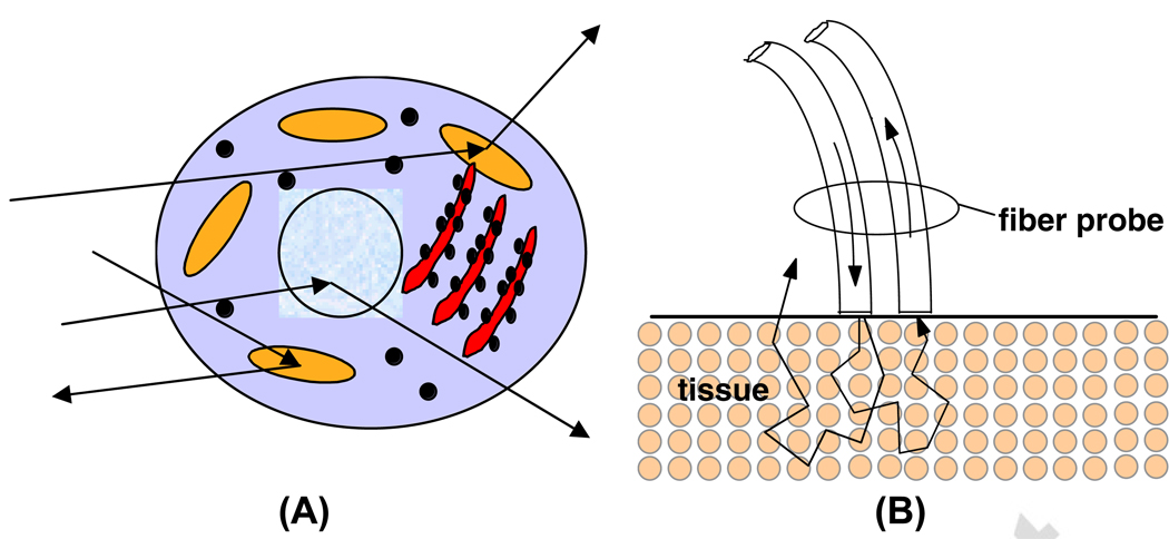
Our group at Boston University (continuing earlier work by Bigio and colleagues, started at Los Alamos National Laboratory)[18, 21, 43–46] developed the method of ESS. ESS, when performed with specific fiberoptic geometries, is sensitive to the absorption spectra of major chromophores (e.g. oxy-/deoxy-hemoglobin) and, more importantly, reports morphological features from superficial tissues. ESS spectra derive from the wavelength-dependent optical scattering efficiency (and the effects of changes in the scattering angular probability) caused by optical index gradients exhibited by cellular and subcellular structures. Unlike Raman and fluorescence spectroscopy, ESS provides largely micro-structural, not biochemical information. Thus, ESS is sensitive to features such as nuclear size, crowding, and chromaticity, chromatin granularity, and mitochondrial and organellar size and density (Figure 1A). Because abnormal tissues are often associated with changes in sub-cellular, nuclear and organellar features, scattering signatures represent the spectroscopic equivalent of a histopathological interpretation. However, the ESS method senses these morphology changes in a semi-quantitative manner, without actually imaging the microscopic structure [47, 48].
Figure 1.
Cartoon illustration of optical scattering from density gradients in cells (A), and a diagram of the optical geometry for the fiber-optic tissue measurements (B). Fiber tips are in optical contact with the tissue surface. Only light that has scattered elastically within the epithelial layer is collected.
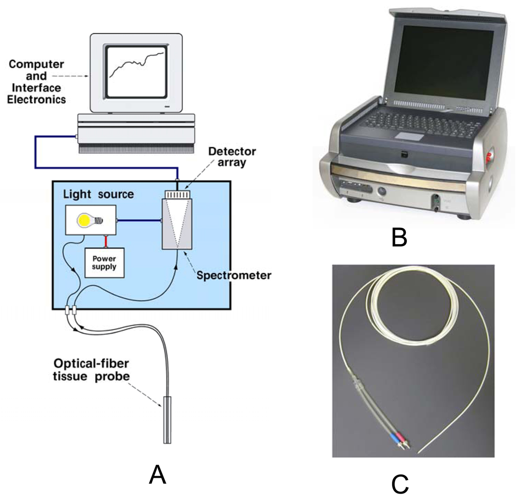
In practice, ESS is a point-spectroscopic measurement taken over a broad wavelength range (320–900 nm) - not an imaging modality – and samples a tissue volume of ≤0.50 mm3. Probes are, typically, used in optical contact with the tissue under examination and have separate illuminating and collecting fibers (Figure 1B). Collected light transmitted to the analyzing spectrometer must first undergo one or more scattering events through a small volume of the tissue before entering the collection fiber(s). No light is collected from surface Fresnel reflection. The standard ESS catheter-type probe consists of a pair of fibers (each with a core diameter of 200 microns) with center-to-center separation of about 250 microns. Due to the small separation of the source and detector fibers, the collected light predominantly samples the mucosal layer, which is typically 300–400 microns thick in the GI tract. Novel tilted-fiber designs can restrict the collected light to even shallower depths [49]. Given the small source-detector separation of the ESS geometry, the method is more sensitive to scattering properties than to absorption [20], although strong absorbers such as oxy- and deoxy-hemoglobin can be quantified. Figure 2 shows a schematic diagram of the primary system components, and photos of the current-generation experimental system. Each measurement takes about 30 milliseconds, and it is possible to perform several measurements per second, limited by the time to move the probe from spot to spot.
Figure 2.
Schematic diagram of the ESS system (A), photos of the ESS instrument (B) and the flexible fiberoptic catheter-type probe used for previous ESS studies (C).
While small-area measurements with ESS are a disadvantage for screening large mucosal regions, surveillance of larger areas can be achieved by making many point measurements in rapid succession. The fact that ESS provides specific information about small areas of tissue provides the rationale for incorporating ESS probes into biopsy forceps tools, especially for gastrointestinal applications. Development and validation studies require precise co-registration of optical measurement sites and surgical biopsies, currently not reliably achieved by existing methods. Presently, fiber probes are passed endoscopically to the mucosa of interest and measurements are taken. The probe is then removed, and a forceps is passed to obtain a pinch biopsy of a best estimate of the ESS measurement spot. This process does not reliably correlate the optical measurement to the physical biopsy for validation studies and unnecessarily extends procedure time.
2.2. Confocal Endomicroscopy
Laser scanning confocal microscopy is an adaptation of light microscopy, whereby focal laser illumination is combined with pinhole limited detection to geometrically reject out-of-focus light. In single-point scanning confocal microscopes, the point is typically scanned in a raster pattern, and measurement of light returning to the detector from successive points is digitized, so that an image of the scanned region can be constructed. Importantly, each resultant image is an “optical section” representing approximately one focal plane within the specimen [50]. The device in this study uses a miniaturized confocal microscope in which a single optical fiber acts as both the illumination point source and the detection pinhole, allowing the small scale required for integration into a biopsy tool [51].
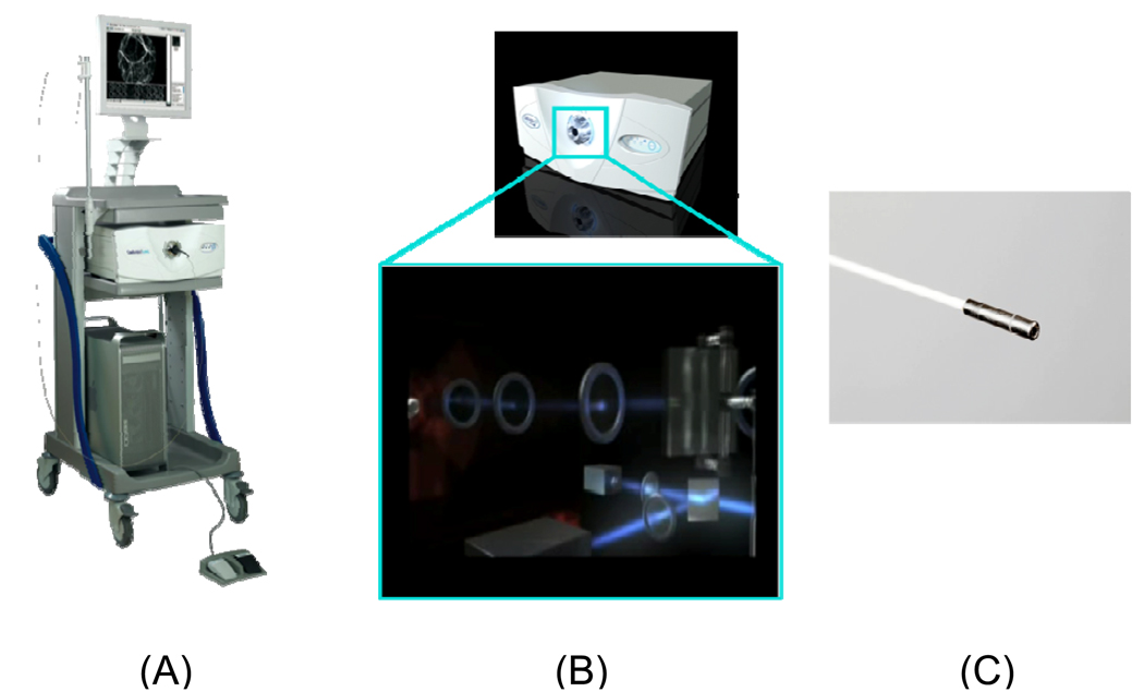
The components of the confocal laser endomicroscope (Cellvizio, Mauna Kea Technologies, Paris, France) are shown in Figure 3. The miniprobe used to integrate into the CLE-guided snare tool was designed for intra-biliary imaging (CholangioFlex™). This probe is 1.2mm in diameter, images at a fixed depth of 55µm, with a maximal field of view of 320µm and lateral resolution of 3.5 µm. [52–54]. The fiber probe consists of a bundle of 10,000 optical fibers with a distal lens, and proximal precision connector. The probe is designed with a semiconductor that oscillates a 488nm laser light at a rate that enables an acquisition frame rate of 12 images per second. The fluorescence signal returning from the tissue is converted into an image using a detector (avalanche photodiode), and software/hardware systems for image correction, stabilization and display [55, 56]. These instruments are encased in an FDA-approved clinical laser-scanning unit.
Figure 3.
Cellvizio confocal laser endomicroscopy system (A). Laser-scanning unit (B). CholangioFlex™ miniprobe (C).
2.3. Integrated Optical Biopsy Tools
The business of endoscopic polyp surveillance has a major efficiency barrier: At present, it is virtually impossible to distinguish a neoplastic from a non-neoplastic polyp by its endoscopic view. As such, the current standard of practice is to remove and examine all polyps histopathologically. While the term “polypectomy” implies a straightforward process, removing polyps using a standard colonoscope, in fact, requires several discrete, time-intensive steps: (1) identification of the polyp, its size, margins and extent; (2) optimal repositioning of the endoscope to effect polypectomy; (3) definitive excision and/or ablation of the polyp; (4) retrieval of polyp tissue and; (5) histopathological processing and interpretation of the biopsy. Small polyps can be removed or electro-ablated in jaw-type forceps and retrieved through the endoscope channel within closed jaws either in toto or piecemeal. Larger polyps, however, require the use of a snare or “garroting” device to excise them. If the tissue fragments are <4 mm, it is often possible to retrieve them by removing the snare device and sucking the tissue into a trap via the scope’s accessory/suction channel. If >4 mm, however, the polyp will not fit into the accessory/suction channel and must be retrieved by (1) removing the snare, (2) passing a grasping device or expandable basket through the lumen, (3) stabilizing the excised tissue at the end of the scope within the grasping device/basket, while (4) withdrawing the entire scope from the patient, and (5) harvesting the tissue. The scope is then (6) reintroduced and (7) once again negotiated to the site of the last polypectomy. It follows that this process can be tedious and time-consuming, especially in patients with multiple large polyps. Thus, depending on the number of larger polyps a patient has, he/she will effectively undergo multiple colonoscopies or passes of the scope, greatly prolonging total procedure time. There is need for a reliable way to identify instantly whether a polyp is neoplastic in situ.
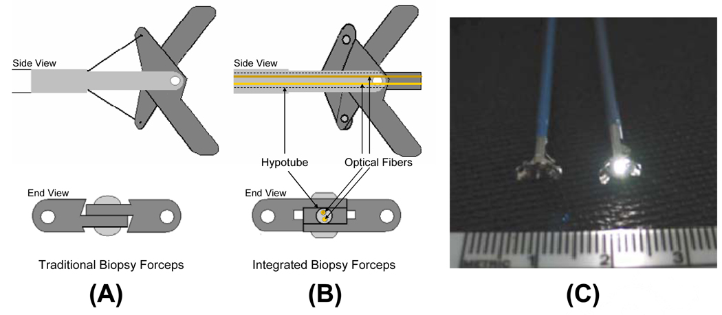
An integrated tool combining an ESS probe with a biopsy forcep/snare is needed, as it would shorten the time required to obtain a biopsy of a measured spot and would greatly increase the spatial correlation of biopsies with the precise spot from which ESS measurements were made. An ESS probe integrated into a biopsy forceps simplifies the process and guarantees accurate co-registration of physical biopsies with ESS measurement spots. Thus, optically-guided biopsy tools represent a major step forward for dysplasia detection in the GI tract. Our group has designed and prototyped two ESS optical biopsy tools that address distinct applications: the ESS optical forceps, and the ESS optical snare. The ESS optical forceps, used for interrogating and biopsying suspect tissue, consists of an ESS probe comprising two-200 µm fibers, installed in a traditional jaw-type biopsy forceps tool, modified with a hollow central channel along its length. The design is such that the probe is actuated to protrude out from between the jaws when open, permitting optical contact with the mucosa while measurements are obtained (Figure 4). In this earliest incarnation the design is essentially similar to a forceps made by SpectraScience, Inc., which uses only one fiber and is intended for fluorescence spectroscopy of the tissue.
Figure 4.
A 2-dimensional diagram of the forcep tip is depicted. The optical forcep is a modified traditional endoscopic jaw-type biopsy forcep (A) with a central channel through which fiberoptic probes can be introduced for tissue measurements (B). A photograph of a clinically-usable unit (C), standard biopsy forceps (left), ESS integrated optical forceps (right).
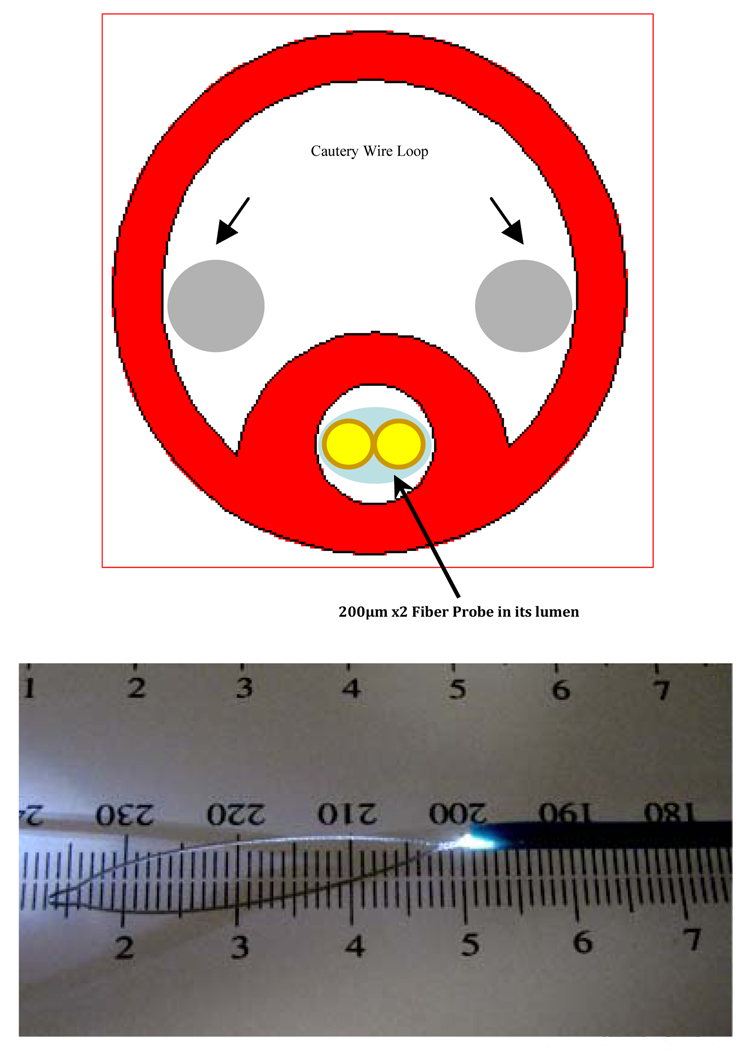
The snare technique is the standard approach used to remove pedunculated polyps at colonoscopy. A nickel-titanium “shape-memory alloy” wire loop is lassoed over the top of the polyp and pulled snugly around the stalk. The stalk is then “garroted” by pulling the wire until the stalk is transected with or without electrocautery. The tissue is retrieved and forwarded to pathology, fixed, sectioned, stained, and assessed for dysplastic or neoplastic tissue within the polyp, down the stalk and to the resection margin. A “smart” snare system could identify the lower border of dysplastic tissue prior to transection of the stalk. The smart snare would have utility for the piecemeal removal of flat and sessile polyps as well, where mucosa bunched up and grasped by the snare could be assessed for dysplastic mucosa and removed. To this end we developed the ESS optical snare, which uses a custom-extruded dual-lumen tubing permitting passage of the cautery snare as well as of the optical probe (Figure 5). Subsequent resections of the surrounding tissue could be guided by ESS measurements to ensure that all dysplastic tissue has been excised and/or ablated. The same approach was used to develop a “smart” snare system using confocal endomicroscopy. In this case the confocal miniprobe is passed through the custom extruded dual-lumen tubing as in the ESS optical snare. This arrangement permits real-time imaging of the mucosal layer of polyps prior to polypectomy or after to confirm complete resection of dysplasia.
Figure 5.
Cross-section and Prototype of an ESS Optical Snare. Custom extruded dual-lumen tubing permits passage of both the cautery wire and 2-fiber ESS probe.
Optically-guided biopsy would enable excision and/or ablation to be performed, while polyp retrieval might be rendered optional, only adenomatous polyps need to be retrieved for histology and consequent risk assesment. Additionally, the reduced number of retrieved samples would yield savings in the cost of histopathology. The rare case of a false-negative by the optical diagnosis would impart no risk to the patient, because it would be excised anyway (and excreted naturally) – simply not retrieved. The improved efficiency and efficacy of the colonoscopy/treatment procedure itself, would impart significant patient benefits, allowing more physician time for careful inspection, and would further reduce the burden on the health care system.
3. Results and Discussion
In this section preliminary results from ongoing IRB-approved clinical studies are presented. Unlike earlier reported results, this endoscopic approach to scattering spectroscopy with integrated tools provides absolute co-registration of optical and physical biopsies. For each of the studies, patients from our existing screening/surveillance pool for colonoscopy, or upper GI endoscopy in the case of Barrett’s Esophagus surveillance, were recruited and consented. Their examinations followed current standard of care, with the only difference being the use of the integrated optical tools whenever endoscopic tissue sampling was indicated according to accepted standards. Optical measurements were then taken prior to biopsy or resection, and the tissue obtained was submitted for standard histopathological diagnosis, and reviewed by three independent pathologists. ESS spectra were correlated to the consensus majority of the histopathology results. Following spectral feature selection by principle component analysis (PCA) [57] a linear support vector machine classifier [58, 59] was trained on the extracted features. Leave-one-out cross-validation was used to obtain performance estimates. This type of pattern recognition based spectral analysis is similar to approaches used in [24, 27, 28] for classification of light scattering spectroscopy data.
3.1. ESS Optical Forceps
The ESS optical forceps was tested for detecting colonic neoplasia during routine colonoscopies. When a polyp was encountered, as determined by the endoscopist, the optical probe in the forceps was put in gentle contact, with jaws open, against the polyp mucosa. Optical readings were then taken before closing the forceps’ jaws to complete the biopsy. By using the ESS optical forceps we are assured that the optical reading will precisely correlate to the location of the biopsy. As seen in Table 1, data from 65 patients has been collected, 96 spectra from 52 neoplastic polyps and 172 spectra from 89 non-neoplastic polyps (normal or hyperplastic histology). From this preliminary dataset, a sensitivity of .83, specificity of .81 and negative predictive value (NPV) of .90 were obtained (Table 2). The negative predictive value, which is the most important statistic for this application, could be increased even further at the expense of a modest reduction in specificity. Thus, integrated ESS optical forceps are able to co-register optical and physical biopsies and analysis to date is highly encouraging for the device's ability to reliably classify neoplastic colonic polyps.
Table 1.
Preliminary data breakdown for each of the studies
| Subjects | Pathology | Number of Polyps |
Number of Spectra |
|
|---|---|---|---|---|
| Forceps – CRC Screening | 65 | Non-Neoplastic Neoplastic |
89 52 |
172 96 |
| Forceps – Barrett’s Screening |
33 | NDBE DBE GCE |
105 16 64 |
|
| Snare – Polypectomy | 10 | Non-Neoplastic Neoplastic |
13 10 |
60 45 |
Table 2.
Performance of the integrated tools on preliminary data
| Pathology | Sensitivity | Specificity | |
|---|---|---|---|
| Forceps – CRC Screening | Neoplastic vs. Non-Neoplastic | .83 | .81 |
| Forceps – Barrett’s Screening |
DBE vs. NDBE | .81 | .88 |
| DBE vs. GCE | .88 | .94 | |
| NDBE vs. GCE | .90 | .83 | |
| Snare – Polypectomy | Neoplastic vs. Non-Neoplastic | .80 | .87 |
The ESS optical forceps have also been used for surveillance of Barrett’s Esophagus. Patients who were scheduled for an upper endoscopy for Barrett’s Esophagus, or those in whom disease has advanced (dysplastic Barrett’s or carcinoma) were candidates for the study. All patients received standard treatment i.e., routine esophageal endoscopy with random multiple physical biopsies using the standard or intensive Seattle Protocol as appropriate. As in the colon study, the optical readings were obtained by first touching the tissue while the jaws of the ESS forceps are open, placing the optical fibers in contact with the mucosa. At the end of the acquisition of optical data, the jaws were closed, thus physically biopsying precisely the same tissue that was read optically. A total of 33 patients have been recruited for the study (Table 1), resulting in 105 ESS spectra of non-dysplastic Barrett’s esophagus (NDBE), 16 spectra of dysplastic Barrett’s esophagus (DBE) and 64 of gastric columnar epithelia (GCE). A sensitivity of .81 and specificity of .88 was obtained for distinguishing DBE from NDBE, .88 and .94 for DBE from GCE, and of .90 and .83 for distinguishing NDBE from GCE (Table 2). These results show promise for using the ESS optical forceps in the screening of Barrett’s esophagus.
3.2. ESS Optical Snare
Recent clinical testing has also begun with the ESS optical snare. In a separate study, subjects were recruited from a pool of patients referred for routine colonoscopy. When polypectomy of larger polyps (>4–5mm) was indicated, an ESS optical snare was used to obtain ESS readings from tissue prior to electrocautery. The tip of the closed optical snare device was placed in contact with polyps and spectra were obtained, followed by electrocautery excision of the polyp. Thus far, from a total of 10 patients, 45 spectra from 13 neoplastic polyps and 60 spectra from 10 non-neoplastic polyps have been taken with the ESS optical snare (Table 1). From this, a sensitivity of .80 and specificity of .87 have been obtained for distinguishing neoplastic from non-neoplastic polyps (Table 2). From these results we can see that the ESS optical snare is a promising technology for diagnosing and snaring larger colonic polyps in real time at colonoscopy.
3.3. Confocal Endomicroscopy Snare
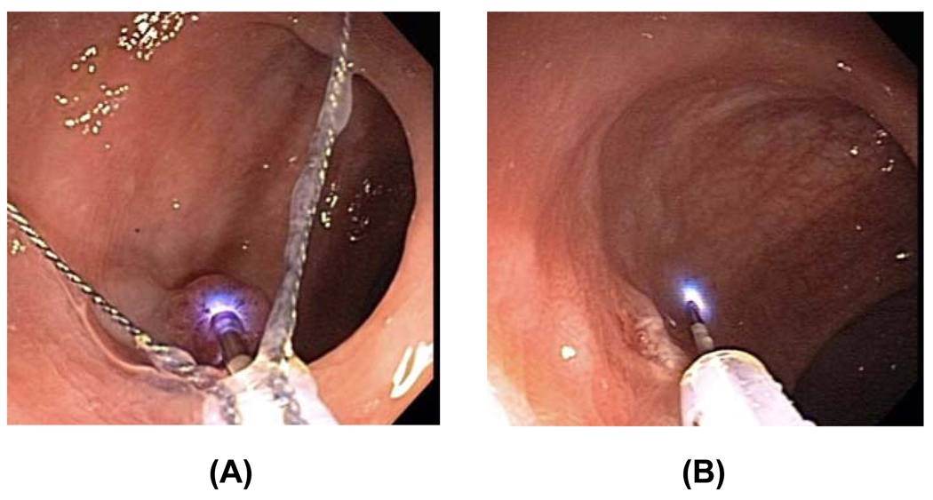
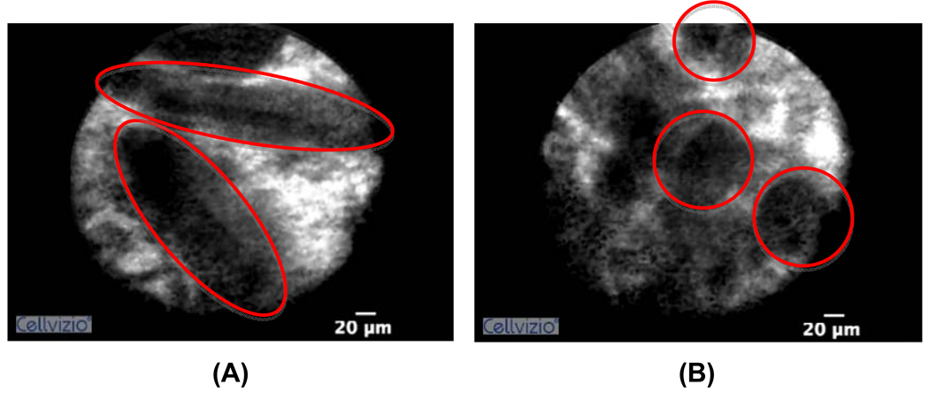
The confocal endomicroscopy snare is currently being used in a clinical study on patients undergoing routine colonoscopy. Once a polyp is identified using standard white-light endoscopy, the patient is subsequently given 1mL of fluorescein intravenously. The confocal snare is inserted through the working channel of the endoscope and used to examine every macroscopically visible lesion. The distal tip of the confocal probe is placed in gentle contact with the identified tissue and the position of the focal plane within the specimen is adjusted using the buttons on the endoscope control body (Figure 6A). The probe was used to confirm complete resection of the polyp (Figure 6B) once polypectomy has been finished. Video loops of each colonic polyp are obtained and stored as digital files using the Cellvizio platform. After image interpretation by an expert, the presence of neoplasia can be observed on the acquired images from the polyp (Figure 7A), as well as its absence once the polyp has been completely removed (Figure 7B).
Figure 6.
Confocal laser endomicroscopy snare. Imaging the polyp before polypectomy (A). Imaging the area after polypectomy to confirm complete resection (B).
Figure 7.
Confocal laser endomicroscopy images. Dysplasia present in the interrogated polyp as shown by highlighted villous structures (A). Image of normal colon mucosa after polypectomy with highlighted crypts (B).
4. Future Work
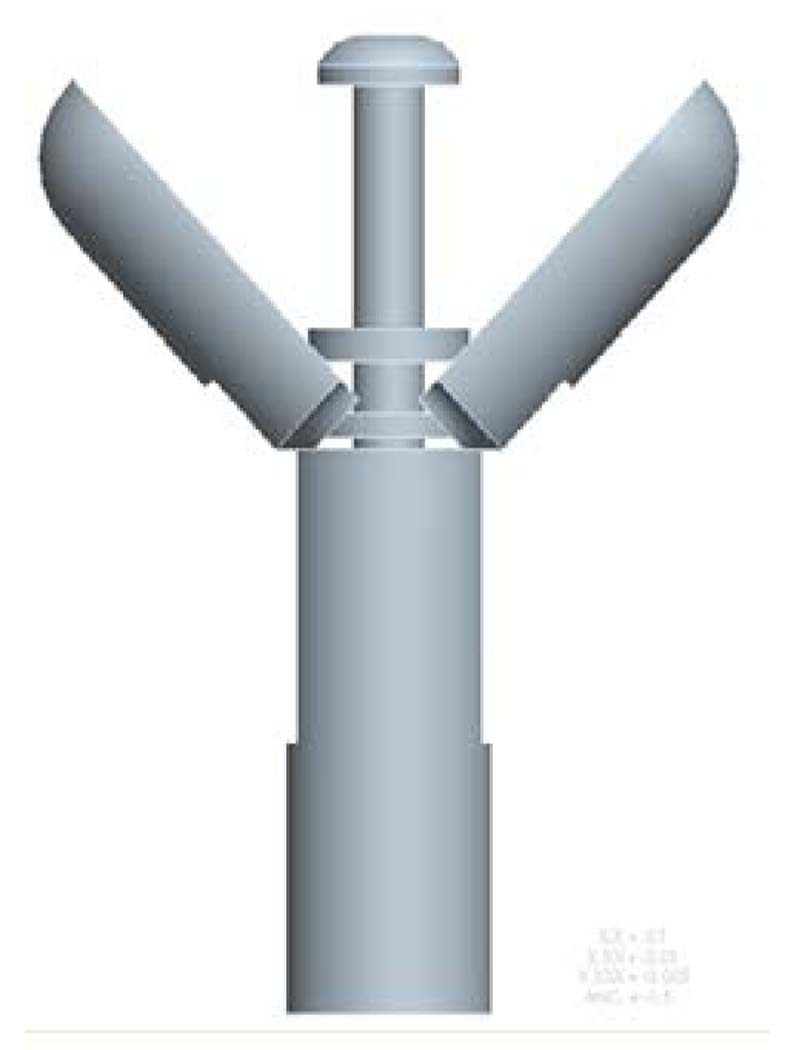
Research is currently being conducted in order to improve the current integrated spectroscopic devices. As stated earlier, each spectroscopic measurement takes about 30 milliseconds, and it is possible to perform several measurements per second, limited by the time to move the probe from spot to spot. With improved system engineering, we envision the performance of >30 measurements/sec that will provide “on-the fly” feedback to the clinician: As such, surveillance of large mucosal areas should be achievable using a rapid succession of point measurements while moving/scanning the probe over the mucosal surface. Currently a next-generation ESS integrated forceps is being developed and prototyped (Figure 8). Its design enables it to be dragged across the mucosa while providing continuous spectral interpretive feedback regarding the mucosa in contact with the tip, analogous to the way a metal detector or Geiger counter provides audible and/or visual feedback to the operator. A smooth mushroom-shaped tip with fibers at the center will permit sliding the probe across large patches mucosa without tears or trauma. Fast, real-time spectral processing will be based on the algorithms obtained as part of the studies.
Figure 8.
Novel CAM-action scanning optical forcep designs. “Mushroom-tipped” ESS fiber probed variations for contact sweeping / scanning large mucosal areas.
5. Conclusions
Beyond validation studies, real-time ESS measurements, as well as confocal laser endomicroscopy, would be clinically useful for increasing the pre-biopsy probability of obtaining neoplastic/dysplastic tissue over benign tissue. Our preliminary results have shown the promise of the different integrated optical tools in different screening/surveillance applications in the GI tract. In the case of colorectal cancer screening, these are tools for polyp classification not polyp detection, thus there would still be dependence on the endoscopists’ ability to detect lesions. ESS and confocal laser endomicroscopy would still be subjected to colonoscopy’s adenoma miss rates, estimated to be 24% in one study [60]. While a real-time, in vivo, classification tool like ESS would enable endoscopists to probe additional polypoid-looking lesions that he/she would otherwise not have thought were significant, the main impact of this kind of tool is the reduction in false positives, which based on current standards of care includes all lesions detected and sent for histopathology assessment that turn our to be benign. It is widely accepted that hyperplastic lesions with no malignant potential could potentially be left in situ if there were an accurate way to classify them in situ [61]. Thus, a real-time, in vivo, classification tool like ESS could help identify such lesions decreasing the number of unnecessarily resected polyps, saving time and cost. In addition, such integrated devices would be invaluable for dysplasia surveillance strategies that currently require large numbers of random biopsies, which include Barrett’s esophagus and colonic dysplasia in inflammatory bowel disease. In contrast to colorectal cancer screening, in Barrett’s esophagus surveillance these tools would serve more as detection tools, as dysplasia in Barrett’s segment is not clearly visible during white light endoscopy. Thus, an optical forceps could guide and refine “random” biopsies, increasing detection yield and decreasing the total number of biopsies required for a given screening session. As such, optical forceps would decrease the morbidity and overall cost of dysplasia surveillance.
Acknowledgements
We would like to thank Dr. Andre Sharon, Dr. Alexis Sauer-Budge, and their team of engineers at the Fraunhofer Center for Manufacturing Innovation in Boston, Ma. for their help and support in prototyping these optically integrated biopsy tools.
Footnotes
Publisher's Disclaimer: This is a PDF file of an unedited manuscript that has been accepted for publication. As a service to our customers we are providing this early version of the manuscript. The manuscript will undergo copyediting, typesetting, and review of the resulting proof before it is published in its final citable form. Please note that during the production process errors may be discovered which could affect the content, and all legal disclaimers that apply to the journal pertain.
References
- 1.Bigio IJ, Mourant JR. Ultraviolet and visible spectroscopies for tissue diagnostics: fluorescence spectroscopy and elastic-scattering spectroscopy. Physics in Medicine and Biology. 1997 May;vol. 42(no. 5):803–814. doi: 10.1088/0031-9155/42/5/005. [DOI] [PubMed] [Google Scholar]
- 2.Bakker Schut TC, Witjes MJ, Sterenborg HJ, Speelman OC, Roodenburg JL, Marple ET, Bruining HA, Puppels GJ. In vivo detection of dysplastic tissue by Raman spectroscopy. Analytical Chemistry. 2000 December 12;vol. 72(no. 24):6010–6018. doi: 10.1021/ac000780u. [DOI] [PubMed] [Google Scholar]
- 3.de Veld DCG, Skurichina M, Witjes MJH, Duin RPW, Sterenborg HJCM, Roodenburg JLN. Clinical study for classification of benign, dysplastic, and malignant oral lesions using autofluorescence spectroscopy. Journal of Biomedical Optics. 2004;vol. 9(no. 5):940–950. doi: 10.1117/1.1782611. [DOI] [PubMed] [Google Scholar]
- 4.Lin W, Yuan X, Yuen P, Wei WI, Sham J, Shi P, Qu J. Classification of in vivo autofluorescence spectra using support vector machines. Journal of Biomedical Optics. 2004;vol. 9(no. 1):180–186. doi: 10.1117/1.1628244. [DOI] [PubMed] [Google Scholar]
- 5.Majumder SK, Ghosh N, Gupta PK. Relevance vector machine for optical diagnosis of cancer. Lasers in Surgery and Medicine. 2005 April;vol. 36(no. 4):323–333. doi: 10.1002/lsm.20160. [DOI] [PubMed] [Google Scholar]
- 6.Majumder SK, Ghosh N, Gupta PK. Support vector machine for optical diagnosis of cancer. Journal of Biomedical Optics. 2005;vol. 10(no. 2):024034. doi: 10.1117/1.1897396. [DOI] [PubMed] [Google Scholar]
- 7.Chang SK, Mirabal YN, Atkinson EN, Cox D, Malpica A, Follen M, Richards-Kortum R. Combined reflectance and fluorescence spectroscopy for in vivo detection of cervical pre-cancer. Journal of Biomedical Optics. 2005;vol. 10(no. 2):024031. doi: 10.1117/1.1899686. [DOI] [PubMed] [Google Scholar]
- 8.Palmer GM, Zhu C, Breslin TM, Xu F, Gilchrist KW, Ramanujam N. Comparison of multiexcitation fluorescence and diffuse reflectance spectroscopy for the diagnosis of breast cancer (March 2003) IEEE Transactions on Biomedical Engineering. 2003 November;vol. 50(no. 11):1233–1242. doi: 10.1109/TBME.2003.818488. [DOI] [PubMed] [Google Scholar]
- 9.Ramanujam N, Mitchell MF, Mahadevan A, Thomsen S, Malpica A, Wright T, Atkinson N, Richards-Kortum R. Development of a multivariate statistical algorithm to analyze human cervical tissue fluorescence spectra acquired in vivo. Lasers in Surgery and Medicine. 1996;vol. 19(no. 1):46–62. doi: 10.1002/(SICI)1096-9101(1996)19:1<46::AID-LSM7>3.0.CO;2-Q. [DOI] [PubMed] [Google Scholar]
- 10.Ramanujam N, Mitchell MF, Mahadevan-Jansen A, Thomsen SL, Staerkel G, Malpica A, Wright T, Atkinson N, Richards-Kortum R. Cervical precancer detection using a multivariate statistical algorithm based on laser-induced fluorescence spectra at multiple excitation wavelengths. Photochemistry and Photobiology. 1996 October;vol. 64(no. 4):720–735. doi: 10.1111/j.1751-1097.1996.tb03130.x. [DOI] [PubMed] [Google Scholar]
- 11.Zhu C, Palmer GM, Breslin TM, Xu F, Ramanujam N. Use of a multiseparation fiber optic probe for the optical diagnosis of breast cancer. Journal of Biomedical Optics. 2005;vol. 10(no. 2):024032. doi: 10.1117/1.1897398. [DOI] [PubMed] [Google Scholar]
- 12.Mirabal YN, Chang SK, Atkinson EN, Malpica A, Follen M, Richards-Kortum R. Reflectance spectroscopy for in vivo detection of cervical precancer. Journal of Biomedical Optics. 2002 October;vol. 7(no. 4):587–594. doi: 10.1117/1.1502675. [DOI] [PubMed] [Google Scholar]
- 13.Utzinger U, Brewer M, Silva E, Gershenson D, Blast RC, Follen M, Richards-Kortum R. Reflectance spectroscopy for in vivo characterization of ovarian tissue. Lasers in Surgery and Medicine. 2001;vol. 28(no. 1):56–66. doi: 10.1002/1096-9101(2001)28:1<56::AID-LSM1017>3.0.CO;2-L. [DOI] [PubMed] [Google Scholar]
- 14.Wallace VP, Bamber JC, Crawford DC, Ott RJ, Mortimer PS. Classification of reflectance spectra from pigmented skin lesions, a comparison of multivariate discriminant analysis and artificial neural networks. Physics in Medicine and Biology. 2000 October;vol. 45(no. 10):2859–2871. doi: 10.1088/0031-9155/45/10/309. [DOI] [PubMed] [Google Scholar]
- 15.Bigio IJ, Bown SG, Briggs G, Kelley C, Lakhani S, Pickard D, Ripley PM, Rose IG, Saunders C. Diagnosis of breast cancer using elastic-scattering spectroscopy: preliminary clinical results. Journal of Biomedical Optics. 2000 April;vol. 5(no. 2):221–228. doi: 10.1117/1.429990. [DOI] [PubMed] [Google Scholar]
- 16.Clark BR, Johnson K, Mackenzie GD, Novelli MR, Selvasekar CR, Thorpe SM, Bown SG, Lovat LB, Bigio IJ. In-vivo detection of pre-cancerous changes in Barrett's esophagus using elastic scattering spectroscopy (ESS) Proceedings of SPIE. 2005;vol. 5862:58620B. [Google Scholar]
- 17.Johnson KS, Chicken DW, Pickard DCO, Lee AC, Briggs G, Falzon M, Bigio IJ, Keshtgar MR, Bown SG. Elastic scattering spectroscopy for intraoperative determination of sentinel lymph node status in the breast. Journal of Biomedical Optics. 2004;vol. 9(no. 6):1122–1128. doi: 10.1117/1.1802191. [DOI] [PubMed] [Google Scholar]
- 18.Mourant JR, Boyer J, Hielscher AH, Bigio IJ. Influence of the scattering phase function on light transport measurements in turbid media performed with small source-detector separations. Optics Letters. 1996 Apr 1;vol. 21(no. 7):546–548. doi: 10.1364/ol.21.000546. [DOI] [PubMed] [Google Scholar]
- 19.Perelman LT, Backman V, Wallace M, Zonios G, Manoharan R, Nusrat A, Shields S, Seiler M, Lima C, Hamano T, Itzkan I, Van Dam J, Crawford JM, Feld MS. Observation of periodic fine structure in reflectance from biological tissue: A new technique for measuring nuclear size distribution. Physical Review Letters. 1998 Jan 19;vol. 80(no. 3):627–630. [Google Scholar]
- 20.Mourant JR, Hielscher AH, Eick AA, Johnson TM, Freyer JP. Evidence of intrinsic differences in the light scattering properties of tumorigenic and nontumorigenic cells. Cancer. 1998 Dec 25;vol. 84(no. 6):366–374. [PubMed] [Google Scholar]
- 21.Mourant JR, Bigio IJ, Boyer J, Conn RL, Johnson T, Shimada T. Spectroscopic diagnosis of bladder cancer with elastic light scattering. Lasers in surgery and medicine. 1995;vol. 17(no. 4) doi: 10.1002/lsm.1900170403. [DOI] [PubMed] [Google Scholar]
- 22.Bigio IJ, Bown SG, Kelly CP, Lovat L, Pickarde D, PM R. Developments in endoscopic technology for oesophageal cancer. J. Royal Coll. Surg. Edinburgh. 2000:267–268. [Google Scholar]
- 23.Lovat L, Bown S. Elastic scattering spectroscopy for detection of dysplasia in Barrett's esophagus. Gastrointest Endosc Clin N Am. 2004 July;vol. 14(no. 3):507–517. doi: 10.1016/j.giec.2004.03.006. ix. [DOI] [PubMed] [Google Scholar]
- 24.Lovat LB, Johnson K, Mackenzie GD, Clark BR, Novelli MR, Davies S, O'Donovan M, Selvasekar C, Thorpe SM, Pickard D, Fitzgerald R, Fearn T, Bigio I, Bown SG. Elastic scattering spectroscopy accurately detects high grade dysplasia and cancer in Barrett's oesophagus. Gut. 2006 Aug;vol. 55(no. 8):1078–1083. doi: 10.1136/gut.2005.081497. [DOI] [PMC free article] [PubMed] [Google Scholar]
- 25.Lovat LB, Johnson K, Novelli MR, O'Donovan M, Davies S, Selvasekar CR, Thorpe S, Bigio IJ, Bown SG. Optical biopsy using elastic scattering spectroscopy can detect high grade dysplasia and cancer in Barrett's esophagus. Gastroenterology. 2004 Apr;vol. 126(no. 4):A22–a22. doi: 10.1136/gut.2005.081497. [DOI] [PMC free article] [PubMed] [Google Scholar]
- 26.Mackenzie GD, Johnson K, Novelli MR, O'Donovan M, Davies S, Selvasekar C, Thorpe S, Bigio IJ, Bown SG, Lovat LB. Optical biopsy using elastic scattering spectroscopy can detect dysplasia and cancer in Barrett's oesophagus, Gut. 2005 Apr;:A15–a15. doi: 10.1136/gut.2005.081497. [DOI] [PMC free article] [PubMed] [Google Scholar]
- 27.Dhar A, Johnson KS, Novelli MR, Bown SG, Bigio IJ, Lovat LB, Bloom SL. Elastic scattering spectroscopy for the diagnosis of colonic lesions: initial results of a novel optical biopsy technique. Gastrointestinal Endoscopy. 2006 February;vol. 63(no. 2):257–261. doi: 10.1016/j.gie.2005.07.026. [DOI] [PubMed] [Google Scholar]
- 28.Ge ZF, Schomacker KT, Nishioka NS. Identification of colonic dysplasia and neoplasia by diffuse reflectance spectroscopy and pattern recognition techniques. Applied Spectroscopy. 1998 Jun;vol. 52(no. 6):833–839. [Google Scholar]
- 29.Zonios G, Perelman LT, Backman V, Manahoran R, Fitzmaurice M, Van Dam J, Feld MS. Diffuse reflectance spectroscopy of human adenomatous colon polyps in vivo. Applied Optics. 1999;vol. 38:6628–6637. doi: 10.1364/ao.38.006628. [DOI] [PubMed] [Google Scholar]
- 30.Wallace MB, Perelman LT, Backman V, Crawford JM, Fitzmaurice M, Seiler M, Badizadegan K, Shields SJ, Itzkan I, Dasari RR, Van Dam J, Feld MS. Endoscopic detection of dysplasia in patients with Barrett's esophagus using light-scattering spectroscopy. Gastroenterology. 2000 Sep;vol. 119(no. 3):677–682. doi: 10.1053/gast.2000.16511. [DOI] [PubMed] [Google Scholar]
- 31.Kara MA, DaCosta RS, Streutker CJ, Marcon NE, Bergman JJ, Wilson BC. Characterization of tissue autofluorescence in Barrett's esophagus by confocal fluorescence microscopy. Dis Esophagus. 2007;vol. 20(no. 2):141–150. doi: 10.1111/j.1442-2050.2007.00660.x. [DOI] [PubMed] [Google Scholar]
- 32.Bouma BE, Tearney GJ, Compton CC, Nishioka NS. High-resolution imaging of the human esophagus and stomach in vivo using optical coherence tomography. Gastrointest Endosc. 2000 Apr;vol. 51(no. 4 Pt 1):467–474. doi: 10.1016/s0016-5107(00)70449-4. [DOI] [PubMed] [Google Scholar]
- 33.Brand S, Poneros JM, Bouma BE, Tearney GJ, Compton CC, Nishioka NS. Optical coherence tomography in the gastrointestinal tract. Endoscopy. 2000 Oct;vol. 32(no. 10):796–803. doi: 10.1055/s-2000-7714. [DOI] [PubMed] [Google Scholar]
- 34.Pitris C, Jesser C, Boppart SA, Stamper D, Brezinski ME, Fujimoto JG. Feasibility of optical coherence tomography for high-resolution imaging of human gastrointestinal tract malignancies. J Gastroenterol. 2000;vol. 35(no. 2):87–92. doi: 10.1007/s005350050019. [DOI] [PubMed] [Google Scholar]
- 35.Poneros JM, Brand S, Bouma BE, Tearney GJ, Compton CC, Nishioka NS. Diagnosis of specialized intestinal metaplasia by optical coherence tomography. Gastroenterology. 2001 Jan;vol. 120(no. 1):7–12. doi: 10.1053/gast.2001.20911. [DOI] [PubMed] [Google Scholar]
- 36.Sivak MV, Jr., Kobayashi K, Izatt JA, Rollins AM, Ung-Runyawee R, Chak A, Wong RC, Isenberg GA, Willis J. High-resolution endoscopic imaging of the GI tract using optical coherence tomography. Gastrointest Endosc. 2000 Apr;vol. 51(no. 4 Pt 1):474–479. doi: 10.1016/s0016-5107(00)70450-0. [DOI] [PubMed] [Google Scholar]
- 37.Wallace M, Van Dam J. Enhanced gastrointestinal diagnosis: light-scattering spectroscopy and optical coherence tomography. Gastrointest Endosc Clin N Am. 2000;vol. 10:71–80. [PubMed] [Google Scholar]
- 38.Kantsevoy SV, Adler DG, Conway JD, Diehl DL, Farraye FA, Kaul V, Kethu SR, Kwon RS, Mamula P, Rodriguez SA, Tierney WM. Confocal laser endomicroscopy. Gastrointest Endosc. 2009 August;vol. 70(no. 2):197–200. doi: 10.1016/j.gie.2009.04.002. [DOI] [PubMed] [Google Scholar]
- 39.Wang TD. Confocal microscopy from the bench to the bedside. Gastrointestinal Endoscopy. 2005 November;vol. 62(no. 5):696–697. doi: 10.1016/j.gie.2005.06.002. [DOI] [PMC free article] [PubMed] [Google Scholar]
- 40.Aisenberg J. Gastrointestinal endoscopy nears the molecular era. Gastrointestinal Endoscopy. 2008 September;vol. 68(no. 3):528–530. doi: 10.1016/j.gie.2008.03.1075. [DOI] [PubMed] [Google Scholar]
- 41.Kiesslich R, Neurath MF. Chromoendoscopy and Other Novel Imaging Techniques. Gastroenterology Clinics of North America. 2006 September;vol. 35(no. 3):605–619. doi: 10.1016/j.gtc.2006.07.004. [DOI] [PubMed] [Google Scholar]
- 42.Kiesslich R, Burg J, Vieth M, Gnaendiger J, Enders M, Delaney P, Polglase A, McLaren W, Janell D, Thomas S, Nafe B, Galle PR, Neurath MF. Confocal laser endoscopy for diagnosing intraepithelial neoplasias and colorectal cancer in vivo. Gastroenterology. 2004 September;vol. 127(no. 3):706–713. doi: 10.1053/j.gastro.2004.06.050. [DOI] [PubMed] [Google Scholar]
- 43.Hielscher AH, Mourant JR, Bigio IJ. Influence of particle size and concentration on the diffuse backscattering of polarized light from tissue phantoms and biological cell suspensions. Applied Optics. 1997 Jan 1;vol. 36(no. 1):125–135. doi: 10.1364/ao.36.000125. [DOI] [PubMed] [Google Scholar]
- 44.Loree TR, Bigio IJ, Spingler G. Medical applications of laser and optical technology. Journal of Laser Applications. 1992;vol. 4(no. 2):58. [Google Scholar]
- 45.Mourant JR, Bigio IJ, Boyer J, Johnson TM, Lacey JA, Bohorfoush AG, Mellow M. Elastic Scattering Spectroscopy as a Diagnostic Tool for Differentiating Pathologies in the Gastrointestinal Tract: Preliminary Testing. Journal of Biomedical Optics. 1996;vol. 1:192. doi: 10.1117/12.231372. [DOI] [PubMed] [Google Scholar]
- 46.Mourant JR, Fuselier T, Boyer J, Johnson TM, Bigio IJ. Predictions and measurements of scattering and absorption over broad wavelength ranges in tissue phantoms. Applied optics. 1997;vol. 36(no. 4):949–957. doi: 10.1364/ao.36.000949. [DOI] [PubMed] [Google Scholar]
- 47.Baak JP, ten Kate FJ, Offerhaus GJ, van Lanschot JJ, Meijer GA. Routine morphometrical analysis can improve reproducibility of dysplasia grade in Barrett's oesophagus surveillance biopsies. J Clin Pathol. 2002 Dec;vol. 55(no. 12):910–916. doi: 10.1136/jcp.55.12.910. [DOI] [PMC free article] [PubMed] [Google Scholar]
- 48.Fang H, Ollero M, Vitkin E, Kimerer LM, Cipolloni PB, Zaman MM, Freedman SD, Bigio IJ, Itzkan I, Hanlon EB, Perelman LT. Noninvasive sizing of subcellular organelles with light scattering spectroscopy. Ieee Journal of Selected Topics in Quantum Electronics. 2003 Mar–Apr;vol. 9(no. 2):267–276. [Google Scholar]
- 49.Reif R, Bigio IJ. Extracting optical properties in small superficial volumes of turbid media using tilted fibers. Proc. SPIE. 2007 Jan;vol. 6446 2007. [Google Scholar]
- 50.Pawley JB. Limitations on optical sectioning in live-cell confocal microscopy. Scanning. 2002;vol. 24(no. 5):241–246. doi: 10.1002/sca.4950240504. [DOI] [PubMed] [Google Scholar]
- 51.Delaney PM, Harris MR. Fiberoptics in confocal microscopy. Handbook of biological confocal microscopy. 1995:515–523. [Google Scholar]
- 52.Cellvizio®: a unique probe-based confocal laser endomicroscopy system. http://www.maunakeatech.com/products/cellvizio_GI/system_description.
- 53.Becker V, Vercauteren T, Hann von Weyhern C, Prinz C, Schmid RM, Meining A. High-resolution miniprobe-based confocal microscopy in combination with video mosaicing (with video) Gastrointestinal Endoscopy. 2007 November;vol. 66(no. 5):1001–1007. doi: 10.1016/j.gie.2007.04.015. [DOI] [PubMed] [Google Scholar]
- 54.von Delius S, Feussner H, Wilhelm D, Karagianni A, Henke J, Schmid RM, Meining A. Transgastric in vivo histology in the peritoneal cavity using miniprobe-based confocal fluorescence microscopy in an acute porcine model. Endoscopy. 2007 May;vol. 39(no. 5):407–411. doi: 10.1055/s-2007-966439. [DOI] [PubMed] [Google Scholar]
- 55.Wallace MB, Fockens P. Probe-Based Confocal Laser Endomicroscopy. Gastroenterology. 2009 May;vol. 136(no. 5):1509–1513. doi: 10.1053/j.gastro.2009.03.034. [DOI] [PubMed] [Google Scholar]
- 56.Yoshida S, Tanaka ST, Hirata M, Mouri R, Kaneko I, Oka S, Yoshihara M, Chayama K. Optical biopsy of GI lesions by reflectance-type laser-scanning confocal microscopy. Gastrointestinal Endoscopy. 2007 July;vol. 66(no. 1):144–149. doi: 10.1016/j.gie.2006.10.054. [DOI] [PubMed] [Google Scholar]
- 57.Duda RO, Hart PE, Stork DG. Pattern classification. New York: John Wiley & Sons; 2001. [Google Scholar]
- 58.Cortes C, Vapnik V. Support-vector networks. Machine Learning. 1995;vol. 20(no. 3):273–297. [Google Scholar]
- 59.Vapnik VN. Statistical learning theory. New York: John Wiley & Sons; 1998. [Google Scholar]
- 60.Rex DK, Cutler CS, Lemmel GT, Rahmani EY, Clark DW, Helper DJ, Lehman GA, Mark DG. Colonoscopic miss rates of adenomas determined by back-to-back colonoscopies. Gastroenterology. 1997 January;vol. 112(no. 1):24–28. doi: 10.1016/s0016-5085(97)70214-2. [DOI] [PubMed] [Google Scholar]
- 61.Ignjatovic A, East JE, Suzuki N, Vance M, Guenther T, Saunders BP. Optical diagnosis of small colorectal polyps at routine colonoscopy (Detect InSpect ChAracterise Resect and Discard; DISCARD trial): a prospective cohort study. Lancet Oncol. 2009 December;vol. 10(no. 12):1171–1178. doi: 10.1016/S1470-2045(09)70329-8. [DOI] [PubMed] [Google Scholar]


