
Figure 8.
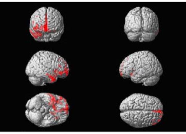
Voxel-based morphometry reveals that the areas of most significant atrophy are in the orbital and medial frontal and right greater than left orbitofrontal regions. The insula was involved as well.
Official websites use .gov
A
.gov website belongs to an official
government organization in the United States.
Secure .gov websites use HTTPS
A lock (
) or https:// means you've safely
connected to the .gov website. Share sensitive
information only on official, secure websites.
Voxel-based morphometry reveals that the areas of most significant atrophy are in the orbital and medial frontal and right greater than left orbitofrontal regions. The insula was involved as well.