Abstract
Deubiquitinating enzymes (DUbs) play important roles in many ubiquitin-dependent pathways, yet how DUbs themselves are regulated is not well understood. Here, we provide insight into the mechanism by which ubiquitination directly enhances the activity of ataxin-3, a DUb implicated in protein quality control and the disease protein in the polyglutamine neurodegenerative disorder, Spinocerebellar Ataxia Type 3. We identify Lys-117, which resides near the catalytic triad, as the primary site of ubiquitination in wild type and pathogenic ataxin-3. Further studies indicate that ubiquitin-dependent activation of ataxin-3 at Lys-117 is important for its ability to reduce high molecular weight ubiquitinated species in cells. Ubiquitination at Lys-117 also facilitates the ability of ataxin-3 to induce aggresome formation in cells. Finally, structure-function studies support a model of activation whereby ubiquitination at Lys-117 enhances ataxin-3 activity independent of the known ubiquitin-binding sites in ataxin-3, most likely through a direct conformational change in or near the catalytic domain.
Keywords: Deubiquitination, Enzymes, Neurodegeneration, Polyglutamine Disease, Ubiquitin, Ubiquitination, Spinocerebellar Ataxia Type 3, Ataxin-3
Introduction
Protein ubiquitination, a central regulator of diverse cellular events, is reversed by deubiquitinating enzymes (DUbs).3 As ubiquitin (Ub) linkage proteases, DUbs serve many functions: processing newly translated Ub to its mature form, editing Ub chains on proteins to facilitate their targeting to specific pathways, and maintaining a ready pool of Ub for conjugation to proteins (1–4). The relevance of DUbs to basic cellular pathways is underscored by their involvement in certain forms of cancer and neurodegenerative disease (4–7).
Despite recent progress in describing DUb function and regulation, general understanding of DUb physiology remains limited, and better knowledge of both their cellular functions and the mechanisms regulating their activity is needed. The human genome encodes ∼90 DUbs grouped into five major families (UCH, USP, MJD, OTU, and JAMM). As with other enzymes, the activity of DUbs must be regulated. DUbs are subject to regulation at many levels including transcription, degradation, substrate-induced conformational change (2–4), and post-translational modifications such as sumoylation and ubiquitination (8–10). Thus far the activity of only one DUb, ataxin-3, has been demonstrated to be directly regulated by its own ubiquitination (10). However, in light of increasing evidence of E3 ligase/DUb interactions (11–13), direct regulation of DUb activity by ubiquitination is an intriguing possibility that may apply more broadly to DUbs.
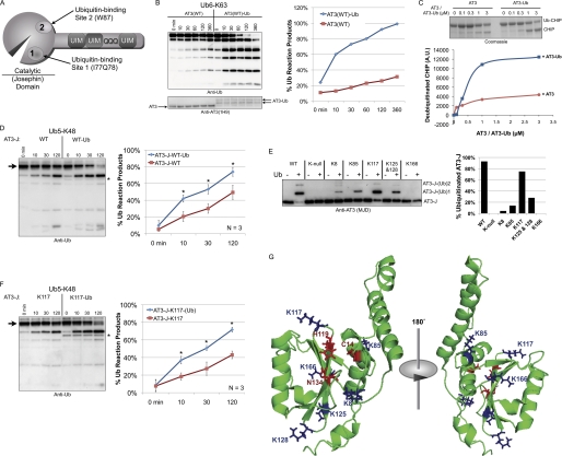
Ataxin-3 (AT3; see Fig. 1A), encoded by the ATXN3 gene in humans, is a member of the MJD family of DUbs (3). AT3 is a polyglutamine (poly(Q)) disease protein. When its poly(Q) tract is abnormally expanded, AT3 causes the neurodegenerative disorder Spinocerebellar Ataxia Type 3 (SCA3) (14–16). In vitro, AT3 preferentially cleaves longer Ub chains and Lys-63-linked chains over Lys-48-linked chains, yet it binds both types of chains (17, 18). Ub-interacting motifs (UIMs) in the C-terminal half of AT3 are required for its preferential cleavage properties and its capacity to bind more avidly to Ub chains containing at least four Ub molecules (18, 19). AT3 is a cytoprotective protein linked to protein quality control. In Drosophila, AT3 suppresses poly(Q)-induced neurodegeneration in a catalytic activity-dependent manner (20). Atxn3 knock-out mouse embryonic fibroblasts (MEFs) are more sensitive than wild type MEFs to heat shock (21). In cells, AT3 assists in the proteasomal targeting of endoplasmic reticulum-associated degradation substrates (22, 23) and induces aggresome formation (24).
FIGURE 1.
Lysine 117 in the Josephin domain is the preferred site of ubiquitination. A, ataxin-3 domain structure. The catalytic (Josephin) domain comprises the catalytic site and two Ub-binding sites. Site 1 is close to the catalytic groove, centered on residues Ile-77 and Gln-78; site 2, distant from the catalytic triad, is centered on Trp-87. The C-terminal half of AT3 contains three UIMs flanking the poly(Q) region. B, left, Ub6-Lys-63 chains were incubated with unmodified, wild type AT3 (AT3(WT)) or ubiquitinated, wild type AT3 (AT3(WT)-Ub), and fractions were collected at the indicated time points. Right, densitometry analysis of data from the left. The results are representative of over 10 similar independent experiments. Lys-63-linked chains were used as the substrate because they are cleaved preferentially to Lys-48-linked Ub chains by full-length AT3 in vitro (18). AT3-Ub species persist over time in DUb reactions because AT3 does not deubiquitinate itself completely (28). C, top, deubiquitination of monoubiquitinated CHIP by AT3 or AT3-Ub. CHIP was monoubiquitinated in vitro, purified, and then mixed with either unmodified AT3 or ubiquitinated AT3 for 10 min. Bottom, densitometry analysis of data from the top. The example is representative of three independently conducted experiments. A.U., arbitrary units. D, left, unmodified Josephin domain (AT3-J) and ubiquitinated AT3-J (prepared as in panel E) were incubated with Ub5-Lys-48 chains, and fractions were collected at the indicated time points. Right, densitometry analyses of the experiment from the left panel and other similar experiments, n = 3. Error bars indicate S.D. Asterisks: p < 0.05. Lys-48-linked chains were used as the substrate in panels D and F because they are cleaved preferentially to Lys-63-linked chains by the Josephin domain (17). E, GST-tagged, Josephin domain constructs (AT3-J) that were wild type (WT), lacking all six lysines (K-null), or retaining only specific lysine residues as indicated were ubiquitinated in vitro by CHIP-Ubch5c, affinity-purified, cleaved from GST, and analyzed by immunoblot. Bands correspond to unmodified and mono- and diubiquitinated AT3-J. These mixtures of ubiquitinated and unmodified AT3-J species were used in DUb reactions in panels D and F. Densitometry analyses of species in the left panel are shown on the right. Results are representative of independent experiments conducted at least three times. F, left, samples prepared as in panel E were incubated with Ub5-Lys-48 chains, and fractions were collected at the indicated time points. Right, densitometry analyses of the experiment from the left and other similar experiments. n = 3. Error bars indicate S.D. Asterisks: p < 0.05. For the left panels in D and F, the arrow denotes the disappearance of the starting substrate (Ub5-K48). The asterisk near blots denotes ubiquitinated AT3-J, which is detectable by anti-Ub antibody. G, solution structure of the Josephin domain highlighting catalytic residues (red) and lysine residues (blue; based on the structure from Ref. 34). AT3-J-K117-(Ub), ubiquitinated AT3-J containing only Lys-117.
The DUb activity of AT3 is directly regulated by its own ubiquitination. Ubiquitinated AT3 (AT3-Ub) cleaves Ub chains much more rapidly than does unmodified AT3 (10). Evidence suggests that AT3 activity is regulated during cell stress in part through ubiquitination. For example, the extent of AT3 ubiquitination increases when the proteasome is impaired, when overall cellular levels of ubiquitin are increased, or when the unfolded protein response is induced (10). Because pathogenic AT3 is more heavily ubiquitinated than wild type AT3 in a mouse model of SCA3 (10), AT3 ubiquitination may also influence disease pathogenesis in SCA3.
Here, we investigate how ubiquitination of AT3 enhances its activity and explore the importance of this post-translational modification to the physiological functions of AT3. Doing so is important because several DUbs are reportedly ubiquitinated, including JosD1, which belongs to the same DUb family as AT3 (8, 25–27, 29);4 consequently, direct regulation of DUb activity through ubiquitination may apply to DUbs other than AT3. Also, greater insight into the AT3 structure-function relationship and the mechanisms by which it is regulated will be important to fully understand the normal functions of AT3 and its dysfunction when the poly(Q) is expanded in the neurodegenerative disease, SCA3. Here, we identify Lys-117 as the preferred ubiquitination site on normal and pathogenic AT3. We determine that ubiquitination at this lysine is sufficient for ubiquitination-dependent activation of AT3, investigate the involvement of five Ub-binding domains on activation, and present evidence that ubiquitination at Lys-117 is important for the cellular functions of AT3.
EXPERIMENTAL PROCEDURES
Constructs
For mammalian cell expression, AT3 constructs were expressed in pCDNA3.1 or in pFLAG6A, whereas HA-Ub was expressed in pRK5. For recombinant protein expression, full-length and truncated AT3 constructs were expressed in pGEX6P1 or pET28a, and CHIP (carboxy terminus of Hsc70-interacting protein) in pGEX6P1. UbcH5c is Addgene plasmid 12643. QuikChange mutagenesis reagents and primer design software (Stratagene) were used for site-directed mutagenesis of AT3 lysine residues to arginine and vice versa. AT3 site 1 and site 2 mutant constructs have been described elsewhere (17).
Cell Culture Transfections and Lysate Preparation
Mammalian cells were transfected using Lipofectamine LTX with Plus (Invitrogen) per the manufacturer's instructions. Cells were harvested in boiling SDS (2%) buffer supplemented with DTT (100 mm) 24–48 h after transfection. Lysates were boiled for 5 min, brought to room temperature, sonicated, centrifuged, and loaded into 10 or 15% SDS-PAGE gels. Where needed, cells were treated with lactacystin (10–15 μm; Boston Biochem), MG132 (15 μm; Boston Biochem), or vehicle (dimethyl sulfoxide (DMSO); Sigma-Aldrich), as indicated in figures and text.
Antibodies
The following antibodies were used: rabbit polyclonal anti-Ub (1:500; Dako); mouse monoclonal anti-AT3 (1:500–1,000; 1H9; generous gift of Dr. Yvon Trottier); rabbit polyclonal anti-MJD (1:40,000; (30)); rabbit polyclonal anti-HA (1:500; Y-11; Santa Cruz Biotechnology); goat polyclonal anti-GST (1:10,000; GE Healthcare); peroxidase-conjugated, goat anti-rabbit, goat anti-mouse, and rabbit anti-goat secondary antibodies (1:15,000; Jackson ImmunoResearch). Fluorescence-conjugated secondary antibodies for immunofluorescence (goat anti-mouse and goat anti-rabbit Alexa Fluor; Invitrogen) were used at 1:2,000.
Western Blotting and Quantification
Western blotting was conducted as described previously (18). Western blots were imaged using a VersaDoc 5000 MP (Bio-Rad). For semiquantification, images collected below saturation on the VersaDoc were quantified with the Quantity One software (Bio-Rad) (10). Student's t test was used for statistical analyses.
Immunofluorescence
Immunofluorescence was conducted as described previously (28). Briefly, cells were fixed in 4% paraformaldehyde in PBS, rinsed and permeabilized with PBS plus 0.1% Triton X-100, blocked in 3% bovine albumin, and stained overnight in primary antibody (mouse anti-AT3 (1H9; 1:500) and/or rabbit anti-Ub (Dako; 1:500)). Nuclei were labeled with DAPI (Invitrogen). Fluorescence was visualized with an Olympus IX-81 compound microscope. Transfected cells with or without aggregates were counted by a blinded investigator.
In Vitro Ubiquitination Reactions and AT3-Ub Preparation
Recombinant protein was prepared as described elsewhere (18, 28). For generation of recombinant AT3-Ub, GST- or His6-tagged AT3 species were incubated for 120 min at 37 °C with CHIP, E1, E2 (Ubch5c), Ub (Sigma-Aldrich), and ATP/MgCl2 in kinase reaction buffer (50 mm Tris, pH 7.5, 50 mm KCl, 0.2 mm DTT). Non-ubiquitinated counterpart AT3 proteins were prepared as above, in the absence of ATP/MgCl2. Next, reactions were incubated with glutathione-Sepharose beads (GE Healthcare) for GST-tagged AT3 or with nickel-nitrilotriacetic acid beads (Qiagen) for His6-AT3, in RIPA buffer (50 mm Tris, 150 mm NaCl, 0.1% SDS, 0.5% deoxycholic acid, 1% Nonidet P-40, pH 7.4) for GST-AT3 or in Buffer A (50 mm Tris, pH 7.5, 150 mm NaCl) for His6-AT3. Beads were rinsed 5–9 times with their respective incubation buffers and another three times with DUb reaction buffer (50 mm HEPES, 0.5 mm EDTA, 1 mm DTT, 0.1 mg/ml ovalbumin, pH 7.5). GST-tagged AT3 was eluted by PreScission protease (GE Healthcare) in DUb reaction buffer, whereas His6-AT3 was eluted with 300 mm imidazole supplemented with 1% DTT in Buffer C (50 mm Tris, pH 7.5, 100 mm NaCl, 20 mm Imidazole, 0.5% TX-100). Protein was quantified using serial dilutions, Coomassie Brilliant Blue staining, and UV spectrophotometer (NanoDrop; Thermo Scientific). Where noted in the figure legends, GST-AT3 was maintained on beads and used immediately in select DUb reactions.
Deubiquitination Reactions
All ubiquitinated and unmodified AT3 species used in DUb reactions underwent the same preparation conditions. Proteins used in DUb reactions were quantified prior to use through Coomassie Brilliant Blue staining and UV spectrophotometry (NanoDrop; Thermo Scientific). 300–400 nm Ub chains (Boston Biochem) were incubated in DUb reaction buffer with 50–150 nm AT3 species for a maximum of 6 h at 37 °C. Fractions were collected in 2% SDS, 100 mm DTT, boiled for 1 min, and loaded into 4–20 or 15% SDS-PAGE gels. Unless otherwise noted in figures and figure legends, AT3 species used in DUb reactions were untagged. In our hands, AT3 does not cleave fluorescent probes such as Ub-AMC; therefore, we have not employed this or other fluorescent probes linked to a single Ub moiety.
Protein Preparation for MS Analyses
For recombinant AT3, GST-tagged AT3 was ubiquitinated in vitro as described above, purified using glutathione-Sepharose beads, rinsed in RIPA buffer (6 rinses) and DUb reaction buffer (3 rinses), and eluted with PreScission protease (GE Healthcare). For AT3-Ub prepared from cells, FLAG-AT3 and HA-Ub were co-transfected in Cos-7 cells in 10-cm plates using Lipofectamine LTX with Plus reagent (Invitrogen). 48 h after transfection, cells were harvested in ice-cold PBS and lysed in RIPA buffer supplemented with SIGMAFAST protease inhibitor cocktail (Sigma-Aldrich). Cells were sonicated and spun at 16,000 × g at 4 °C, and FLAG-tagged AT3 was purified using anti-FLAG antibody-bound beads (M2; Sigma-Aldrich) for 3 h at 4 °C. Beads were rinsed with RIPA + protease inhibitor (three quick rinses and five 5-min rinses). Bound protein was eluted with 3× FLAG peptide (Sigma-Aldrich) in RIPA buffer. Eluted protein was mixed with NuPAGE LDS sample denaturing buffer (Invitrogen) and oxidizing reagent (Invitrogen), incubated at 70 °C for 10 min, and electrophoresed in 4–12% NuPAGE Gels (Invitrogen). Coomassie Brilliant Blue staining was performed using mass spectrometry-compatible NOVEX Coomassie Blue colloidal staining (Invitrogen) as instructed by the manufacturer.
Protein Identification by LC-MS/MS
Protein identification and ubiquitination sites on AT3 were conducted based on previously described protocols (31, 32). Briefly, protein bands corresponding to unmodified and modified AT3 were excised and destained with 30% methanol for 4 h. Upon reduction (10 mm DTT) and alkylation (65 mm 2-chloroacetamide or iodoacetamide, with similar results) of the cysteines, proteins were digested overnight with sequencing grade, modified trypsin (Promega). Resulting peptides were resolved on a nano-capillary reverse phase column (Picofrit column, New Objective) using a 1% acetic acid/acetonitrile gradient at 300 nl/min and directly introduced into a linear ion-trap mass spectrometer (LTQ Orbitrap XL, Thermo Fisher). Data-dependent MS/MS spectra on the five most intense ions from each full MS scan were collected (relative Collision Energy ∼35%). Proteins were identified by searching the data against Human International Protein Index database (version 3.5) appended with decoy (reverse) sequences using the X!Tandem/Trans-Proteomic Pipeline (TPP) software suite. All peptides and proteins with a PeptideProphet and ProteinProphet probability score of >0.9 (false discovery rate <2%) were considered positive identifications and manually verified.
RESULTS
Lysine 117 of AT3 Is Preferentially Ubiquitinated in Vitro
To understand how AT3 ubiquitination enhances its DUb activity, we first investigated ubiquitination sites on AT3 in vitro. AT3 interacts with CHIP (33), an E3 Ub ligase that functions with heat shock proteins to target misfolded proteins for proteasomal degradation. Thus, we used CHIP as the E3 ligase for AT3 in vitro, with Ubch5c serving as the E2. As shown previously, the ability of AT3 to cleave Lys-63-linked Ub chains (a preferred Ub chain substrate for AT3 in vitro (18)) was markedly enhanced when AT3 was ubiquitinated by CHIP-Ubch5c (Fig. 1B) (10). Ubiquitinated AT3 also removed Ub from monoubiquitinated CHIP more readily than did unmodified AT3 (Fig. 1C).
Ubiquitination of the isolated Josephin domain of AT3 enhances its DUb activity, although not nearly as robustly as with full-length AT3 (Fig. 1D) (10). Accordingly, we first investigated which of the six lysine residues in the Josephin domain are preferentially ubiquitinated by CHIP-Ubch5c. We generated a Josephin construct (denoted AT3-J) in which all lysines were mutated to the similar, non-ubiquitinatable amino acid arginine (K-null) and a series of AT3-J constructs containing single lysine residues. As shown in Fig. 1E, lysine at position 117 (Lys-117) of AT3-J was the preferred site for in vitro ubiquitination by CHIP-Ubch5c. Moreover, ubiquitination at Lys-117 alone was sufficient to enhance DUb activity of the isolated Josephin domain (Fig. 1F). The solution structure of the Josephin domain (34) indicates that Lys-117 resides near the catalytic triad (Fig. 1G).
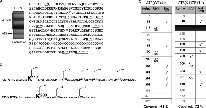
To determine whether full-length AT3 is also preferentially ubiquitinated at Lys-117, we generated recombinant, full-length AT3, ubiquitinated it with CHIP-Ubch5c (Fig. 2A), and subjected it to liquid chromatography-tandem mass spectrometry (LC-MS/MS) analyses. Full-length AT3 also proved to be preferentially ubiquitinated at Lys-117 (Fig. 2, B and C). Ubiquitination also occurred less frequently at Lys-190, Lys-200, Lys-206, and Lys-291, the latter of which resides next to the poly(Q) tract (Fig. 2A). When Lys-117 was mutated to arginine, Lys-200 was ubiquitinated more frequently (Fig. 2, B and C). These results show that Lys-117 is the preferred site for ubiquitination of AT3 by CHIP-Ubch5c in vitro.
FIGURE 2.
Lys-117 is the preferred ubiquitination site for full-length AT3. A, left, GST-tagged, wild type AT3 (AT3(WT)) was ubiquitinated in vitro by CHIP-Ubch5c, purified, and cleaved from GST. The picture shows a Coomassie Blue stain of the AT3 bands analyzed by LC-MS/MS. Right, amino acid sequence of AT3. Underlined and bolded letters indicate lysine residues and arginine residues, respectively. Trypsin digests for MS analyses occur at the Lys and Arg residues. B, top, summary of LC-MS/MS analyses of ubiquitinated, wild type AT3 (AT3(WT)-Ub), which showed that AT3 is predominantly ubiquitinated at Lys-117. Ubiquitination was also observed less frequently at Lys-190, Lys-200, Lys-206, and Lys-291 among different runs. LC-MS/MS analyses were conducted on three different samples, with Lys-117 being the predominantly ubiquitinated species in each run. Bottom, for AT3 with Lys-117 mutated to arginine and ubiquitinated in vitro (AT3(K117R)-Ub), Lys-200 became the predominant site of ubiquitination. C, sample of data collected by LC-MS/MS. Fractions indicate number of ubiquitinated (Ub'd) lysine hits per total number of peptides identified for that run.
Ubiquitination of Lys-117 Is Sufficient to Increase DUb Activity of AT3
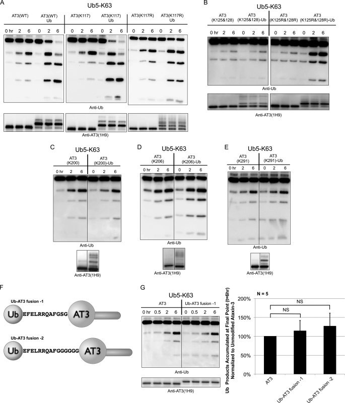
We next determined whether ubiquitination of Lys-117 is sufficient for activation of full-length AT3. Recombinant AT3 lacking all lysines except Lys-117 cleaved ubiquitin chains more rapidly when ubiquitinated (Fig. 3A). Thus, ubiquitination at Lys-117 is sufficient to enhance the catalytic activity of full-length AT3, similarly to ubiquitinated, wild type AT3. When Lys-117 was mutated to arginine (AT3(K117R)), we still observed ubiquitination-dependent activation of AT3, although not as robustly as with AT3 lacking all lysines except Lys-117 or with wild type AT3 (Fig. 3A). Thus, ubiquitination at one or more additional sites (e.g. Lys-190, Lys-200, Lys-206, or Lys-291; Fig. 2) is capable of enhancing AT3 DUb activity. However, when AT3 constructs possessing single lysine residues were ubiquitinated in vitro and tested for activation, we observed inefficient ubiquitination and no obvious increase in activity toward Ub chains by the mixtures of unmodified and ubiquitinated species of AT3 (Fig. 3, B–E).
FIGURE 3.
Ubiquitination at Lys-117 is sufficient for activation of AT3. A, wild type AT3 (AT3(WT)), AT3 containing Lys-117 but no other lysines (AT3(K117)), or AT3 mutated only at Lys-117 (AT3(K117R)) was ubiquitinated in vitro, purified, and then incubated with Ub5-Lys-63 chains to monitor DUb activity when compared with unmodified counterparts. Fractions were collected at the indicated time points. Examples are representative of five independent experiments each. B, AT3 containing Lys-125 and Lys-128 but no other lysine residues (AT3(K125&128)) or AT3 mutated only at Lys-125 and Lys-128 (AT3(K125R&128R)) was ubiquitinated in vitro and then incubated with Ub5-Lys-63 chains to monitor DUb activity when compared with unmodified counterparts. Fractions were collected at the indicated time points. Examples are representative of five independent experiments. C–E, AT3 containing only the individual lysines indicated were ubiquitinated in vitro, and DUb activity toward Ub5-Lys-63 chains was compared with the unmodified AT3 counterparts. Examples are representative of independent experiments conducted at least three times each. AT3(K200), AT3 containing Lys-200 but no other lysine residues; AT3(K206)-Ub, ubiquitinated AT3 containing Lys-206 but no other lysine residues; AT3(K206), AT3 containing Lys-206 but no other lysine residues; AT3(K291), AT3 containing Lys-291 but no other lysine residues; AT3(K291)-Ub, ubiquitinated AT3 containing Lys-291 but no other lysine residues. F, Ub-AT3 fusion constructs used in DUb reactions. G, left, DUb activity of unmodified AT3 or an Ub-AT3 fusion toward Ub5-Lys-63 chains was assessed for the indicated time points. Right, densitometry analyses of DUb reaction products at the final time point (6 h) from the left panel and other similar experiments. Shown are means normalized to unmodified AT3, ± S.D.; n = 5. Results are not significantly different (NS) from one another (p > 0.1). AT3(K200)-Ub, ubiquitinated AT3 with only Lys-200 present.
To investigate whether quantitative ubiquitination of AT3 outside of Lys-117 can enhance DUb activity, we generated and tested the activity of two linear Ub-AT3 fusion proteins, one containing a longer, glycine-rich linker between Ub and AT3 (Fig. 3F and supplemental Fig. 1). DUb activity of AT3 was not enhanced with either Ub fusion (Fig. 3G). These results establish that ubiquitination at Lys-117 is sufficient for activation of AT3 and that ubiquitination of AT3 must occur at a specific site to enhance its activity.
Ubiquitination of Wild Type and Poly(Q)-expanded AT3 Occurs Preferentially at Lys-117 in Cells
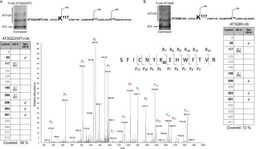
Ubiquitinated AT3 purified from cells shows enhanced DUb activity (10). To determine which lysine residues of wild type AT3 are ubiquitinated in cells, we co-expressed FLAG-tagged AT3 with Ub in Cos-7 cells, immunopurified ubiquitinated AT3, and subjected it to LC-MS/MS. Similar to recombinant AT3, AT3 isolated from cells was ubiquitinated predominantly at Lys-117 (Fig. 4A).
FIGURE 4.
Normal and poly(Q)-expanded AT3 are both preferentially ubiquitinated at Lys-117 in cells. FLAG-tagged, wild type AT3 (panel A; FLAG-AT3Q22(WT)) or expanded AT3 (panel B; FLAG-AT3Q80) was co-expressed with Ub in Cos-7 cells. Cells were harvested 24–48 h after transfection, and FLAG-AT3 was immunopurified with anti-FLAG antibody. For both panels, top, Coomassie Blue staining of the bands analyzed by LC-MS/MS and ubiquitinated species that were identified. Bottom, example of data collected by LC-MS/MS. Fractions indicate the number of ubiquitinated (Ub'd) lysine hits per total number of peptides identified for that run. The MS/MS spectrum in panel A is representative of cellular AT3 ubiquitinated on Lys-117 (see “Experimental Procedures” for details). The inset shows the ubiquitinated peptide (amino acids 111–124) encompassing the modified Lys-117. Observed b and y ion series are indicated. Diglycine signature, characteristic of the ubiquitinated tryptic peptides, is denoted by Kgg. LC-MS/MS analyses were conducted on two different samples each, and Lys-117 was consistently the predominantly ubiquitinated species for both wild type and pathogenic AT3.
We also determined the lysine residues at which pathogenic (poly(Q)-expanded) AT3 (AT3Q80) is ubiquitinated in cells. Expanded AT3 was also predominantly ubiquitinated at Lys-117 (Fig. 4B). Unlike with AT3Q22(WT), however, ubiquitination of AT3Q80 at Lys-200 was not observed, although the peptide sequence encompassing Lys-200 was identified numerous times from ubiquitinated AT3Q80. Thus, although wild type and pathogenic AT3 are both primarily ubiquitinated at Lys-117 in cells, there are differences between wild type and pathogenic AT3 in ubiquitination at other sites, which may relate to disease pathogenesis in SCA3.
Testing the Role of Ub-binding Domains on Ubiquitination-dependent Activation
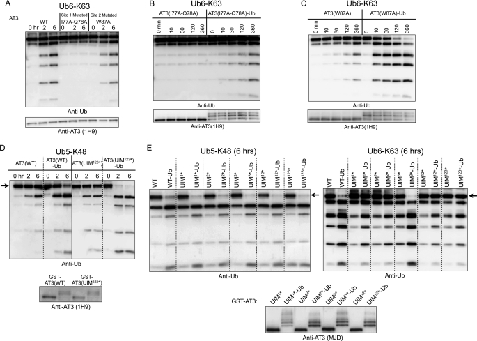
Five independent Ub-binding domains have been identified in AT3: three well established UIMs in the C-terminal region (35–38), mutations in which abolish the ability of AT3 to bind Ub chains with higher affinity (10, 18), and two recently described Ub-binding sites in the catalytic Josephin domain, site 1 and site 2 (39) (Fig. 1A). Site 1 is near the catalytic triad, centered on two adjacent amino acids, Ile-77 and Gln-78. AT3 with Ile-77 and Gln-78 mutated to alanine residues shows no cleavage of Lys-63-linked Ub chains during the time frame of our in vitro assays (up to 6 h, Fig. 5A) (17). Site 2, centered on Trp-87, is physically remote from the catalytic site. Mutating this residue to alanine (W87A) does not noticeably alter AT3 activity toward Lys-63-linked Ub chains (Fig. 5A). Mutations of sites 1 and 2 to alanine are expected to reduce their ability to bind Ub (17).
FIGURE 5.
Ubiquitin-binding domains on AT3 are not necessary for ubiquitination-dependent activation. A, Ub6-Lys-63 chains were incubated with recombinant wild type AT3 (WT), AT3 with site 1 mutated (I77A/Q78A), or AT3 with site 2 mutated (W87A). Fractions were collected at the indicated time points. B and C, unmodified AT3 or ubiquitinated AT3 with site 1 (B) or site 2 (C) mutated was incubated with Ub6-Lys-63 chains, and fractions were collected as indicated. Examples in panels A–C are representative of at least four independent experiments each. D, GST-tagged, wild type AT3 (AT3(WT)) or AT3 with all three UIMs mutated (UIM123*) was ubiquitinated in vitro, and DUb activity toward Lys-48-linked, penta-Ub (Ub5-Lys-48) chains was compared with the activity of the respective, unmodified AT3 counterparts. E, unmodified or in vitro ubiquitinated, GST-tagged wild type AT3 (WT) or AT3 with single UIMs mutated (UIM*) was incubated with Ub5-Lys-48 chains (left) or Ub6-Lys-63 chains (right). Only the final time point (6 h) is shown for each reaction (an intermediate time point is shown in supplemental Fig. 2). The arrows in panels D and E indicate loss of the main substrate band during reactions. Anti-AT3 blots in panels A–E show the enzymes used in each reaction. Examples in panels D and E are representative of experiments repeated at least three times each. AT3(I77a-Q78A), AT3 with site 1 mutated; AT3(I77A-Q78A)-Ub, ubiquitinated AT3 with site 1 mutated; AT3(W87A), AT3 with site 2 mutated; AT3(W87A)-Ub, ubiquitinated AT3 with site 2 mutated.
We investigated whether either site 1 or site 2 is involved in AT3 activation by ubiquitination. When site 2 is mutated, ubiquitination of AT3 still markedly enhanced its activity toward Ub chains (Fig. 5C), similar to wild type AT3, indicating that site 2 is not necessary for activation. Remarkably, however, site 1-mutated AT3, which normally shows no DUb activity (Fig. 5A), did show slight cleavage of Ub chains when it was ubiquitinated (Fig. 5B). AT3 with site 1 residues mutated to alanine may retain subtle DUb activity that is not detectable in our standard assays but that, upon AT3 ubiquitination, is amplified and thus observable by Western blotting (Fig. 5B). This result further suggests that site 1 is also not strictly necessary for Ub-dependent activation of AT3, although it is a critical determinant of overall AT3 activity.
The UIMs of AT3 are known to modulate its cleavage preference toward Lys-63 Ub chains; although the isolated Josephin domain preferentially cleaves Lys-48-linked Ub, full-length AT3 preferentially cleaves Lys-63-linked Ub chains (17, 18). UIMs also modulate the cleavage properties of ubiquitinated AT3; AT3-Ub in which all three UIMs are mutated shows less robust activation toward Lys-63-linked chains than does wild type AT3-Ub (10). UIMs, however, are not strictly necessary for ubiquitination-dependent activation because ubiquitinated AT3 with all three UIMs mutated still shows enhanced DUb activity, particularly toward Lys-48-linked chains (Fig. 5D). Thus, although the three UIMs collectively are not required for ubiquitination-dependent activation, they do modulate the ability of ubiquitinated AT3 to cleave Lys-63 chains.
Because it is unknown how individual UIMs affect cleavage properties of AT3, we tested the importance of each UIM on the ability of ubiquitinated AT3 to cleave Lys-48- versus Lys-63-linked chains. Ubiquitinated AT3 mutated in UIM1, UIM2, or both UIM1 and UIM2 showed only modestly enhanced activity toward Lys-63 chains but continued to show markedly enhanced activity toward Lys-48 chains (Fig. 5E and supplemental Fig. 2). However, ubiquitinated AT3 in which only UIM3 is mutated behaved essentially identically to ubiquitinated, wild type AT3, showing enhanced activity toward both Lys-63-linked and Lys-48-linked Ub chains. We conclude that UIM1 and UIM2 each confer upon ubiquitinated AT3 an increased ability to cleave Lys-63-linked Ub chains, whereas UIM3 is dispensable for this activity.
In summary, none of the five identified Ub-binding sites on AT3 appear strictly necessary for ubiquitination-dependent activation of this DUb. These results also support the documented role of UIMs in modulating AT3 cleavage of Lys-63-linked chains (10, 18).
Ubiquitination Modulates Cellular Properties of AT3
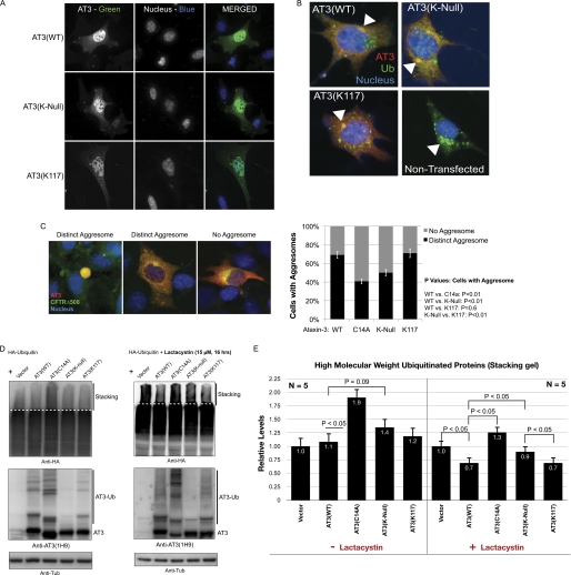
AT3 has been implicated in various protein quality control pathways (20–23, 35, 36, 40). To begin exploring the functional role of AT3 ubiquitination in cells, we first investigated whether lysines on AT3 are important for its subcellular localization under normal cellular conditions. To eliminate confounding effects from endogenous AT3, we employed Atxn3 knock-out MEFs (21) transiently transfected with empty vector or vector expressing wild type AT3 (AT3(WT)), lysine-less AT3 (AT3(K-Null)), or AT3 with only Lys-117 present (AT3(K117)). In MEFs expressing plasmids at low levels, AT3 localized to both the cytoplasm and the nucleus but was enriched in the nucleus (Fig. 6A). Lysine-less AT3 and Lys-117-only AT3 showed a similar subcellular localization, indicating that lysines on AT3 do not affect its localization in fibroblasts (Fig. 6A).
FIGURE 6.
AT3 ubiquitination is important for its cellular functions. A, Atxn3 knock-out MEFs were transiently transfected with plasmids encoding wild type AT3 (AT3(WT)), AT3 with no lysines present (AT3(K-Null)), or AT3 with only Lys-117 present (AT3(K117)). Cells were fixed 48 h after transfection and immunolabeled with anti-AT3 monoclonal 1H9 antibody and polyclonal anti-Ub antibody. B, Atxn3 knock-out MEFs were transfected as in panel A. Cells were treated with the proteasome inhibitor lactacystin (15 μm) for 10 h before fixation and immunolabeling as indicated. The arrowheads indicate perinuclear, ubiquitin-positive aggresomes that are also AT3-immunoreactive in cells expressing the indicated AT3 constructs. C, Atxn3 knock-out MEFs were co-transfected with GFP-CFTRΔ508 and AT3 constructs as indicated. 48 h after transfection, cells were treated for 4 h with the proteasome inhibitor MG132 (15 μm), fixed, stained with monoclonal anti-AT3 antibody (1H9), and visualized. Left, types of GFP-CFTRΔ508 aggregates observed in MEFs. Right, quantification of data from three similar, independent experiments. Error bars indicate S.D. Only cells co-expressing GFP-CFTRΔ508 and AT3 were counted. D, Cos-7 cells were transfected as indicated, treated with or without the proteasomal inhibitor lactacystin (15 μm for 16 h) at 48 h after transfection, and analyzed by immunoblot. The area above the dotted lines is the stacking portion of the gel and the focus of our analyses. AT3(C14A), catalytically inactive AT3. E, densitometry analyses of ubiquitinated species in the stacking gel portion from panel D and other similar experiments. n = 5. Error bars indicate S.E.
AT3 promotes aggresome formation in mammalian cells (24). Also, in postmortem brain tissue from various neurodegenerative diseases, non-pathogenic AT3 colocalizes to inclusions formed by various disease proteins (41–44). We thus investigated AT3 localization to ubiquitinated aggregates by transfecting Atxn3 knock-out MEFs with different forms of AT3 and inhibiting the proteasome, which induces aggresome production. Large, Ub-positive aggresomes in MEFs were also immunoreactive for AT3 (Fig. 6B). Thus, AT3 localizes to Ub-positive aggresomes independent of lysine residues.
To determine whether ubiquitination of AT3 contributes to its role in aggresome formation (24), we co-expressed the aggregation-prone protein, CFTRΔ508, with various AT3 constructs in Atxn3 knock-out MEFs. Upon proteasome inhibition, GFP-tagged CFTRΔ508 aggregated in a subset of MEFs (Fig. 6C and data not shown). Catalytically inactive and non-ubiquitinatable AT3 both led to fewer cells with CFTRΔ508 aggresomes than did wild type AT3 and AT3 containing only Lys-117 (Fig. 6C). All AT3 constructs co-localized with GFP-CFTRΔ508 aggresomes, and we did not observe differences in aggresome localization among different AT3 constructs (data not shown). Similar results were obtained with another protein, GFP-iNOS (inducible nitric oxide synthase), upon proteasome inhibition (data not shown). Together, these results suggest that ubiquitination-dependent activation of AT3 enhances its ability to promote aggresome formation.
AT3 expression leads to lower levels of ubiquitinated proteins in cells and mouse tissue including brain (18, 45). To examine whether ubiquitination of AT3 contributes to this function, we co-expressed Ub and different forms of AT3 in Cos-7 cells, which were then treated with or without the proteasome inhibitor, lactacystin (Fig. 6D). In unstressed cells, expression of catalytically inactive AT3 (AT3(C14A)) led to significantly higher levels of high molecular weight (HMW) Ub species in the stacking portion of the gel (Fig. 6D, above the dotted line; and 6E). Expression of non-ubiquitinatable AT3 also trended toward higher levels of HMW but did not reach statistical significance (Fig. 6E). Upon proteasome inhibition, however, expression of non-ubiquitinatable AT3 led to statistically significant higher levels of HMW Ub species in the stacking gel (Fig. 6, D and E). The presence of a single lysine, the preferred ubiquitination site Lys-117, was sufficient to prevent this increase in HMW Ub species. Wild type and lysine-less AT3 cleave Ub chains similarly in vitro (supplemental Fig. 3). Therefore, the higher levels of HMW species in the presence of lysine-less AT3 are likely not due to lower overall DUb activity. Instead, our results suggest that ubiquitination at Lys-117 enhances the ability of AT3 to regulate the level of HMW Ub species in cells.
DISCUSSION
Enzymes are regulated in many ways including through post-translational modification. Direct regulation of the activity of a DUb by its very substrate, ubiquitin, was recently shown to occur with AT3, a DUb implicated in protein quality control. In this study, we sought to determine how ubiquitination directly enhances the activity of AT3 and to define the functional consequences of activation. We found that both normal and pathogenic, poly(Q)-expanded AT3 are preferentially ubiquitinated at a specific lysine residue, Lys-117, and that ubiquitination at this site alone is sufficient for activation. Site-specific ubiquitination of AT3 is important for the ability of this DUb to promote aggresome formation and to reduce levels of HMW Ub species in cells.
Mechanistic Insight into AT3 Activation by Ubiquitination
To gain insight into the mechanism by which ubiquitination activates AT3, we identified the preferred ubiquitination site on AT3 through site-directed mutagenesis and mass spectrometry. Lysine at position 117 proved to be the preferred ubiquitination site in vitro and in cells (Figs. 1–4). Importantly, ubiquitination at Lys-117 was sufficient for AT3 activation, and ubiquitination at no other single lysine led to activation (Fig. 3).
Several unrelated DUbs require substrate-induced conformational changes to become active. Substrate binding can properly align the catalytic triad, as in USP7 (46), or reorder structures that otherwise cover or disable access to the catalytic site, as in UCH-L3 and probably OTU (47–49). Unlike in these DUbs, the catalytic triad of AT3 is believed to exist in an active form in the absence of Ub binding (34). Ub binding to site 1 or site 2 of the catalytic domain of AT3 does not result in structural rearrangement of the catalytic site (39). Several observations are important when considering the possible molecular mechanism by which ubiquitination activates AT3. 1) Ubiquitination of AT3 at Lys-117 is sufficient for activation; 2) Lys-117 lies close to the catalytic triad (34); 3) none of the five Ub-binding sites on AT3 appear strictly necessary for activation; and 4) we previously showed that ubiquitination-dependent activation of AT3 does not occur in trans (10). Consequently, ubiquitination-dependent activation of AT3 may stem from a conformational change in or near the catalytic domain. For example, Ub conjugated to Lys-117 could increase physical exposure of the catalytic site, increasing its access to isopeptide bonds. Alternatively, ubiquitination at Lys-117 could alter the positioning of the catalytic residues, increasing their ability to attack isopeptide bonds. Structural studies will be required to fully understand the mechanism by which ubiquitination activates AT3.
The CHIP-Ubch5c complex did not robustly ubiquitinate any lysines other than Lys-117 (Figs. 1–3 and data not shown). However, because AT3 lacking Lys-117 showed some enhancement of activity when ubiquitinated, ubiquitination at additional lysines of AT3 has the capacity to enhance its DUb activity (Fig. 3). To activate AT3, ubiquitination must occur at a specific site (or sites) because Ub-AT3 fusions did not cleave Ub chains any more efficiently than unmodified AT3 (Fig. 3). Moreover, AT3 mixtures containing ubiquitinated species of single lysines other than Lys-117 did not show enhanced activity, underscoring the importance of Lys-117 in activation of AT3 by ubiquitination (Fig. 3).
Our in vitro studies addressed AT3 ubiquitination by CHIP. In principle, lysines in AT3 beyond Lys-117 could be preferentially ubiquitinated by other E3 ligases with which AT3 interacts, including E4B and Hrd1 (22, 50). Ubiquitination at sites other than Lys-117 could alter properties of AT3 beyond its catalytic activity, including the interactions of AT3 with the proteasome or protein partners (e.g. Valosin Containing Protein, tubulin, HDAC6, and Rad23 (24, 28, 34, 37, 51)).
Functional Importance of AT3 Activation by Ubiquitination
It is difficult to assess AT3 function in cells because no clearly defined substrates are known. However, utilizing published reports on the cellular functions of AT3, we assessed some of its properties. AT3 has been implicated in various aspects of protein quality control; it assists with proteasomal targeting of substrates (22, 23, 40), is involved in aggresome formation (24), and reduces levels of ubiquitinated species in cells and brain (18, 45). The extent of ubiquitination of AT3 increases during certain types of stress, including proteasomal inhibition (10). Here, we presented evidence that expression of non-ubiquitinatable AT3 leads to higher steady-state levels of HMW Ub species in cells when the proteasome is inhibited and that AT3 ubiquitination at Lys-117 helps promote aggresome formation (Fig. 6). We suggest that AT3 activation by ubiquitination under conditions of stress helps re-establish protein homeostasis by enhancing editing of Ub chains on substrates, thereby facilitating the targeting of substrates to Ub-dependent pathways including proteasomal degradation and by assisting with sequestration of misfolded proteins. Future work will assess this hypothesis using animal and cellular models of protein homeostasis.
Relevance of AT3 Ubiquitination to Disease Pathogenesis in SCA3
An abnormal poly(Q) expansion in AT3 causes SCA3. We previously showed that pathogenic AT3 is more heavily ubiquitinated than normal AT3 in a mouse model of SCA3 and that pathogenic AT3 is also activated by ubiquitination (10). As with normal AT3, Lys-117 is the major ubiquitination site in poly(Q)-expanded AT3 (Fig. 4). AT3 protects neurons from toxic proteins in an activity-dependent manner, and poly(Q)-expanded AT3 retains some of this neuroprotective function (20). We therefore would anticipate that ubiquitination of pathogenic AT3 in SCA3 disease brain, which likely occurs primarily at Lys-117, would increase the catalytic activity of expanded AT3 and enhance its neuroprotective functions in SCA3.
A major focus in the poly(Q) disease field is defining the ways that poly(Q) expansion alters functional properties of the various disease proteins. To date, there are no reports of differences in the ubiquitin binding and cleavage properties of pathogenic AT3 versus normal AT3 (10, 18, 35). Here, we did detect a modest difference in the pattern of ubiquitination of pathogenic versus normal AT3. Although Lys-117 remains the major site of ubiquitination in pathogenic AT3, one alternative site, Lys-200, is no longer ubiquitinated (Fig. 4). Whether this difference is relevant to SCA3 pathogenesis is uncertain. Should ubiquitination at Lys-200 prove to be important for AT3 function, its absence in poly(Q)-expanded AT3 might affect disease initiation or progression.
Summary
Multiple mechanisms exist to ensure that enzymes generally act only under certain circumstances. In DUbs, this is achieved partly by non-catalytic domains that drive specific interactions, partly by protein turnover, partly by substrate-induced conformational changes, and partly by post-translational modifications. The work presented here provides structure-function insight into ubiquitination-dependent activation of the DUb and disease protein AT3 and establishes the importance of this post-translational modification to cellular functions of AT3. Our finding of differences in ubiquitination sites between normal and pathogenic AT3 may also offer a clue toward understanding disease pathogenesis in SCA3 and the role of AT3 in protein quality control under basal and stressed conditions.
Supplementary Material
Acknowledgments
We thank Dr. Randy Pittman for scientific input, Dr. Yvon Trottier for 1H9 antibody, Dr. Ted Dawson for the HA-Ub plasmid, and Dr. Rachel Klevit for the Ubch5c plasmid.
This work was supported, in whole or in part, by National Institutes of Health Grants K99NS064097 (Pathway to Independence Award to S. V. T.), R01 NS038712 (to H. L. P.), and NRSA 1F32NS064596 (to K. M. S.) through the NINDS.

The on-line version of this article (available at http://www.jbc.org) contains supplemental Figs. 1–3.
S. V. Todi and H. L. Paulson, unpublished observations.
- DUb
- deubiquitinating enzyme
- Ub
- ubiquitin
- AT3
- ataxin-3
- AT3-Ub
- ubiquitinated AT3
- CHIP
- carboxyl terminus of Hsc70-interacting protein
- HMW
- high molecular weight species
- SCA3
- Spinocerebellar Ataxia Type 3
- UIM
- ubiquitin-interacting motif
- MEF
- mouse embryonic fibroblast
- RIPA
- radioimmune precipitation
- CFTR
- cystic fibrosis transmembrane conductance regulator.
REFERENCES
- 1.Amerik A. Y., Hochstrasser M. (2004) Biochim. Biophys Acta 1695, 189–207 [DOI] [PubMed] [Google Scholar]
- 2.Komander D., Clague M. J., Urbé S. (2009) Nat. Rev. Mol. Cell. Biol. 10, 550–563 [DOI] [PubMed] [Google Scholar]
- 3.Nijman S. M., Luna-Vargas M. P., Velds A., Brummelkamp T. R., Dirac A. M., Sixma T. K., Bernards R. (2005) Cell 123, 773–786 [DOI] [PubMed] [Google Scholar]
- 4.Reyes-Turcu F. E., Ventii K. H., Wilkinson K. D. (2009) Annu. Rev. Biochem. 78, 363–397 [DOI] [PMC free article] [PubMed] [Google Scholar]
- 5.Fischer J. A. (2003) Int. Rev. Cytol. 229, 43–72 [DOI] [PubMed] [Google Scholar]
- 6.Jiang Y. H., Beaudet A. L. (2004) Curr. Opin. Pediatr. 16, 419–426 [DOI] [PubMed] [Google Scholar]
- 7.Wilson S. M., Bhattacharyya B., Rachel R. A., Coppola V., Tessarollo L., Householder D. B., Fletcher C. F., Miller R. J., Copeland N. G., Jenkins N. A. (2002) Nat. Genet. 32, 420–425 [DOI] [PubMed] [Google Scholar]
- 8.Denuc A., Bosch-Comas A., Gonzàlez-Duarte R., Marfany G. (2009) PLoS One 4, e5571. [DOI] [PMC free article] [PubMed] [Google Scholar]
- 9.Meulmeester E., Kunze M., Hsiao H. H., Urlaub H., Melchior F. (2008) Mol. Cell. 30, 610–619 [DOI] [PubMed] [Google Scholar]
- 10.Todi S. V., Winborn B. J., Scaglione K. M., Blount J. R., Travis S. M., Paulson H. L. (2009) EMBO J. 28, 372–382 [DOI] [PMC free article] [PubMed] [Google Scholar]
- 11.Marfany G., Denuc A. (2008) Biochem. Soc Trans. 36, 833–838 [DOI] [PubMed] [Google Scholar]
- 12.Sowa M. E., Bennett E. J., Gygi S. P., Harper J. W. (2009) Cell 138, 389–403 [DOI] [PMC free article] [PubMed] [Google Scholar]
- 13.Ventii K. H., Wilkinson K. D. (2008) Biochem. J. 414, 161–175 [DOI] [PMC free article] [PubMed] [Google Scholar]
- 14.Kawaguchi Y., Okamoto T., Taniwaki M., Aizawa M., Inoue M., Katayama S., Kawakami H., Nakamura S., Nishimura M., Akiguchi I., et al. (1994) Nat. Genet. 8, 221–228 [DOI] [PubMed] [Google Scholar]
- 15.Stevanin G., Cancel G., Didierjean O., Dürr A., Abbas N., Cassa E., Feingold J., Agid Y., Brice A. (1995) Am. J. Hum. Genet. 57, 1247–1250 [PMC free article] [PubMed] [Google Scholar]
- 16.Stevanin G., Cassa E., Cancel G., Abbas N., Dürr A., Jardim E., Agid Y., Sousa P. S., Brice A. (1995) J. Med. Genet. 32, 827–830 [DOI] [PMC free article] [PubMed] [Google Scholar]
- 17.Nicastro G., Todi S. V., Karaca E., Bonvin A. M., Paulson H. L., Pastore A. (2010) PLoS One 5, e12430. [DOI] [PMC free article] [PubMed] [Google Scholar]
- 18.Winborn B. J., Travis S. M., Todi S. V., Scaglione K. M., Xu P., Williams A. J., Cohen R. E., Peng J., Paulson H. L. (2008) J. Biol. Chem. 283, 26436–26443 [DOI] [PMC free article] [PubMed] [Google Scholar]
- 19.Sims J. J., Cohen R. E. (2009) Molecular Cell. 33, 775–783 [DOI] [PMC free article] [PubMed] [Google Scholar]
- 20.Warrick J. M., Morabito L. M., Bilen J., Gordesky-Gold B., Faust L. Z., Paulson H. L., Bonini N. M. (2005) Mol. Cell. 18, 37–48 [DOI] [PubMed] [Google Scholar]
- 21.Reina C. P., Zhong X., Pittman R. N. (2010) Hum. Mol. Genet. 19, 235–249 [DOI] [PMC free article] [PubMed] [Google Scholar]
- 22.Wang Q., Li L., Ye Y. (2006) J. Cell Biol. 174, 963–971 [DOI] [PMC free article] [PubMed] [Google Scholar]
- 23.Zhong X., Pittman R. N. (2006) Hum. Mol. Genet. 15, 2409–2420 [DOI] [PubMed] [Google Scholar]
- 24.Burnett B. G., Pittman R. N. (2005) Proc. Natl. Acad. Sci. U.S.A. 102, 4330–4335 [DOI] [PMC free article] [PubMed] [Google Scholar]
- 25.Fernández-Montalván A., Bouwmeester T., Joberty G., Mader R., Mahnke M., Pierrat B., Schlaeppi J. M., Worpenberg S., Gerhartz B. (2007) FEBS J. 274, 4256–4270 [DOI] [PubMed] [Google Scholar]
- 26.Meray R. K., Lansbury P. T., Jr. (2007) J. Biol. Chem. 282, 10567–10575 [DOI] [PubMed] [Google Scholar]
- 27.Shen C., Ye Y., Robertson S. E., Lau A. W., Mak D. O., Chou M. M. (2005) J. Biol. Chem. 280, 35967–35973 [DOI] [PubMed] [Google Scholar]
- 28.Todi S. V., Laco M. N., Winborn B. J., Travis S. M., Wen H. M., Paulson H. L. (2007) J. Biol. Chem. 282, 29348–29358 [DOI] [PubMed] [Google Scholar]
- 29.Wada K., Kamitani T. (2006) Biochem. Biophys. Res. Commun. 342, 253–258 [DOI] [PubMed] [Google Scholar]
- 30.Paulson H. L., Das S. S., Crino P. B., Perez M. K., Patel S. C., Gotsdiner D., Fischbeck K. H., Pittman R. N. (1997) Ann. Neurol. 41, 453–462 [DOI] [PubMed] [Google Scholar]
- 31.Keller A., Nesvizhskii A. I., Kolker E., Aebersold R. (2002) Anal. Chem. 74, 5383–5392 [DOI] [PubMed] [Google Scholar]
- 32.Nesvizhskii A. I., Keller A., Kolker E., Aebersold R. (2003) Anal. Chem. 75, 4646–4658 [DOI] [PubMed] [Google Scholar]
- 33.Jana N. R., Dikshit P., Goswami A., Kotliarova S., Murata S., Tanaka K., Nukina N. (2005) J. Biol. Chem. 280, 11635–11640 [DOI] [PubMed] [Google Scholar]
- 34.Nicastro G., Menon R. P., Masino L., Knowles P. P., McDonald N. Q., Pastore A. (2005) Proc. Natl. Acad. Sci. U.S.A. 102, 10493–10498 [DOI] [PMC free article] [PubMed] [Google Scholar]
- 35.Burnett B., Li F., Pittman R. N. (2003) Hum. Mol. Genet. 12, 3195–3205 [DOI] [PubMed] [Google Scholar]
- 36.Chai Y., Berke S. S., Cohen R. E., Paulson H. L. (2004) J. Biol. Chem. 279, 3605–3611 [DOI] [PubMed] [Google Scholar]
- 37.Doss-Pepe E. W., Stenroos E. S., Johnson W. G., Madura K. (2003) Mol. Cell. Biol. 23, 6469–6483 [DOI] [PMC free article] [PubMed] [Google Scholar]
- 38.Shoesmith Berke S. J., Chai Y., Marrs G. L., Wen H., Paulson H. L. (2005) J. Biol. Chem. 280, 32026–32034 [DOI] [PubMed] [Google Scholar]
- 39.Nicastro G., Masino L., Esposito V., Menon R. P., De Simone A., Fraternali F., Pastore A. (2009) Biopolymers 91, 1203–1214 [DOI] [PubMed] [Google Scholar]
- 40.do Carmo Costa M., Bajanca F., Rodrigues A. J., Tomé R. J., Corthals G., Macedo-Ribeiro S., Paulson H. L., Logarinho E., Maciel P. (2010) PLoS One 5, e11728. [DOI] [PMC free article] [PubMed] [Google Scholar]
- 41.Donaldson K. M., Li W., Ching K. A., Batalov S., Tsai C. C., Joazeiro C. A. (2003) Proc. Natl. Acad. Sci. U.S.A. 100, 8892–8897 [DOI] [PMC free article] [PubMed] [Google Scholar]
- 42.Fujigasaki H., Uchihara T., Koyano S., Iwabuchi K., Yagishita S., Makifuchi T., Nakamura A., Ishida K., Toru S., Hirai S., Ishikawa K., Tanabe T., Mizusawa H. (2000) Exp. Neurol. 165, 248–256 [DOI] [PubMed] [Google Scholar]
- 43.Seilhean D., Takahashi J., El Hachimi K. H., Fujigasaki H., Lebre A. S., Biancalana V., Dürr A., Salachas F., Hogenhuis J., de Thé H., Hauw J. J., Meininger V., Brice A., Duyckaerts C. (2004) Acta Neuropathol. 108, 81–87 [DOI] [PubMed] [Google Scholar]
- 44.Takahashi J., Tanaka J., Arai K., Funata N., Hattori T., Fukuda T., Fujigasaki H., Uchihara T. (2001) J. Neuropathol Exp. Neurol. 60, 369–376 [DOI] [PubMed] [Google Scholar]
- 45.Schmitt I., Linden M., Khazneh H., Evert B. O., Breuer P., Klockgether T., Wuellner U. (2007) Biochem. Biophys. Res. Commun. 362, 734–739 [DOI] [PubMed] [Google Scholar]
- 46.Hu M., Li P., Li M., Li W., Yao T., Wu J. W., Gu W., Cohen R. E., Shi Y. (2002) Cell 111, 1041–1054 [DOI] [PubMed] [Google Scholar]
- 47.Johnston S. C., Larsen C. N., Cook W. J., Wilkinson K. D., Hill C. P. (1997) EMBO J. 16, 3787–3796 [DOI] [PMC free article] [PubMed] [Google Scholar]
- 48.Johnston S. C., Riddle S. M., Cohen R. E., Hill C. P. (1999) EMBO J. 18, 3877–3887 [DOI] [PMC free article] [PubMed] [Google Scholar]
- 49.Nanao M. H., Tcherniuk S. O., Chroboczek J., Dideberg O., Dessen A., Balakirev M. Y. (2004) EMBO Rep. 5, 783–788 [DOI] [PMC free article] [PubMed] [Google Scholar]
- 50.Matsumoto M., Yada M., Hatakeyama S., Ishimoto H., Tanimura T., Tsuji S., Kakizuka A., Kitagawa M., Nakayama K. I. (2004) EMBO J. 23, 659–669 [DOI] [PMC free article] [PubMed] [Google Scholar]
- 51.Wang G., Sawai N., Kotliarova S., Kanazawa I., Nukina N. (2000) Hum. Mol. Genet. 9, 1795–1803 [DOI] [PubMed] [Google Scholar]
Associated Data
This section collects any data citations, data availability statements, or supplementary materials included in this article.


