Figure 2.
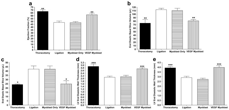
Cardiac parameters measured by MRI four weeks after treatment. (a) Cardiac ejection fraction. Both the VEGF myoblast and thoracotomy control groups displayed significantly higher ejection fractions than the ligation and myoblast only groups (P<0.01). There was no significant difference in ejection fraction between the thoracotomy control group and the VEGF165-transfected myoblast group. (b) End diastolic volume (EDV). EDV significantly increased in the ligation and myoblast only groups compared to both thoracotomy controls and VEGF165-transfected myoblasts (P<0.01). There was no significant increase in EDV between thoracotomy controls and the VEGF myoblast group. (c) End systolic volume (ESV). ESV significantly increased in the ligation and myoblast only groups compared to both thoracotomy controls and VEGF165-transfected myoblasts (P<0.05). There was no significant increase in ESV between thoracotomy controls and the VEGF myoblast group. (d) End diastolic wall thickness (EDWT) four weeks post treatment. The EDWT significantly decreased in the ligation and myoblast only groups compared to both thoracotomy controls and VEGF165-transfected myoblasts (P<0.001). There was no significant decrease in EDWT between thoracotomy controls and the VEGF myoblast group. (e) End systolic wall thickness (ESWT). ESWT significantly decreased in the ligation and myoblast only groups compared to both thoracotomy controls and VEGF165- transfected myoblasts (P<0.001). There was no significant decrease in ESWT between thoracotomy controls and the VEGF myoblast group. All data are presented as mean ± SEM (n=5 thoracotomy, n=7 ligation, n=7 myoblast only, and n=7 VEGF myoblast group).

