Figure 4.
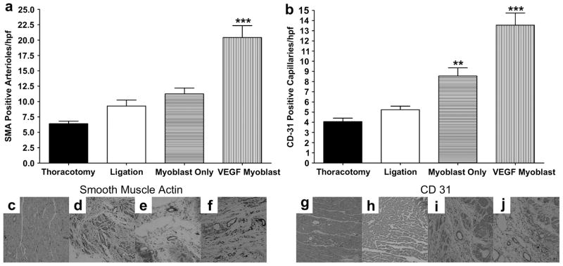
Representative immunohistochemistry stains for SM α-actin and CD31 in the infarct border zone, 4 weeks after myocardial infarction. (c,g) Thoracotomy only, (d,h) ligation only, (e,i) untransfected myoblast injection and (f,j) VEGF165-transfected myoblast injection group; (a). Average number of smooth muscle actin (SMA)-positive arterioles from five random high-power fields (hpf) per animal from the ischemic border zone of the infarct. The number of arterioles in the VEGF165-transfected myoblast group was significantly greater than all other groups P<0.001. (b) Average number of CD31-positive capillaries from five random high-power fields (hpf) per animal from the ischemic border zone of the infarct. The number of capillaries in the VEGF165-transfected myoblast group was significantly greater than all other groups P<0.001. The average number of capillaries in the myoblast only group was significantly higher than both thoracotomy and ligation groups; P<0.01 and P<0.05 respectively. All data are presented as mean ± SEM (n=5 thoracotomy, n=7 ligation, n=7 myoblast only, and n=7 VEGF myoblast group).

