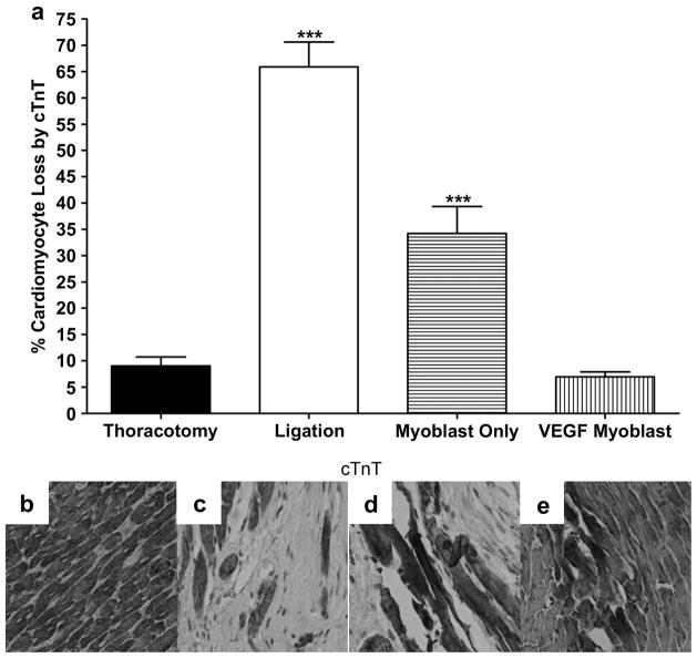
Figure 5.
The loss of cardiomyocytes, 4 weeks after surgery as indicated by loss of immunohistochemical staining for cTnT; (a) Loss of cardiomyocytes from the infarct border zone 4 weeks after surgery identified from cTnT staining and represented as percent cardiomyocytes lost per hpf. Five random hpf were counted per animal. The loss of cardiomyocytes was significantly prevented in both the myoblast only group (P<0.001 vs ligation) and the VEGF myoblast group (P<0.001 vs ligation and myoblast only). Additionally, the ligation and myoblast only groups were statistically greater than the thoracotomy control (P<0.001), whereas the VEGF myoblast treatment group was not statistically different from thoracotomy control. Representative micrographs of cTnT staining for (b) thoracotomy control group, (c) ligation, (d) myoblast only injection group and (e) VEGF165-transfected myoblast injection group. With cell death, myofibers lose their usual normal brownish staining. All data are presented as mean ± SEM (n=5 thoracotomy, n=7 ligation, n=7 myoblast only, and n=7 VEGF myoblast group).

