Abstract
Multislice computed tomography (CT) angiography has been increasingly used in the detection and diagnosis of coronary artery disease because of its rapid technical evolution from the early generation of 4-slice CT scanners to the latest models such as 64-slice, 256-slice and 320-slice CT scanners. Technical developments of multislice CT imaging enable improved diagnostic value in the detection of coronary artery disease, and this indicates that multislice CT can be used as a reliable less-invasive alternative to invasive coronary angiography in selected patients. In addition, multislice CT angiography has played a significant role in the prediction of disease progression and cardiac events. Despite promising results reported in the literature, multislice CT has the disadvantage of having a high radiation dose which could contribute to the radiation-induced malignancy. A variety of strategies have been currently undertaken to reduce the radiation dose associated with multislice CT coronary angiography while in the meantime acquiring diagnostic images. In this article, the author will review the technical developments, radiation dose associated with multislice CT coronary angiography, and strategies to reduce radiation dose. The diagnostic and prognostic value of multislice CT angiography in coronary artery disease is briefly discussed, and future directions of multislice CT angiography in the diagnosis of coronary artery disease will also be highlighted.
Keywords: Coronary artery disease, Computed tomography, Diagnostic value, Radiation dose, Radiation risk
INTRODUCTION
Coronary artery disease (CAD) is the leading cause of death in Western countries. Conventional coronary angiography is the gold standard technique for diagnosis of CAD, due to its superior spatial and temporal resolution. The diagnostic value of conventional coronary angiography has been challenged by the emergence and fast growing use of a less invasive imaging technique, multislice computerized tomography (MSCT) angiography[1-3]. The diagnostic accuracy of MSCT angiography in CAD has been significantly augmented with the increased performance of MSCT from early generation of the 4-slice CT to 16-slice, 64-slice, dual-source CT and the latest models such as 256-slice and 320-slice CT scanners[2-8]. This is mainly demonstrated by the improved spatial and temporal resolution from the latest MSCT scanners such as 64 or more slice scanners. In particular, MSCT angiography has been reported to demonstrate a very high negative predictive value (more than 95%), indicating that it can be used as a reliable technique for excluding patients suspected of CAD, thereby reducing the need for invasive coronary angiography.
While the number of MSCT examinations in cardiac imaging continues to increase, the potential risk of radiation exposure associated with CT should not be ignored, given the fact that CT is a high-radiation imaging modality. The radiation risks arising from cardiac CT angiography have raised serious concerns in the medical field as CT is associated with a non-negligible life attributable risk of cancer[9]. Therefore, the benefit of the use of multislice CT angiography in the diagnostic workup and patient management must be weighed against the potential risks related to radiation exposure. In this review article, I will introduce the technical developments of MSCT in cardiac imaging, focusing on the diagnostic and prognostic value of MSCT angiography in CAD, followed by the strategies currently available to address radiation dose reduction. Future directions, including justification of use of MSCT in cardiac imaging, will be highlighted.
MULTISLICE CT ANGIOGRAPHY IN CARDIAC IMAGING-TECHNICAL EVOLUTION OF CT SCANNERS
Traditionally, electron-beam CT (EBCT) with high temporal resolution (50-100 ms) makes this technique well suited for imaging the coronary tree, with capability of evaluating tiny abnormalities such as coronary calcium deposits and plaques, even with the motion of a rapidly beating heart. However, the inferior spatial resolution (1.5 mm) of EBCT limits its diagnostic value in the detection of CAD as the coronary artery is a very small structure ranging from 1.5 mm to 3.0 mm in diameter, thus, image quality of both normal and abnormal coronary arteries is degraded to a greater extent. The introduction of MSCT scanners in 1998 represented a significant technical improvement in the CT imaging technique because imaging of the heart was made possible with MSCT as multiple images could be acquired in a single breath-hold within a very short time[10].
The significant application of 4-slice CT in clinical practice is in cardiac imaging with the aid of an electrocardiographic (ECG)-gating technique. The ability of 4-slice CT (gantry rotation time, 500 ms with temporal resolution of 250 ms) to image the coronary arteries and detect CAD less invasively has attracted much attention from physicians in the medical field, and this is represented by the increasing number of publications on 4-slice CT coronary angiography in the literature[2,3,11,12]. Earlier studies with 4-slice CT were promising as a less-invasive technique in cardiac imaging, although the diagnostic value was insufficient to replace conventional coronary angiography in the diagnosis of CAD[13]. With experience gathered, it was found that the image quality of coronary arteries was impaired in many cases with 4-slice CT due to limited spatial and temporal resolution, and the unassessable segments could be more than 20% in 4-slice studies[13]. Thus 4-slice CT is unsuitable for imaging patients with heart rate higher than 60 bpm.
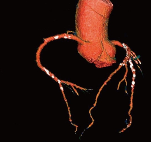
With the introduction of 16-slice and 64-slice CT, image quality in coronary MSCT has become more consistent with improved results[14-18]. With gantry rotation times down to 330 ms for 64-slice CT, temporal resolution for cardiac ECG-gated imaging was further markedly improved, allowing evaluation of both the main coronary artery and its side branches, including the distal artery segments. In contrast to early generations, 64-slice CT showed improved diagnostic accuracy in CAD because of improved spatial and temporal resolution. Isotropic volume data (0.6 mm × 0.6 mm × 0.6 mm) is possible with 64-slice CT, and the scanning time is reduced to less than 15 s, allowing a decreased breath-hold time, better utilization of contrast medium with fewer enhancements of adjacent structures, and a lower dose of applied contrast medium[15-18]. An improvement in image quality has also been reported in the visualization of all coronary artery branches, with high sensitivity and specificity achieved[15-18] (Figure 1).
Figure 1.
Three-dimensional volume rendering of the left and right coronary arteries and their branches acquired with 64-slice computed tomography angiography in a 61-year-old diagnosed with coronary artery disease. Extensive calcification is present in the coronary artery wall which is shown as the white dots.
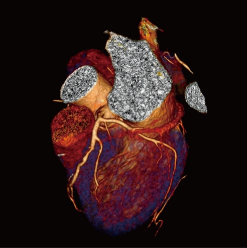
Further technical improvement in cardiac imaging was achieved with the development of dual-source CT (DSCT) due to its high temporal resolution of 83 ms, thus increasing visualization of coronary arteries in patients with high heart rate by reducing motion artifacts[19,20]. Studies have shown that the cardiac CT image quality of the coronary artery is independent of heart rate, and high diagnostic accuracy is achieved with DSCT[19,20] (Figure 2). There is no doubt that DSCT represents another technical development with its superior temporal resolution which contributes specifically to the assessment of patients with high heart rate.
Figure 2.
Three-dimensional volume rendering of the left coronary artery is acquired with dual-source computed tomography angiography in a 47-year-old woman suspected of coronary artery disease. Left anterior descending and left circumflex are clearly demonstrated without any sign of lumen stenosis or calcification.
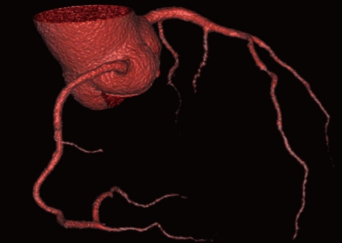
Expansion of MSCT systems from 64-slice to the latest models of 256-slice and 320-slice systems has allowed whole heart coverage in one gantry rotation with a slice thickness of 0.5 mm[6-8]. With 320-slice CT, 16 cm of craniocaudal coverage can be obtained in a single heartbeat, with excellent image quality and demonstration of the entire coronary arteries, and therefore, the entire cardiac volume data can be acquired within 1 s without of the need for patient movement during the scan[7,8] (Figure 3). Table 1 lists the developments of MSCT scanners from 4-slice to 320-slice CT in terms of spatial and temporal resolution when compared to the gold standard technique, conventional coronary angiography.
Figure 3.
Three-dimensional volume rendering of the coronary arteries and side branches are clearly visualized with use of 320-slice computed tomography angiography in a 58-year-old man presenting with chest pain. Volumetric data were acquired within a single heartbeat with excellent image quality.
Table 1.
Developments in multislice computed tomography scanners in terms of spatial and temporal resolution when compared to electron-beam computed tomography and invasive coronary angiography
| Imaging modalities | Spatial resolution (mm) | Temporal resolution (ms) |
| EBCT | 1.5-3.0 | 50-100 |
| 4-slice CT | 1.0-1.25 | 250 |
| 16-slice CT | 0.75 | 165-188 |
| 64-slice single-source CT | 0.5-0.6 | 165 |
| 64-slice dual-source CT | 0.5-0.6 | 75-83 |
| 256/320-slice CT | 0.5-0.6 | 135-175 |
| Invasive coronary angiography | 0.2 | 20 |
CT: Computed tomography; EBCT: Electron-beam CT.
MULTISLICE CT ANGIOGRAPHY IN CAD-RADIATION DOSE
Despite the above-mentioned promising results of MSCT angiography in CAD, MSCT has the disadvantage of requiring a high radiation dose. Conventional coronary angiography is effective at a radiation dose from 3 to 9 mSv, while MSCT coronary angiography delivers a radiation dose as high as 20 mSv according to early studies[21,22]. Moreover, the average dose per site was reported to demonstrate significant variability ranging from 5 to 30 mSv[23]. Thus, reduction of the radiation dose in MSCT coronary angiography is of paramount importance to ensure that it is a safe technique for use in clinical application.
There are a number of strategies that have been undertaken to reduce the radiation dose from cardiac MSCT, and the most commonly used approaches include: adjustment of tube voltage (kVp) and tube current (mAs), increasing pitch value and choosing different ECG-gating methods (prospective vs retrospective gating). Effective reduction of radiation dose can be achieved by changing or selecting appropriate parameters without compromising diagnostic image quality.
MULTISLICE CT ANGIOGRAPHY IN CAD-RADIATION DOSE REDUCTION STRATEGIES
ECG-controlled tube current modulation
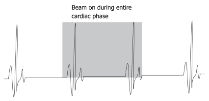
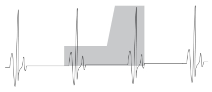
One of the most effective approaches for dose reduction is adjustment of the tube current according to ECG signal, which is defined as ECG-controlled tube current modulation. Traditionally, cardiac MSCT angiography is performed using a retrospective ECG-gating technique, which indicates that the volume data are acquired during the entire cardiac cycle within a single breath-hold helical scan (Figure 4). This ensures acquisition of volumetric data during systolic and diastolic cardiac cycles, thus reconstruction of images at the diastolic phase allows us to generate images with the least motion artifacts. However, image reconstruction of the volume data only occurs in a specific phase of the cardiac cycle (end systole or mid diastole). Thus, not all of the data is used for diagnostic purposes, but the patient is exposed to X-rays during the entire cardiac cycle. This implies that the tube current can be adjusted in different cardiac phases so that high-quality diagnostic images of coronary arteries during the reconstruction window, and low-quality, higher noise images of the cardiac chamber and cardiac valves during the rest of cardiac cycle can be acquired (Figure 5).
Figure 4.
Diagram showing retrospective electrocardiogram-gating without tube current modulation in multislice computed tomography coronary angiography. An X-ray beam is turned on during the entire cardiac cycle without adjusting the tube current.
Figure 5.
Diagram showing retrospective electrocardiogram-gating with the use of tube current modulation in multislice computed tomography coronary angiography. Normal tube current is applied only during the image reconstruction phase (late diastolic phase), while the tube current is reduced significantly during the systolic phase.
ECG-controlled tube current modulation represents the most significant improvement in minimizing radiation exposure from CT technology and is the only one dedicated to cardiac imaging. It has been reported that radiation dose can be reduced by 30%-50% through modulation of the tube current output to decrease the dose given during the systolic phase[24,25]. The estimated radiation dose reduction is similar to or less than that of a conventional coronary angiography examination with use of this dose-saving strategy[24,26].
Automatic exposure control
Automatic exposure control (AEC) is regarded as another effective approach to reduce radiation dose for coronary cardiac CT examination. AEC takes this into consideration by automatically adjusting the tube current in the x, y plane (angular modulation) or along the scanning direction (z-axis modulation) or both (combined modulation) according to the anatomic geometry of the body region to be scanned to obtain diagnostic image quality while lowering radiation dose[27,28]. Deetjen et al[25] studied different groups of patients undergoing 16-slice and 64-slice coronary angiography examinations and showed significant reduction of radiation dose (42.8%) with AEC. This is consistent with a recent report involving a comparison of 4 different CT manufacturers with different AEC systems[29]. Söderberg et al[29] found that the magnitude of dose saving was considerable, ranging from 35% to 60%, with the use of AEC systems with similar performance of tube current modulation dynamics among the different CT scanners.
Adjustment of tube voltage
Lowering the tube voltage is widely undertaken in clinical practice to reduce radiation dose, since radiation dose varies with the square of the kVp. Modern CT scanners include tube voltages of 120 or 140 kVp, reflecting the settings most often resulting in adequate image quality. However, cardiac CT acquisition with 100 kVp, or even lower, is possible and has been recommended as an effective means to reduce radiation dose in cardiac CT imaging[30,31]. Previous studies have shown that decreasing the X-ray tube voltage from 120 to 100 kVp or 80 kVp resulted in up to a 70% reduction in radiation exposure for a constant tube current using 16-slice and 64-slice CT, with increased image noise and unchanged contrast-to-noise ratio[30,31]. Studies utilizing dual-source CT compared a 100 kVp protocol to the routine 120 kVp for cardiac CT, and demonstrated a 25%-54% reduction in radiation dose, depending on the tube current time product[32,33].
It should be emphasized that changing tube voltage needs to be correlated with patient’s body mass index (BMI). Lowering tube voltage from 120 to 100 kVp can be performed when the patient’s BMI is less than 25 kg/m2. Reduction of the tube voltage to 80 kVp should be considered in children and slim young adults with BMI below 20 kg/m2. It has been reported that the tube voltage can be lowered to 80 kVp without impairing image quality while the radiation dose was reduced by up to 80% in normal weight patients[34]. Therefore, tube voltage can be adjusted in cardiac CT angiography without affecting diagnostic image quality, and this should be applied whenever possible in clinical practice.
High pitch value
It is well-known that radiation dose is inversely proportional to the pitch value. A high pitch (1.0-2.0) is recommended for helical CT angiography with the aim of reducing radiation dose without affecting image quality. However, for MSCT coronary angiography, a very low pitch (0.2-0.4) is routinely used to produce volume coverage without gaps in each phase of the cardiac cycle with multiple overlapping regions, thus resulting in high radiation exposure. Increasing pitch to a higher value is made possible with the development of a second generation of dual-source CT scanner[35-39], which enables high detector coverage with the use of two 128-section detectors. This dual-source CT system allows coronary CT angiography to be performed at high pitch value of up to 3.4 with significant reduction of radiation dose. By combining high pitch and large detector coverage, the acquisition time of coronary CT angiography is reduced from the previous 5-10 s to a quarter of a second, allowing depiction of the entire heart within a single heartbeat. More than 90% of all coronary segments were assessable with dual-source CT coronary angiography at a high pitch of 3.4, resulting in a radiation dose less than 1 mSv[35,36]. The new scan mode with a temporal resolution of 75 ms is regarded as an extremely attractive alternative to invasive coronary angiography because of the very low radiation dose and high image quality.
Prospective ECG-triggering
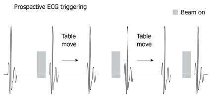
Prospective ECG-triggering with axial non-helical scan was used a long time ago with electron-beam CT for calcium scoring, but was proposed recently for cardiac imaging, and this imaging protocol is increasingly reported in the literature because of its very low radiation dose[40-48]. The principle of prospective ECG-triggering is different from that of retrospective ECG-gating as the former is used to acquire data by selectively turning the X-ray tube on only in the selected cardiac phase, triggered by the ECG signal, and turning off during the rest of the R-R cycle (Figure 6). This is also referred to as sequential or step-and-shoot acquisition with prospective triggering, and the effective pitch is 1.0. The main advantage of this scanning protocol is the lower radiation dose as X-ray exposure only occurs during the selected cardiac phase rather than throughout the entire cardiac cycle. Therefore, a significant reduction in radiation dose can be achieved from prospective ECG-triggering, which is the most attractive side of this scanning protocol compared to retrospective ECG-gating.
Figure 6.
The diagram shows prospective electrocardiogram-triggering with X-ray beam on during a portion of the cardiac cycle, while in the remaining cardiac phase, the X-ray beam is turned off.
Studies using prospective ECG-triggering showed significant dose reduction when compared to the conventional retrospective ECG-gating, with up to 90% dose reduction achieved in some studies[46,47]. Even if the published results concerning image quality have generally been satisfactory, there is concern about the negative effect of low-dose scanning protocols which may affect image quality of cardiac CT examination. A “staged” protocol was recently introduced by researchers to address the situation when image quality may be suboptimal[49]. Pflederer et al[49] evaluated the staged low-dose methods for coronary CT angiography compared to standard protocols and reported an 86% reduction of the effective dose with use of a prospective triggering approach. However, a higher number of unevaluable coronary segments and impaired image quality was noticed in this group. An additional standard sequence was added to the patients with non-diagnostic images and the overall radiation dose reduction still remained significant (1.5-3.4 mSv for low-dose protocols vs 9.8 mSv for the standard protocol). Thus, the staged strategy with an initial low-dose CT protocol should be encouraged to be used as a dose-saving algorithm.
DIAGNOSTIC VALUE OF MULTISLICE CT ANGIOGRAPHY IN CAD
Over the last decade much interest has been focused on imaging and diagnosis of CAD with MSCT as it is a less invasive and faster scanning technique with extended z-axis coverage when compared to single slice CT. Earlier studies with 4-slice CT showed moderate diagnostic accuracy with pooled sensitivity and specificity of 78% and 93%, respectively because the spatial and temporal resolution is limited, resulting in a high number of unassessable segments[10]. With the introduction of 16-slice CT, the diagnostic value of MSCT angiography in CAD has been much improved, with more coronary segments assessable. Studies using 16-slice CT with acquisition and rotation times of < 400 ms have reported sensitivities between 83% and 98% and specificities between 96% and 98%[14,15]. Despite improvement in both spatial and temporal resolution, isotropic volume data is still not possible with 16-slice CT. Moreover, the temporal resolution is insufficient to ensure acquisition of diagnostic images in patients with high heart rate.
MSCT examination times were reduced with further improvement in scanning techniques with 64-slice CT because of improved spatial and temporal resolution compared with 16-slice and 4-slice CT. Acquisition of isotropic volume data is possible with 64-slice CT, thus detection of main and side coronary artery branches is significantly improved when compared to earlier generations of MSCT scanners. Several meta-analyses of 64-slice CT studies reported sensitivities and specificities ranging from 86% to 99% and 88% to 97%, respectively[50-53]. These studies indicated that MSCT, especially with 64-or more CT, has high diagnostic accuracy for detection of CAD and could be used as an effective alternative to invasive coronary angiography in selected patients.
One of the difficulties for MSCT cardiac imaging is that image quality highly depends on heart rate. Despite technical improvements in MSCT scanners with subsequent improvement in temporal resolution, the assessable segments and diagnostic accuracy are still higher in patients with a lower heart rate, and lower and deteriorated in patients with a higher heart rate. Therefore, an aggressive approach such as the use of β-blockers in patients with a heart rate more than 70 bpm has become a part of the routine procedure prior to CT scanning. With the advent of dual-source CT, temporal resolution has been increased to 83 ms, indicating that image quality is less dependent on the heart rate, thus potentially obviating the need for β-blockers before scanning.
In a recent study, Donnino et al[54] compared dual-source with single-source CT for assessment of diagnostic image quality of coronary artery examinations. Their results showed a significant improvement in image quality with dual-source CT over single-source CT, in the absence of pre-examination use of β-blockers, and higher heart rate in the dual-source group. Reports by others also supported the improved diagnostic value of dual-source CT in cardiac imaging[55,56]. Dual-source CT was reported to be superior to single-source 64-slice CT for the detection of CAD with a sensitivity and specificity of 100% for both in a small group of patients[56], whereas the 64-slice scanner achieved a sensitivity of 100%, but a reduced specificity of 90%.
Latest models such as 256-slice and 320-slice CT scanners allow for longer z-axis coverage ranging from 12.8 to 16 cm on one gantry rotation, so it is possible to achieve full cardiac coverage in one gantry rotation within a very short period, thus eliminating the restrictions and limitations associated with 64-slice CT scanners[57,58]. de Graaf et al[57] reported that a high diagnostic value, especially a 100% negative predictive value (including the non-diagnostic images) and diagnostic accuracy of 95%, was achieved with 320-slice CT angiography for detection of significant coronary stenosis in a patient-based analysis. This indicated that 320-slice CT angiography is a highly sensitive modality for the detection of significant CAD. Pasricha et al[58] compared image quality of 320-slice CT in patients with atrial fibrillation with that acquired from the group with sinus rhythm. In this study, 96% of the coronary segments were assessable with sufficient quality for diagnosis in patients with atrial fibrillation, and this showed the potential application of 320-slice CT in this patient group. Although 320-slice CT shows very promising results, more data are needed to confirm its diagnostic accuracy in CAD.
PROGNOSTIC VALUE OF MULTISLICE CT ANGIOGRAPHY IN CAD
MSCT is not only able to evaluate the coronary luminal changes, but also visualize the coronary artery wall morphology, identify and characterize coronary plaques, especially the non-stenotic plaques that may be undetected by conventional coronary angiography. Thus, MSCT could be used as a non-invasive technique to provide prognostic information in patients suspected of having CAD.
MSCT also allows for non-invasive detection of plaque morphology and composition (calcified vs non-calcified atherosclerotic plaques), as well as the assessment of the extent of vascular remodeling[59]. Atherosclerotic plaque size and geometry play an important role in the natural progression of the disease process and may have important clinical predictive value. Schmid et al[59] in their study concluded that a significant increase in the amount of noncalcified plaque was observed with 64-slice CT over a mean interval follow-up of 17 mo, and their results indicated that MSCT may be used as a tool to study the progression of coronary atherosclerosis.
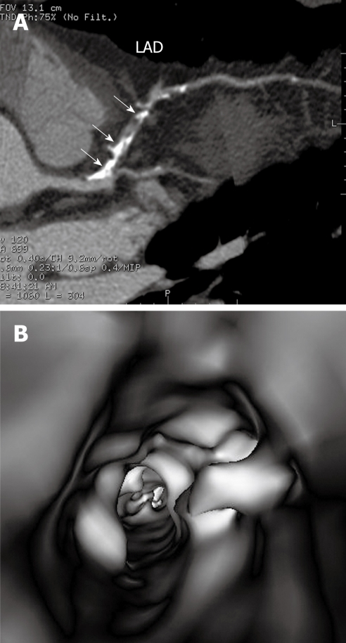
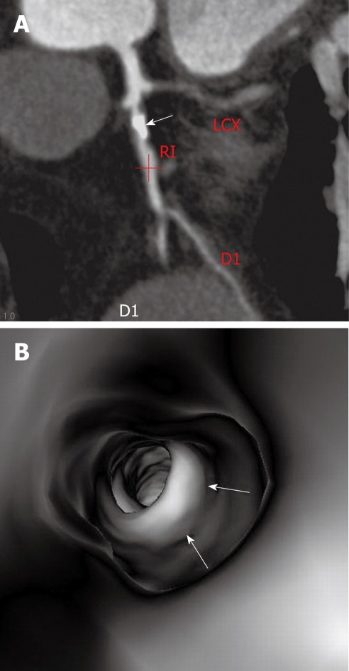
Coronary artery plaque is characterized into 3 types based on the CT attenuation[60]: non-calcified plaques refer to plaques having lower density compared with the contrast-enhanced vessel lumen (Figure 7A); calcified plaques indicate plaques with high density (Figure 7B); mixed plaques refer to plaques with non-calcified and calcified components within a single plaque or within a segment of the coronary artery (Figure 7C). Accurate identification of the type of plaques as well as demonstration of coronary luminal changes is important for prediction of disease progress based on 2D and 3D CT visualizations. In contrast to conventional 2D or 3D extraluminal visualizations, the 3D virtual intravascular endoscopy has been reported to provide additional information about the intraluminal appearance of coronary plaques (Figure 8), and corresponding luminal changes due to the presence of plaques in the artery wall[61]. Virtual intravascular endoscopy also helps to confirm the degree of coronary stenosis as severe calcification sometimes results in a false positive sign of lumen occlusion on 2D views (Figure 9).
Figure 7.
Characterization of coronary plaques. RCA-right coronary artery. A: Curved planar reformatted image acquired with 64-slice computed tomography angiography shows a non-calcified plaque at the mid-segment of the right coronary artery in a 57-year-old man suspected of coronary artery disease; B: Calcified plaques (arrows) are found at the proximal and middle segments of the right coronary artery; C: Mixed type plaques are shown in the left anterior descending (LAD) artery in a 59-year-old man suspected of coronary artery disease; the long arrow indicates the calcified plaque, while the short arrows indicate non-calcified components.
Figure 8.
Virtual endoscopy visualization of coronary plaques. A: Extensive calcified plaques in the left anterior descending (LAD) are observed on curved planar reformatted view with more than 70% luminal stenosis in a 52-year-old man with chest pain; B: Corresponding virtual intravascular endoscopy visualization demonstrates irregular intraluminal appearance with significant stenosis of the coronary artery.
Figure 9.
Virtual endoscopy confirmation of plaque stenosis. A: A calcified plaque (arrow) is present in the left anterior descending with more than 90% lumen stenosis in a 51-year-old man presenting with symptoms of chest pain; B: Corresponding virtual intravascular endoscopy shows intraluminal protrusion caused by the plaque, but the luminal stenosis is less than 70%; arrows indicate the intraluminal appearance of calcified plaque. RI: Ramus intermedius; LCX: left circumflex; D1: Diagonal branch.
Preliminary reports have shown that MSCT angiography is able to provide independent prognostic information for predicting cardiac events and mortality in patients with known or suspected CAD at short-term follow-up. Gilard et al[62] reported that a normal MSCT in a population of 141 patients with suspected CAD was associated with a low rate of cardiac events (mortality 0%, myocardial infarction 0.7%, coronary angiography 3.5%) at 1-year follow-up. Similar results were reported by other studies demonstrating that obstructive plaque, particularly in the left main or left anterior descending arteries, had the highest risk (up to 34% cardiac event rate), while in contrast, a normal coronary CT angiography was associated with a 0% event rate[63-65]. However, these studies have been largely limited by small sample sizes, single centre evaluations and short follow-up periods. Two recent studies based on a large cohort of patients offered independent and additional prognostic information of MSCT angiography for the prediction of incidence of cardiac events[66,67].
Hadamitzky et al[66] in their recent report assessed the prognostic value of MSCT angiography in the prediction of cardiac events in asymptomatic patients. They retrospectively analyzed 451 asymptomatic patients with 16-slice and 64-slice CT angiography during a median follow-up of 27.5 mo. Their study confirmed that MSCT angiography could be reliably used to predict further cardiac events, despite the low cardiac event rate in asymptomatic patients. Min et al[67] in a large cohort of 5330 patients without known CAD determined the prognostic value of MSCT angiography by measuring the left ventricular ejection fraction (LVEF) in addition to the traditional criterion of presence of obstructive CAD. Their study demonstrated that MSCT angiography successfully identified individuals at higher risk of all-cause death at a follow-up of 2.3 years. The presence of obstructive CAD in an increasing number of coronary arteries indicated a particularly poor prognosis. In addition, measures of LVEF were found to add incremental prognostic values above and beyond CAD detection with patients without obstructive CAD or with normal LVEF and a low risk of death.
MULTISLICE CT ANGIOGRAPHY IN CAD-FUTURE DIRECTIONS
Future directions of MSCT angiography in the diagnosis of CAD lie in 3 main aspects: improvement of temporal resolution, reduction of radiation dose and judicious use of MSCT. The MSCT scanning technique has improved significantly over the last decade, with acquisition of isotropic volume data in patients with high heart rate. However, the current temporal resolution of MSCT imaging (75-83 ms with dual-source CT, 135-175 ms with 256-slice and 320-slice CT) is still inferior to that of invasive coronary angiography (20 ms), thus further technical improvement in temporal resolution is necessary so that MSCT can be applied in more patients, especially for those with high or irregular heart rate. Control of high heart rate with the use of β-blockers is still commonly performed in many cardiac MSCT examinations, including scans with the use of 320-slice CT, and further improvement in temporal resolution will contribute to the elimination of the aggressive procedure of heart rate control in patients with heart rate more than 70 bpm.
As already discussed above, radiation exposure associated with MSCT angiography is relatively high and poses a potential risk of radiation-induced malignancy. Although dose-saving strategies have been recommended to reduce the radiation dose from MSCT angiography, CT scanning protocols across institutions are widely variable and could contribute to the high radiation exposure to patients. Two recent studies highlighted the importance of standardization of common CT scans including cardiac CT imaging, as well as the cancer risk associated with the radiation[68,69]. Smith-Bindman et al[68], in their prospective study involving 4 institutions, collected data on radiation doses for the most common CT scans and found a significant variation in radiation dose, a mean 13-fold variation between the highest and lowest dose for each CT type studied (range, 6- to 22-fold difference across study types). They estimated that 1 in every 270 40-year-old women undergoing CT coronary angiography will develop cancer from the procedure. In another study, Berrington de González et al[69] estimated that CT scans performed in 2007 could have led to 29 000 excess cancers, which will appear in the next 20 to 30 years and by the authors’ estimates, at a 50% mortality rate, will cause approximately 15 000 deaths annually.
There is no doubt that, with increasing technological improvements, MSCT will continue to play an important role in the diagnosis of CAD. Judicious use of MSCT in cardiac imaging is essential to maximize its clinical applications while minimizing the potential risk of radiation exposure. This is particularly important for young individuals, especially women, for whom alternative diagnostic modalities that do not involve the use of ionizing radiation should be considered, such as echocardiography, or magnetic resonance imaging[23]. Physicians need to be aware of the potential risk of radiation dose associated with CT cardiac imaging. It has been reported that 47% of radiologists and 9% of emergency department physicians believed that there was an increased risk of cancer associated with CT scans[70]. Thus, there is an urgent need for physicians to educate themselves and increase their awareness about ionizing radiation from CT and its associated risks[71]. The benefit-to-risk ratio for imaging patients suspected of CAD must be driven by the benefit and appropriateness of the cardiac MSCT examination requested by the cardiologists. The main purpose of utilizing MSCT imaging is to address specific medical questions without allowing concerns about radiation exposure to dissuade cardiologists or their patients from obtaining or undergoing the needed MSCT examination[72].
CONCLUSION
Multislice CT angiography, as a less-invasive imaging modality has demonstrated high diagnostic value in the detection and diagnosis of CAD. In particular, the very high negative predictive value of MSCT angiography allows it to be used as a reliable technique for screening purposes. With continued technical improvements in the scanning technique, MSCT will play an increasing role in the detection and characterization of coronary plaques. Moreover, MSCT is regarded as a reliable modality for prediction of disease prognosis in patients suspected of CAD.
Serious concerns have recently been raised about the radiation dose from MSCT angiography in clinical practice, as it has been confirmed to be related to a potential risk of inducing cancer. MSCT coronary angiography should be performed with dose-saving strategies whenever possible to reduce the radiation exposure to patients. Multislice CT scanning protocols in cardiac imaging should be standardized across institutions with the aim of reducing dose variation across patients and facilities. Utilization of coronary MSCT angiography must be considered with care as to whether it leads to an overall benefit or whether the radiation risk may be greater than the benefit expected from the CT examinations. Physicians, especially cardiologists, should be aware of the potential risk from CT scans, and consider reducing unnecessary CT examinations or replacing CT with other alternative modalities, such as ultrasound or magnetic resonance imaging.
Footnotes
Peer reviewers: Sang-Hak Lee, Associate Professor, Cardiology Division, Department of Internal Medicine, Severance Cardiovascular Hospital, Yonsei University College of Medicine, 134 Shinchon-dong, Seodaemun-gu, Seoul, 120-752, South Africa; Seung-Woon Rha, MD, PhD, FACC, FAHA, FESC, FSCAI, FAPSIC, Cardiovascular Center, Korea University Guro Hospital, 80, Guro-dong, Guro-gu, Seoul, 152-703, South Korea
S- Editor Cheng JX L- Editor Cant MR E- Editor Zheng XM
References
- 1.McCollough CH, Zink FE. Performance evaluation of a multi-slice CT system. Med Phys. 1999;26:2223–2230. doi: 10.1118/1.598777. [DOI] [PubMed] [Google Scholar]
- 2.Nieman K, Oudkerk M, Rensing BJ, van Ooijen P, Munne A, van Geuns RJ, de Feyter PJ. Coronary angiography with multi-slice computed tomography. Lancet. 2001;357:599–603. doi: 10.1016/S0140-6736(00)04058-7. [DOI] [PubMed] [Google Scholar]
- 3.Achenbach S, Giesler T, Ropers D, Ulzheimer S, Derlien H, Schulte C, Wenkel E, Moshage W, Bautz W, Daniel WG, et al. Detection of coronary artery stenoses by contrast-enhanced, retrospectively electrocardiographically-gated, multislice spiral computed tomography. Circulation. 2001;103:2535–2538. doi: 10.1161/01.cir.103.21.2535. [DOI] [PubMed] [Google Scholar]
- 4.Kuettner A, Trabold T, Schroeder S, Feyer A, Beck T, Brueckner A, Heuschmid M, Burgstahler C, Kopp AF, Claussen CD. Noninvasive detection of coronary lesions using 16-detector multislice spiral computed tomography technology: initial clinical results. J Am Coll Cardiol. 2004;44:1230–1237. doi: 10.1016/j.jacc.2004.05.079. [DOI] [PubMed] [Google Scholar]
- 5.Leber AW, Knez A, von Ziegler F, Becker A, Nikolaou K, Paul S, Wintersperger B, Reiser M, Becker CR, Steinbeck G, et al. Quantification of obstructive and nonobstructive coronary lesions by 64-slice computed tomography: a comparative study with quantitative coronary angiography and intravascular ultrasound. J Am Coll Cardiol. 2005;46:147–154. doi: 10.1016/j.jacc.2005.03.071. [DOI] [PubMed] [Google Scholar]
- 6.Chao SP, Law WY, Kuo CJ, Hung HF, Cheng JJ, Lo HM, Shyu KG. The diagnostic accuracy of 256-row computed tomographic angiography compared with invasive coronary angiography in patients with suspected coronary artery disease. Eur Heart J. 2010;31:1916–1923. doi: 10.1093/eurheartj/ehq072. [DOI] [PubMed] [Google Scholar]
- 7.Rybicki FJ, Otero HJ, Steigner ML, Vorobiof G, Nallamshetty L, Mitsouras D, Ersoy H, Mather RT, Judy PF, Cai T, et al. Initial evaluation of coronary images from 320-detector row computed tomography. Int J Cardiovasc Imaging. 2008;24:535–546. doi: 10.1007/s10554-008-9308-2. [DOI] [PubMed] [Google Scholar]
- 8.Dewey M, Zimmermann E, Deissenrieder F, Laule M, Dübel HP, Schlattmann P, Knebel F, Rutsch W, Hamm B. Noninvasive coronary angiography by 320-row computed tomography with lower radiation exposure and maintained diagnostic accuracy: comparison of results with cardiac catheterization in a head-to-head pilot investigation. Circulation. 2009;120:867–875. doi: 10.1161/CIRCULATIONAHA.109.859280. [DOI] [PubMed] [Google Scholar]
- 9.Einstein AJ, Henzlova MJ, Rajagopalan S. Estimating risk of cancer associated with radiation exposure from 64-slice computed tomography coronary angiography. JAMA. 2007;298:317–323. doi: 10.1001/jama.298.3.317. [DOI] [PubMed] [Google Scholar]
- 10.Kalendar WA. Computed tomography: fundamentals, system technology, image quality, applications. Munich: MCD Verlag; 2000. pp. 35–81. [Google Scholar]
- 11.Kopp AF, Schroeder S, Kuettner A, Baumbach A, Georg C, Kuzo R, Heuschmid M, Ohnesorge B, Karsch KR, Claussen CD. Non-invasive coronary angiography with high resolution multidetector-row computed tomography. Results in 102 patients. Eur Heart J. 2002;23:1714–1725. doi: 10.1053/euhj.2002.3264. [DOI] [PubMed] [Google Scholar]
- 12.Nieman K, Rensing BJ, van Geuns RJ, Munne A, Ligthart JM, Pattynama PM, Krestin GP, Serruys PW, de Feyter PJ. Usefulness of multislice computed tomography for detecting obstructive coronary artery disease. Am J Cardiol. 2002;89:913–918. doi: 10.1016/s0002-9149(02)02238-5. [DOI] [PubMed] [Google Scholar]
- 13.Sun Z, Jiang W. Diagnostic value of multislice computed tomography angiography in coronary artery disease: a meta-analysis. Eur J Radiol. 2006;60:279–286. doi: 10.1016/j.ejrad.2006.06.009. [DOI] [PubMed] [Google Scholar]
- 14.Kuettner A, Beck T, Drosch T, Kettering K, Heuschmid M, Burgstahler C, Claussen CD, Kopp AF, Schroeder S. Diagnostic accuracy of noninvasive coronary imaging using 16-detector slice spiral computed tomography with 188 ms temporal resolution. J Am Coll Cardiol. 2005;45:123–127. doi: 10.1016/j.jacc.2004.10.050. [DOI] [PubMed] [Google Scholar]
- 15.Mollet NR, Cademartiri F, Krestin GP, McFadden EP, Arampatzis CA, Serruys PW, de Feyter PJ. Improved diagnostic accuracy with 16-row multi-slice computed tomography coronary angiography. J Am Coll Cardiol. 2005;45:128–132. doi: 10.1016/j.jacc.2004.09.074. [DOI] [PubMed] [Google Scholar]
- 16.Ong TK, Chin SP, Liew CK, Chan WL, Seyfarth MT, Liew HB, Rapaee A, Fong YY, Ang CK, Sim KH. Accuracy of 64-row multidetector computed tomography in detecting coronary artery disease in 134 symptomatic patients: influence of calcification. Am Heart J. 2006;151:1323.e1–1323.e6. doi: 10.1016/j.ahj.2005.12.027. [DOI] [PubMed] [Google Scholar]
- 17.Pugliese F, Mollet NR, Runza G, van Mieghem C, Meijboom WB, Malagutti P, Baks T, Krestin GP, deFeyter PJ, Cademartiri F. Diagnostic accuracy of non-invasive 64-slice CT coronary angiography in patients with stable angina pectoris. Eur Radiol. 2006;16:575–582. doi: 10.1007/s00330-005-0041-0. [DOI] [PubMed] [Google Scholar]
- 18.Raff GL, Gallagher MJ, O'Neill WW, Goldstein JA. Diagnostic accuracy of noninvasive coronary angiography using 64-slice spiral computed tomography. J Am Coll Cardiol. 2005;46:552–557. doi: 10.1016/j.jacc.2005.05.056. [DOI] [PubMed] [Google Scholar]
- 19.Achenbach S, Ropers D, Kuettner A, Flohr T, Ohnesorge B, Bruder H, Theessen H, Karakaya M, Daniel WG, Bautz W, et al. Contrast-enhanced coronary artery visualization by dual-source computed tomography--initial experience. Eur J Radiol. 2006;57:331–335. doi: 10.1016/j.ejrad.2005.12.017. [DOI] [PubMed] [Google Scholar]
- 20.Johnson TR, Nikolaou K, Wintersperger BJ, Leber AW, von Ziegler F, Rist C, Buhmann S, Knez A, Reiser MF, Becker CR. Dual-source CT cardiac imaging: initial experience. Eur Radiol. 2006;16:1409–1415. doi: 10.1007/s00330-006-0298-y. [DOI] [PubMed] [Google Scholar]
- 21.Sun Z. Multislice CT angiography in cardiac imaging: prospective. ECG-gating or retrospective ECG-gating? Biomed Imaging Interv J. 2010;6:e4. doi: 10.2349/biij.6.1.e4. [DOI] [PMC free article] [PubMed] [Google Scholar]
- 22.Sun Z, Ng KH. Multislice CT angiography in cardiac imaging. Part III: radiation risk and dose reduction. Singapore Med J. 2010;51:374–380. [PubMed] [Google Scholar]
- 23.Hausleiter J, Meyer T, Hermann F, Hadamitzky M, Krebs M, Gerber TC, McCollough C, Martinoff S, Kastrati A, Schömig A, et al. Estimated radiation dose associated with cardiac CT angiography. JAMA. 2009;301:500–507. doi: 10.1001/jama.2009.54. [DOI] [PubMed] [Google Scholar]
- 24.Jakobs TF, Becker CR, Ohnesorge B, Flohr T, Suess C, Schoepf UJ, Reiser MF. Multislice helical CT of the heart with retrospective ECG gating: reduction of radiation exposure by ECG-controlled tube current modulation. Eur Radiol. 2002;12:1081–1086. doi: 10.1007/s00330-001-1278-x. [DOI] [PubMed] [Google Scholar]
- 25.Deetjen A, Möllmann S, Conradi G, Rolf A, Schmermund A, Hamm CW, Dill T. Use of automatic exposure control in multislice computed tomography of the coronaries: comparison of 16-slice and 64-slice scanner data with conventional coronary angiography. Heart. 2007;93:1040–1043. doi: 10.1136/hrt.2006.103838. [DOI] [PMC free article] [PubMed] [Google Scholar]
- 26.Abada HT, Larchez C, Daoud B, Sigal-Cinqualbre A, Paul JF. MDCT of the coronary arteries: feasibility of low-dose CT with ECG-pulsed tube current modulation to reduce radiation dose. AJR Am J Roentgenol. 2006;186:S387–S390. doi: 10.2214/AJR.05.0216. [DOI] [PubMed] [Google Scholar]
- 27.Kalra MK, Naz N, Rizzo SM, Blake MA. Computed tomography radiation dose optimization: scanning protocols and clinical applications of automatic exposure control. Curr Probl Diagn Radiol. 2005;34:171–181. doi: 10.1067/j.cpradiol.2005.06.002. [DOI] [PubMed] [Google Scholar]
- 28.Mulkens TH, Bellinck P, Baeyaert M, Ghysen D, Van Dijck X, Mussen E, Venstermans C, Termote JL. Use of an automatic exposure control mechanism for dose optimization in multi-detector row CT examinations: clinical evaluation. Radiology. 2005;237:213–223. doi: 10.1148/radiol.2363041220. [DOI] [PubMed] [Google Scholar]
- 29.Söderberg M, Gunnarsson M. Automatic exposure control in computed tomography--an evaluation of systems from different manufacturers. Acta Radiol. 2010;51:625–634. doi: 10.3109/02841851003698206. [DOI] [PubMed] [Google Scholar]
- 30.Hausleiter J, Meyer T, Hadamitzky M, Huber E, Zankl M, Martinoff S, Kastrati A, Schömig A. Radiation dose estimates from cardiac multislice computed tomography in daily practice: impact of different scanning protocols on effective dose estimates. Circulation. 2006;113:1305–1310. doi: 10.1161/CIRCULATIONAHA.105.602490. [DOI] [PubMed] [Google Scholar]
- 31.Park EA, Lee W, Kang JH, Yin YH, Chung JW, Park JH. The image quality and radiation dose of 100-kVp versus 120-kVp ECG-gated 16-slice CT coronary angiography. Korean J Radiol. 2009;10:235–243. doi: 10.3348/kjr.2009.10.3.235. [DOI] [PMC free article] [PubMed] [Google Scholar]
- 32.Pflederer T, Rudofsky L, Ropers D, Bachmann S, Marwan M, Daniel WG, Achenbach S. Image quality in a low radiation exposure protocol for retrospectively ECG-gated coronary CT angiography. AJR Am J Roentgenol. 2009;192:1045–1050. doi: 10.2214/AJR.08.1025. [DOI] [PubMed] [Google Scholar]
- 33.Leschka S, Stolzmann P, Schmid FT, Scheffel H, Stinn B, Marincek B, Alkadhi H, Wildermuth S. Low kilovoltage cardiac dual-source CT: attenuation, noise, and radiation dose. Eur Radiol. 2008;18:1809–1817. doi: 10.1007/s00330-008-0966-1. [DOI] [PubMed] [Google Scholar]
- 34.Alkadhi H, Stolzmann P, Scheffel H, Desbiolles L, Baumüller S, Plass A, Genoni M, Marincek B, Leschka S. Radiation dose of cardiac dual-source CT: the effect of tailoring the protocol to patient-specific parameters. Eur J Radiol. 2008;68:385–391. doi: 10.1016/j.ejrad.2008.08.015. [DOI] [PubMed] [Google Scholar]
- 35.Leschka S, Stolzmann P, Desbiolles L, Baumueller S, Goetti R, Schertler T, Scheffel H, Plass A, Falk V, Feuchtner G, et al. Diagnostic accuracy of high-pitch dual-source CT for the assessment of coronary stenoses: first experience. Eur Radiol. 2009;19:2896–2903. doi: 10.1007/s00330-009-1618-9. [DOI] [PubMed] [Google Scholar]
- 36.Achenbach S, Marwan M, Schepis T, Pflederer T, Bruder H, Allmendinger T, Petersilka M, Anders K, Lell M, Kuettner A, et al. High-pitch spiral acquisition: a new scan mode for coronary CT angiography. J Cardiovasc Comput Tomogr. 2009;3:117–121. doi: 10.1016/j.jcct.2009.02.008. [DOI] [PubMed] [Google Scholar]
- 37.Ertel D, Lell MM, Harig F, Flohr T, Schmidt B, Kalender WA. Cardiac spiral dual-source CT with high pitch: a feasibility study. Eur Radiol. 2009;19:2357–2362. doi: 10.1007/s00330-009-1503-6. [DOI] [PubMed] [Google Scholar]
- 38.Achenbach S, Marwan M, Ropers D, Schepis T, Pflederer T, Anders K, Kuettner A, Daniel WG, Uder M, Lell MM. Coronary computed tomography angiography with a consistent dose below 1 mSv using prospectively electrocardiogram-triggered high-pitch spiral acquisition. Eur Heart J. 2010;31:340–346. doi: 10.1093/eurheartj/ehp470. [DOI] [PubMed] [Google Scholar]
- 39.Lell M, Marwan M, Schepis T, Pflederer T, Anders K, Flohr T, Allmendinger T, Kalender W, Ertel D, Thierfelder C, et al. Prospectively ECG-triggered high-pitch spiral acquisition for coronary CT angiography using dual source CT: technique and initial experience. Eur Radiol. 2009;19:2576–2583. doi: 10.1007/s00330-009-1558-4. [DOI] [PubMed] [Google Scholar]
- 40.Hsieh J, Londt J, Vass M, Li J, Tang X, Okerlund D. Step-and-shoot data acquisition and reconstruction for cardiac x-ray computed tomography. Med Phys. 2006;33:4236–4248. doi: 10.1118/1.2361078. [DOI] [PubMed] [Google Scholar]
- 41.Husmann L, Valenta I, Gaemperli O, Adda O, Treyer V, Wyss CA, Veit-Haibach P, Tatsugami F, von Schulthess GK, Kaufmann PA. Feasibility of low-dose coronary CT angiography: first experience with prospective ECG-gating. Eur Heart J. 2008;29:191–197. doi: 10.1093/eurheartj/ehm613. [DOI] [PubMed] [Google Scholar]
- 42.Shuman WP, Branch KR, May JM, Mitsumori LM, Lockhart DW, Dubinsky TJ, Warren BH, Caldwell JH. Prospective versus retrospective ECG gating for 64-detector CT of the coronary arteries: comparison of image quality and patient radiation dose. Radiology. 2008;248:431–437. doi: 10.1148/radiol.2482072192. [DOI] [PubMed] [Google Scholar]
- 43.Hirai N, Horiguchi J, Fujioka C, Kiguchi M, Yamamoto H, Matsuura N, Kitagawa T, Teragawa H, Kohno N, Ito K. Prospective versus retrospective ECG-gated 64-detector coronary CT angiography: assessment of image quality, stenosis, and radiation dose. Radiology. 2008;248:424–430. doi: 10.1148/radiol.2482071804. [DOI] [PubMed] [Google Scholar]
- 44.Earls JP, Berman EL, Urban BA, Curry CA, Lane JL, Jennings RS, McCulloch CC, Hsieh J, Londt JH. Prospectively gated transverse coronary CT angiography versus retrospectively gated helical technique: improved image quality and reduced radiation dose. Radiology. 2008;246:742–753. doi: 10.1148/radiol.2463070989. [DOI] [PubMed] [Google Scholar]
- 45.Xu L, Yang L, Zhang Z, Li Y, Fan Z, Ma X, Lv B, Yu W. Low-dose adaptive sequential scan for dual-source CT coronary angiography in patients with high heart rate: Comparison with retrospective ECG gating. Eur J Radiol. 2009:Epub ahead of print. doi: 10.1016/j.ejrad.2009.06.003. [DOI] [PubMed] [Google Scholar]
- 46.Pontone G, Andreini D, Bartorelli AL, Cortinovis S, Mushtaq S, Bertella E, Annoni A, Formenti A, Nobili E, Trabattoni D, et al. Diagnostic accuracy of coronary computed tomography angiography: a comparison between prospective and retrospective electrocardiogram triggering. J Am Coll Cardiol. 2009;54:346–355. doi: 10.1016/j.jacc.2009.04.027. [DOI] [PubMed] [Google Scholar]
- 47.Klass O, Jeltsch M, Feuerlein S, Brunner H, Nagel HD, Walker MJ, Brambs HJ, Hoffmann MH. Prospectively gated axial CT coronary angiography: preliminary experiences with a novel low-dose technique. Eur Radiol. 2009;19:829–836. doi: 10.1007/s00330-008-1222-4. [DOI] [PubMed] [Google Scholar]
- 48.Bischoff B, Hein F, Meyer T, Krebs M, Hadamitzky M, Martinoff S, Schömig A, Hausleiter J. Comparison of sequential and helical scanning for radiation dose and image quality: results of the Prospective Multicenter Study on Radiation Dose Estimates of Cardiac CT Angiography (PROTECTION) I Study. AJR Am J Roentgenol. 2010;194:1495–1499. doi: 10.2214/AJR.09.3543. [DOI] [PubMed] [Google Scholar]
- 49.Pflederer T, Jakstat J, Marwan M, Schepis T, Bachmann S, Kuettner A, Anders K, Lell M, Muschiol G, Ropers D, et al. Radiation exposure and image quality in staged low-dose protocols for coronary dual-source CT angiography: a randomized comparison. Eur Radiol. 2010;20:1197–1206. doi: 10.1007/s00330-009-1645-6. [DOI] [PubMed] [Google Scholar]
- 50.Vanhoenacker PK, Heijenbrok-Kal MH, Van Heste R, Decramer I, Van Hoe LR, Wijns W, Hunink MG. Diagnostic performance of multidetector CT angiography for assessment of coronary artery disease: meta-analysis. Radiology. 2007;244:419–428. doi: 10.1148/radiol.2442061218. [DOI] [PubMed] [Google Scholar]
- 51.Sun Z, Lin C, Davidson R, Dong C, Liao Y. Diagnostic value of 64-slice CT angiography in coronary artery disease: a systematic review. Eur J Radiol. 2008;67:78–84. doi: 10.1016/j.ejrad.2007.07.014. [DOI] [PubMed] [Google Scholar]
- 52.Abdulla J, Abildstrom SZ, Gotzsche O, Christensen E, Kober L, Torp-Pedersen C. 64-multislice detector computed tomography coronary angiography as potential alternative to conventional coronary angiography: a systematic review and meta-analysis. Eur Heart J. 2007;28:3042–3050. doi: 10.1093/eurheartj/ehm466. [DOI] [PubMed] [Google Scholar]
- 53.Mowatt G, Cook JA, Hillis GS, Walker S, Fraser C, Jia X, Waugh N. 64-Slice computed tomography angiography in the diagnosis and assessment of coronary artery disease: systematic review and meta-analysis. Heart. 2008;94:1386–1393. doi: 10.1136/hrt.2008.145292. [DOI] [PubMed] [Google Scholar]
- 54.Donnino R, Jacobs JE, Doshi JV, Hecht EM, Kim DC, Babb JS, Srichai MB. Dual-source versus single-source cardiac CT angiography: comparison of diagnostic image quality. AJR Am J Roentgenol. 2009;192:1051–1056. doi: 10.2214/AJR.08.1198. [DOI] [PubMed] [Google Scholar]
- 55.Busch S, Nikolaou K, Johnson T, Rist C, Knez A, Reiser M, Becker C. [Quantification of coronary artery stenoses: comparison of 64-slice and dual source CT angiography with cardiac catheterization] Radiologe. 2007;47:295–300. doi: 10.1007/s00117-007-1476-x. [DOI] [PubMed] [Google Scholar]
- 56.Scheffel H, Alkadhi H, Plass A, Vachenauer R, Desbiolles L, Gaemperli O, Schepis T, Frauenfelder T, Schertler T, Husmann L, et al. Accuracy of dual-source CT coronary angiography: First experience in a high pre-test probability population without heart rate control. Eur Radiol. 2006;16:2739–2747. doi: 10.1007/s00330-006-0474-0. [DOI] [PMC free article] [PubMed] [Google Scholar]
- 57.de Graaf FR, Schuijf JD, van Velzen JE, Kroft LJ, de Roos A, Reiber JH, Boersma E, Schalij MJ, Spanó F, Jukema JW, et al. Diagnostic accuracy of 320-row multidetector computed tomography coronary angiography in the non-invasive evaluation of significant coronary artery disease. Eur Heart J. 2010;31:1908–1915. doi: 10.1093/eurheartj/ehp571. [DOI] [PubMed] [Google Scholar]
- 58.Pasricha SS, Nandurkar D, Seneviratne SK, Cameron JD, Crossett M, Schneider-Kolsky ME, Troupis JM. Image quality of coronary 320-MDCT in patients with atrial fibrillation: initial experience. AJR Am J Roentgenol. 2009;193:1514–1521. doi: 10.2214/AJR.09.2319. [DOI] [PubMed] [Google Scholar]
- 59.Schmid M, Achenbach S, Ropers D, Komatsu S, Ropers U, Daniel WG, Pflederer T. Assessment of changes in non-calcified atherosclerotic plaque volume in the left main and left anterior descending coronary arteries over time by 64-slice computed tomography. Am J Cardiol. 2008;101:579–584. doi: 10.1016/j.amjcard.2007.10.016. [DOI] [PubMed] [Google Scholar]
- 60.Pundziute G, Schuijf JD, Jukema JW, Boersma E, de Roos A, van der Wall EE, Bax JJ. Prognostic value of multislice computed tomography coronary angiography in patients with known or suspected coronary artery disease. J Am Coll Cardiol. 2007;49:62–70. doi: 10.1016/j.jacc.2006.07.070. [DOI] [PubMed] [Google Scholar]
- 61.Sun Z, Dimpudus FJ, Nugroho J, Adipranoto JD. CT virtual intravascular endoscopy assessment of coronary artery plaques: a preliminary study. Eur J Radiol. 2010;75:e112–e119. doi: 10.1016/j.ejrad.2009.09.007. [DOI] [PubMed] [Google Scholar]
- 62.Gilard M, Le Gal G, Cornily JC, Vinsonneau U, Joret C, Pennec PY, Mansourati J, Boschat J. Midterm prognosis of patients with suspected coronary artery disease and normal multislice computed tomographic findings: a prospective management outcome study. Arch Intern Med. 2007;167:1686–1689. doi: 10.1001/archinte.167.15.1686. [DOI] [PubMed] [Google Scholar]
- 63.Gaemperli O, Valenta I, Schepis T, Husmann L, Scheffel H, Desbiolles L, Leschka S, Alkadhi H, Kaufmann PA. Coronary 64-slice CT angiography predicts outcome in patients with known or suspected coronary artery disease. Eur Radiol. 2008;18:1162–1173. doi: 10.1007/s00330-008-0871-7. [DOI] [PubMed] [Google Scholar]
- 64.Aldrovandi A, Maffei E, Palumbo A, Seitun S, Martini C, Brambilla V, Zuccarelli A, Tarantini G, Weustink AC, Mollet NR, et al. Prognostic value of computed tomography coronary angiography in patients with suspected coronary artery disease: a 24-month follow-up study. Eur Radiol. 2009;19:1653–1660. doi: 10.1007/s00330-009-1344-3. [DOI] [PubMed] [Google Scholar]
- 65.Carrigan TP, Nair D, Schoenhagen P, Curtin RJ, Popovic ZB, Halliburton S, Kuzmiak S, White RD, Flamm SD, Desai MY. Prognostic utility of 64-slice computed tomography in patients with suspected but no documented coronary artery disease. Eur Heart J. 2009;30:362–371. doi: 10.1093/eurheartj/ehn605. [DOI] [PubMed] [Google Scholar]
- 66.Hadamitzky M, Meyer T, Hein F, Bischoff B, Martinoff S, Schömig A, Hausleiter J. Prognostic value of coronary computed tomographic angiography in asymptomatic patients. Am J Cardiol. 2010;105:1746–1751. doi: 10.1016/j.amjcard.2010.01.354. [DOI] [PubMed] [Google Scholar]
- 67.Min JK, Lin FY, Dunning AM, Delago A, Egan J, Shaw LJ, Berman DS, Callister TQ. Incremental prognostic significance of left ventricular dysfunction to coronary artery disease detection by 64-detector row coronary computed tomographic angiography for the prediction of all-cause mortality: results from a two-centre study of 5330 patients. Eur Heart J. 2010;31:1212–1219. doi: 10.1093/eurheartj/ehq020. [DOI] [PubMed] [Google Scholar]
- 68.Smith-Bindman R, Lipson J, Marcus R, Kim KP, Mahesh M, Gould R, Berrington de González A, Miglioretti DL. Radiation dose associated with common computed tomography examinations and the associated lifetime attributable risk of cancer. Arch Intern Med. 2009;169:2078–2086. doi: 10.1001/archinternmed.2009.427. [DOI] [PMC free article] [PubMed] [Google Scholar]
- 69.Berrington de González A, Mahesh M, Kim KP, Bhargavan M, Lewis R, Mettler F, Land C. Projected cancer risks from computed tomographic scans performed in the United States in 2007. Arch Intern Med. 2009;169:2071–2077. doi: 10.1001/archinternmed.2009.440. [DOI] [PMC free article] [PubMed] [Google Scholar]
- 70.Lee CI, Haims AH, Monico EP, Brink JA, Forman HP. Diagnostic CT scans: assessment of patient, physician, and radiologist awareness of radiation dose and possible risks. Radiology. 2004;231:393–398. doi: 10.1148/radiol.2312030767. [DOI] [PubMed] [Google Scholar]
- 71.Shiralkar S, Rennie A, Snow M, Galland RB, Lewis MH, Gower-Thomas K. Doctors' knowledge of radiation exposure: questionnaire study. BMJ. 2003;327:371–372. doi: 10.1136/bmj.327.7411.371. [DOI] [PMC free article] [PubMed] [Google Scholar]
- 72.Sun Z, Ng KH, Vijayananthan A. Is utilisation of computed tomography justified in clinical practice? Part I: application in the emergency department. Singapore Med J. 2010;51:200–206. [PubMed] [Google Scholar]


