Abstract
Contrast enhanced ultrasonography (CEUS) has improved both the detection and characterization of focal liver lesions. It is now possible to evaluate in real time the perfusion of focal liver lesions in the arterial, portal and late contrast phases, and thus to characterize focal liver lesions with high diagnostic accuracy. As a result, CEUS has taken a central diagnostic role in the evaluation of focal liver lesions that are indeterminate upon computed tomography (CT) and magnetic resonance imaging. The combined use of second generation contrast agents and low mechanical index techniques is essential for the detection of liver metastases, and it now allows the examination of the entire liver in both the portal and late phases. Several studies have shown that using CEUS instead of conventional ultrasonography without contrast agents significantly improves sensitivity in detection of liver metastases. Furthermore, the detection rate with CEUS seems to be similar to that of CT. This review describes the clinical role of CEUS in detecting liver metastases, including details about examination techniques, features of metastases observed with CEUS, and clinical results and guidelines.
Keywords: Ultrasound, Contrast-enhanced ultrasound, Liver neoplasms, Metastasis, Ultrasound contrast agent
INTRODUCTION
The liver is a very common site for the spread of malignancy. Between 15%-25% of patients with colorectal cancer have synchronous liver metastases, and a similar proportion develop metachronous liver metastases after colorectal resection with curative intent[1-3]. Also, patients with gastric, pancreatic, breast and lung cancer have been shown to have a high frequency of liver metastases[4,5]. The frequent involvement of the liver is probably due to its inherent characteristics, such as its blood supply from both the portal vein and the hepatic artery, the high volume of blood flow, its major role in biochemical activities and its anatomy, which provides several different possibilities for tumor cells to become trapped. These factors all create an ideal environment for the rapid growth of malignant cells in the liver[6].
Early detection of liver metastases in patients with known malignancy is important for determining therapeutic strategy, and crucial to the prognosis for survival. In some patients with preoperatively detected liver metastases, synchronous therapy of the primary tumor and liver metastases is a possibility. Also, the detection of liver metastases in some patients with known malignancy influences the use of adjuvant preoperative irradiation or chemo-irradiation. Finally, it is important to detect small liver metastases because the chances of radical treatment are related to the size and number of liver metastases. It is therefore crucial to have a preoperative imaging modality with a high sensitivity for the detection of liver metastases. Moreover, detection of all metastases and their localization is essential for the optimization of the therapeutic strategy, which may include liver surgery and radiofrequency ablation (RFA).
Furthermore, a high specificity in the preoperative imaging is required. The prevalence of solid benign liver tumors has been reported to be more than 20% in autopsy series[7,8]. In patients with malignancy, 25%-50% of lesions under 20 mm in size are benign[9,10], and about 80% of lesions less than 10 mm in size are benign[11].
Conventional transabdominal ultrasonography is still used in the detection of liver metastases, even though its sensitivity is known to be relatively low (53%-77%)[12-15]. The technique’s sensitivity depends on the size of the metastasis, and it is only 20% effective for metastases smaller than 10 mm[13]. In addition, the echogenicity of the metastases is important. Isoechoic metastases are difficult to detect because they exhibit the same or similar acoustic behavior as the surrounding normal liver tissue (Figure 1), while hyperechoic metastases can mimic hemangiomas[16]. Finally, it is well known that the sensitivity of US is reduced in patients with obesity, a high lying diaphragm, interposition of the intestine, tissue-composition or even lack of co-operation.
Figure 1.
US, CEUS and CT visualizations of the same liver metastasis (indicated by an arrow). The lesion is not recognizable using US, but is visible by CEUS and CT. A: US; B: CEUS; C: CT.
With the abovementioned sensitivity of US in mind, the reported sensitivity of contrast enhanced CT (58%-85%)[12,17-20] and MRI (70%-98%)[19,21] is clearly superior.
However, during the 1990s, diagnostic ultrasound entered a new era with the introduction of microbubble contrast agents. Based on contrast-specific gray-scale US techniques, which are very sensitive to the non-linear signals from the microbubbles, the dynamic detection of tissue flow in both macro- and microvasculature was improved. This resulted in better detection of focal liver lesions. Several studies have shown that US techniques using intravenous contrast media (CEUS) have clearly improved sensitivity in detecting liver metastases to 80%-90% (Table 1), which is comparable with the best reported CT results. Some studies have found that CEUS improves sensitivity by more than 50%, and is especially helpful for metastases smaller than 10 mm[22,23].
Table 1.
Results of studies comparing CEUS and US in the detection of liver metastases
| Study | n | Study group1 | Gold standard | Type of analysis |
Sensitivity |
Specificity |
||
| US | CEUS | US | CEUS | |||||
| Piscaglia et al[33], 2007 | 109 | UK | CT, FNA, follow up | P-by-P2 | 0.77 | 0.95 (23%)3 | ||
| Konopke et al[23], 2007 | 100 | K | IOUS | P-by-P | 0.56 | 0.84 (50%) | 0.93 | 0.84 |
| Larsen et al[38], 2007 | 365 | UK | FNA, CT, IOUS | P-by-P | 0.69 | 0.80 (16%) | 0.98 | 0.98 |
| Janica et al[40], 2007 | 51 | S or K | CT, FNA, follow up | P-by-P | 0.63 | 0.90 (43%) | ||
| Dietrich et al[32] | 131 | UK | CT, MRI, FNA, follow up | P-by-P | 0.81 | 0.91 (12%) | ||
| Quaia et al[31], 2006 | 253 | S or K | FNA, CT, MRI, IOUS | P-by-P | 0.40 | 0.83 (107%) | 0.63 | 0.84 |
| Konopke et al[39], 2005 | 56 | S or K | IOUS, FNA, CT | P-by-P | 0.53 | 0.86 (62%) | 0.89 | 0.89 |
| Oldenburg et al[15], 2005 | 40 | S | CT, MRI | L-by-L4 | 0.69 | 0.90 (30%) | ||
| Albrecht et al[51], 2003 | 123 | S or K | CT (MRI, IOUS, FNA) | P-by-P | 0.94 | 0.98 (4%) | 0.60 | 0.88 |
| Esteban et al[36], 2002 | 27 | K | CT | L-by-L | Found 9.3 metastases pr. patient | Found 18.8 metastases pr. patient | ||
| Solbiati et al[52], 2001 | 32 | K | CT | L-by-L | Found in 21 out of 32 patients 10-94 more metastases than US | |||
| Bertanik et al[53], 2001 | 28 | K | CT | L-by-L | 0.59 | 0.97 (64%) | ||
| Albrecht et al[22], 2001 | 62 | S or K | CT, MRI, IOUS, FNA | P-by-P | 0.92 | 0.97 (5%) | ||
| Harvey et al[37], 2000 | 11 | K | CT | L-by-L | Found 9.0 metastases pr. patient | Found 21.8 metastases pr. patient | ||
Included patients with known (K), suspected (S) or unknown (UK) liver metastases;
Patient-by-patient analysis;
Figures in brackets are percent changes in sensitivity;
Lesion-by-lesion analysis.
CONTRAST AGENTS
This paper does not focus on the history of ultrasound contrast agents or their differences. In the case of liver metastasis detection, it is sufficient to emphasize the clear advantages of second generation contrast agents because they allow continuous real-time imaging of all the vascular contrast phases in the liver, and the scanning time (not more than 5 min) makes it possible to systematically scan the entire liver in the portal and late contrast phases. This is essential for liver metastasis detection[24,25]. In the following sections, the role of CEUS involving a second generation contrast agent in the detection of liver metastases will be reviewed.
BASIC PRINCIPLES
Performing CEUS for the detection of liver metastases always begins with a careful conventional B-mode US to assess the morphology of the lesions (i.e. fatty sparring, hemangioma or cyst), and the liver in general, including the assessment of diffuse parenchymal changes, such as steatosis or cirrhosis.
Then, before beginning the CEUS of the liver, it is important to position the patient correctly, because due to the low MI, the penetration is limited to 12-14 cm. In order to overcome this limitation, some authors recommend placing the patient on the left side instead of the normal supine position, because in this position the liver moves forward toward the transducer at the anterior abdominal wall and usually improves the penetration by one to two cm[26].
When using Sonovue® (sulfur hexafluoride with phospholipid shell) (Bracco, Milan, Italy) for liver metastasis detection, a 2.4 mL bolus is given through a 20-gauge (minimum diameter) intravenous catheter and a three-way stopcock, which is followed by a flush with 5-10 mL of saline. Because of the specific blood supply to the liver, three phases of contrast enhancement appear, namely, the arterial, due to the supply from hepatic artery (10-20 s to 25-35 s after injection), the portal (30-45 s to 120 s), and the late (> 120 s) phases[27].
Video frames of the entire liver are recorded in all three contrast phases, but the portal and late phases are of greatest interest when detecting liver metastases. Finally, the examination can be evaluated on workstations.
FEATURES OF METASTASES ON CEUS
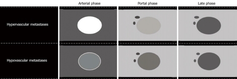
Both hypovascular and hypervascular liver metastases have a predominantly arterial blood supply, but the degree of arterial perfusion is variable, and the contrast enhancement in the arterial phase is related to this variation. Hypovascular metastases, which have relatively low arterial perfusion, are usually seen in patients with adenocarcinoma or squamous cell carcinoma, most likely related to colorectal cancer, gastric cancer, pancreatic cancer or ovarian cancer. In nearly all hypovascular metastases, contrast enhancement of varying degrees is seen in the arterial phase, typically in the periphery (rim enhancement) (Figure 2). However, some authors describe a more diffuse enhancement, especially when the tumor is small[28].
Figure 2.
Hypovascular liver metastases visualized with US and CEUS. The same hypovascular liver metastasis is visualized by US and CEUS in both the arterial and portal phases. In the arterial phase a slight rim enhancement is seen (arrows). The non-enhancing area in the center represents a necrosis, and is demonstrated in both the arterial and late phases. A: B-mode US; B: Arterial phase; C: Late phase.
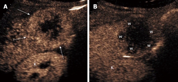
Hypervascular metastases, which frequently arise from neuroendocrine tumors, malignant melanoma, and sarcoma, as well as from renal, breast, or thyroid cancer, have a very high arterial perfusion and display a diffuse or inhomogeneous enhancement in the arterial phase. These metastases present a hyper-reflective signal compared to the surrounding normal liver parenchyma (Table 2 and Figure 3).
Table 2.
Enhancement patterns of liver metastases
| Tumor entity | Arterial phase | Portal phase | Delayed phase |
| Hypovascular metastasis | |||
| Typical features | Rim enhancement | Hypo-enhancement | Hypo-/non-enhancement |
| Additional features | Complete enhancement | Non-enhancement areas | |
| Non-enhancement areas (necrosis) | |||
| Hypervascular metastasis | |||
| Typical features | Hyper-enhancement, complete | Hypo-enhancement | Hypo-/non-enhancement |
| Additional features | Chaotic vessels | ||
| Cystic metastasis | |||
| Typical features | Hyper-enhancement nodular/rim component | Hypo-enhancement | Hypo-enhancement |
Figure 3.
Hypervascular liver metastases visualized by CEUS. The patient is a woman, 77 years old, with renal cancer and hypervascular liver metastases in segment 6 as visualized with CEUS. In the arterial phase, the metastases present a hyper-reflective signal when compared to the surrounding normal liver parenchyma (arrows). The non- enhanced area in the center represents necrosis. In the late phase there is clearly a washout of contrast in the metastases (w). The kidneys are visualized below the liver (k). A: Arterial phase; B: Late phase.
In the portal and delay phases, both hyper- and hypovascular metastases appear as dark defects, while the enhancement persists in normal liver parenchyma (Figure 4). The metastases do not retain the contrast agent like the normal liver parenchyma. This rapid and complete “washout” in the metastases can be explained by a consistently lower fractional vascular volume when compared to normal liver parenchyma[29], and also by the absence of portal supply to the neoplastic lesions. The “washout” phenomenon in CEUS must not be confused with the “washout” of contrast agents seen on CT or MRI. The ultrasound contrast agent remains exclusively intravascular, which is not the case for the contrast agents used in CT and MRI.
Figure 4.
Typical contrast enhancement pattern of a hepatic metastasis by CEUS.
Using CEUS, it is possible to differentiate cystic metastases from a non-neoplastic complex of cysts by demonstrating vascular flow in the cyst wall or in mural nodules[28].
The optimal time to scan for all types of metastases is from about 90 s to 5 min. This is the time when the contrast between the enhanced normal liver and the non-enhanced metastases is most pronounced, but some metastases can be detected in the arterial phase, especially hypervascular metastases.
RESULTS OF CLINICAL STUDIES
General considerations
One of the main problems in comparing the sensitivity of CEUS in liver metastasis detection to other imaging modalities is the choice of a gold standard. Theoretically, all detected liver lesions must be histologically proven, but for ethical and technical reasons, this is not possible. Due to a lack of verification, many studies on the topic have important limitations. Not all patients with liver metastases are histologically verified, and in cases where histology is obtained, it is usually only performed on one lesion per patient, even if multiple lesions are present. This is especially problematic in lesion-by-lesion analysis. However, in some studies, the preoperative CEUS findings have been compared with the histopathological specimens after liver resection, which is clearly the ideal gold standard[30]. Such results are correct for the particular specimen, but this practice does not exclude non-visualized metastases in the remainder of the liver.
In cases where the gold standard is based only on imaging modalities, like CT or MRI, the true prevalence of liver metastases remains unknown, since some of the smaller liver metastases will remain undetected and will not be included in the population of metastases[21]. Even the use of intraoperative ultrasonography (IOUS) as a gold standard cannot guarantee 100% reliability, primarily because it is unable to detect micrometastases[23]. Another problem is that many studies use CT (sometimes together with histology, MRI, and follow-up)[31-34] as the gold standard and then compare the sensitivity of CEUS and CT, which inevitably leads to an overestimate of CT’s sensitivity. However, bearing these studies’ limitations in mind, the results of the clinical studies concerning CEUS versus US and CEUS versus CT for the assessment of liver metastases are described in the next section.
Contrast enhanced US versus US
Several studies have shown that CEUS detects more liver metastases than US (Table 1). The first published studies were mostly based on lesion-by-lesion analysis. All of these studies showed that CEUS has better sensitivity in detecting liver metastases than US[35-37]. However, it is well known that it is often difficult to compare the same liver lesions by US and CEUS, despite their assignment to a Couinaud segment. Thus, when many liver lesions are present and are analyzed on a lesion-by-lesion basis, there is a potential source of error[37,38]. Nevertheless, these promising results have been confirmed in several studies based on a patient-by-patient analysis[23,32,33,38-40]. Most of these studies showed that the detection of liver metastases was significantly improved, by between 5% and 62%, with one study showing a 107% improvement. However, this study also found that US had a remarkably low sensitivity (0.40)[31]. The impact of CEUS on therapeutic strategies also seems to be significant. In one study involving[40] patients who underwent laparatomy for liver resection, the preoperative US findings would have led to an extension of the resections in 16 cases (40%). In a significant number of cases (9/40 patients, 22.5%), preoperative CEUS findings changed the surgical strategy[30]. Furthermore, in patients treated with chemotherapy, CEUS had significantly greater sensitivity than US as determined by both patient-by-patient analysis (79.5% vs 63.2%) and lesion-by-lesion analysis (82.0% vs 60.3%)[30]. Another study showed that the origin of the metastases seems not to influence the rate of detection in either US or CEUS (colorectal liver metastases were compared to metastases from cancers originating in the pancreas, kidneys, stomach, ovaries, breast, gallbladder and lungs)[23].
Contrast enhanced intraoperative US (CE-IOUS) versus intraoperative US (IOUS)
Two studies have found a further value for CE-IOUS, describing additional findings of colorectal liver metastases using contrast agents as compared to conventional IOUS[41,42]. Additional liver metastases were found in 19% and 13% of the patients, respectively, and in one of the studies, CE-IOUS altered the surgical plans in 30% of cases[41]. On the other hand, a recently published study involving 39 patients with 137 identified malignant lesions concluded that the use of CE-IOUS in addition to preoperative contrast enhanced CT and IOUS did not improve the ability to characterize previously detected lesions. This study also showed that only in a small number of patients did CE-IOUS facilitate the detection of new liver metastases or have implications on surgical strategy[43]. The differences in these results could be explained by the varying levels of skill with the techniques used in the studies, but further studies on the role of CE-IOUS seem to be needed.
Contrast enhanced US versus CT
The detection rate for liver metastases by CEUS seems to be similar to the best reported results by CT and MRI[15,31-33,39,44]. However, some of the studies must be evaluated with caution, due to the use of unclear gold standards[31] and because CEUS is sometimes compared to somewhat out-of-date CT equipment[31,33,39]. The technical advantages achieved with modern multidetector CT (MDCT) might show that the results from studies using single-slice CT and thick scan slices are no longer valid when compared to modern MDCT. None of the existing studies found significant differences between CEUS and CT with regard to sensitivity in detecting liver metastases (Table 3), but most studies found the sensitivity of CEUS to be slightly higher[33,39,40].
Table 3.
Sensitivity of CEUS and CT in detecting liver metastases; an overview of studies
| Study | n | Study group1 | Type of CT | Analysis | Gold standard |
Sensitivity |
Statistic | |
| CEUS | CT | |||||||
| Quaia et al[31], 2006 | 253 | K or S | 1-slice CT | P-by-P3 | CT, FNA, Follow-up, MRI, IOUS | 0.83 | 0.89 | NS2 |
| Larsen et al[34] , 2007 | 365 | S | 4-slice CT | P-by-P | CT, IOUS, CEUS, FNA, surgery resection, follow-up | 0.8 | 0.89 | NS |
| Dietrich et al[32] | 131 | UK | Multislice CT in most cases4 | P-by-P | CT, MRI, FNA, follow up | 0.91 | 0.89 | NS |
| Piscaglia et al[33], 2007 | 109 | S | 1-or 4-slice CT | P-by-P | CT, US, FNA, Follow-up | 0.95 | 0.91 | NS |
| Janica et al[40], 2007 | 51 | K or S | Not described | L-by-L5 | FNA, surgical resection, CT and follow-up | 0.9 | 0.78 | NS |
| Konopke et al[39], 2005 | 56 | S or K | 1-, 4- or 16-slice CT | P-by-P | IOUS, FNA, CT | 0.86 | 0.76 | NS |
Included patients with known (K), suspected (S) or unknown (UK) liver metastases;
Not significant;
Patient-by-patient analysis;
Details not described.
Lesion-by-lesion analysis.
Contrast enhanced US versus MRI or positron emission tomography (PET)
To date, there have been no studies comparing the effectiveness of CEUS and MRI [with liver-specific contrast agents like SPIO (superparamagnetic iron oxide) or Gd-EOB-DTBA (gadolinium ethoxybenzyl diethylenetriaminepentaacetic acid)] or PET in detecting liver metastases. However, in some studies, MRI is used in combination with other imaging modalities as the gold standard (Tables 1 and 3).
LIMITATIONS OF CEUS
In general, if an examination of the liver by US is insufficient, then examination by CEUS will also be insufficient. The limitations that apply to US are the same as those that apply to CEUS, so the quality of the examination still depends on the skill of the operator. In addition, as has been noted previously, CEUS has limited ability to observe certain parts of the liver, especially in obese patients and/or in cases of steatosis. Further limitations are related to the acoustic window and movement artifacts.
It is not possible to simultaneously examine multiple lesions in the arterial and early portal phases[45]. The characterization of suspected liver lesions can also be complicated by difficulties in interpreting results in cases of hypervascular metastases and hemangiomas with incomplete filling.
In addition, in patients with many cysts, metastases can be missed because it can be difficult to differentiate some of them from cysts in the late contrast phase, since both appear hypoechoic. Conversely, cysts can be misinterpreted as metastases. Also in cases with few and relatively small cysts, which have not been detected by US, the cysts can be misinterpreted as small metastases. They can usually be distinguished from metastases by their characteristically increased transmission[46]. However, it is recommended that cysts be confirmed by US before CEUS[38,47].
THE CLINICAL ROLE OF CEUS IN THE DETECTION OF LIVER METASTASES
The European Federation of Societies for Ultrasound in Medicine and Biology (EFSUMB) in Guidelines and good clinical practice recommendations for contrast enhanced ultrasound (CEUS) - update 2008[46] recommends that CEUS be used in the following cases: (1) All liver ultrasound scans to rule out liver metastases or abscess, unless conventional ultrasound shows clear evidence of these lesions; (2) In selected cases, when clinically relevant for treatment planning by helping to assess the number and location of liver metastases as a complement to contrast enhanced CT and/or contrast enhanced MRI; and (3) For surveillance of oncology patients in whom CEUS has previously been useful. These guidelines are based on literature surveys including results from several clinical trials, some of which have been mentioned earlier in this paper. However, it is important to emphasize that CEUS is complementary to CT/MRI in the preoperative staging before liver resection. It cannot replace the other imaging modalities in the preoperative work-up or in the follow-up of patients with liver metastases during chemotherapy, since CT and MRI give more comprehensive information about the liver and all other organs. Even the PACS-systems have been improved, so it is easier to manage digital cine-loops, and while some institutions have introduced the practice of doing a standard sweep of the entire liver when performing CEUS, there are still problems with reproducible image documentation. This limits the ability of CEUS to clearly show small changes over time. In most cancer centers, including ours, CT and MRI are therefore preferred for follow-up imaging. On the other hand, CEUS is a very useful and important non-invasive imaging modality for resolving problems, such as patients with or without known malignancies where CT has demonstrated an uncharacteristic appearance. It is also necessary to emphasize that, in patients who are known or strongly suspected of having a malignancy, an adequate US examination for the detection of liver metastases includes CEUS, even if the baseline US is normal. Numerous studies have demonstrated that CEUS clearly and significantly improved sensitivity and specificity in the detection of liver metastases (Table 1).
The economic advantage of the use of CEUS for characterization of focal liver lesions seems to be clear. Compared with CT and MRI, CEUS provides significant cost savings, both for a national health service and for hospitals[48,49]. However, there are still some barriers to using CEUS, like the costs of CEUS compared to US and the fees paid by health insurance companies to perform CEUS[50].
CONCLUSION
The use of second generation ultrasound contrast agents in combination with low MI contrast-specific US techniques has clearly improved US imaging of the liver, including the dynamic examination of focal liver lesions. Contrast enhanced US has improved the detection of liver metastases when compared to US itself, and it seems to have a diagnostic performance and accuracy similar to that of CT.
Footnotes
Peer reviewer: Seong Woo Jeon, Assistant Professor, Internal Medicine, Kyungpook National University Hospital, 50 Samduk-2Ga, Chung-gu, Daegu 700-721, South Korea
S- Editor Zhang HN L- Editor Herholdt A E- Editor Liu N
References
- 1.Tong D, Russell AH, Dawson LE, Wisbeck W. Second laparotomy for proximal colon cancer. Sites of recurrence and implications for adjuvant therapy. Am J Surg. 1983;145:382–386. doi: 10.1016/0002-9610(83)90207-6. [DOI] [PubMed] [Google Scholar]
- 2.Gilbert JM, Jeffrey I, Evans M, Kark AE. Sites of recurrent tumour after 'curative' colorectal surgery: implications for adjuvant therapy. Br J Surg. 1984;71:203–205. doi: 10.1002/bjs.1800710311. [DOI] [PubMed] [Google Scholar]
- 3.Russell AH, Tong D, Dawson LE, Wisbeck W. Adenocarcinoma of the proximal colon. Sites of initial dissemination and patterns of recurrence following surgery alone. Cancer. 1984;53:360–367. doi: 10.1002/1097-0142(19840115)53:2<360::aid-cncr2820530232>3.0.co;2-u. [DOI] [PubMed] [Google Scholar]
- 4.Pickren JW, Tsuka Y, Lane WW. Liver metastases. In: Weiss L, Gilbert HA, editors. Liver metastasis. Boston: GK Hall Medical Publischers; 1982. pp. 2–18. [Google Scholar]
- 5.Cosgrove DO. Malignant liver disease. In: Meire HB, Cosgrove DO, Dewbury KC, Farrant P, editors. Clinical ultrasound: a comprehensive text, 2nd ed. London: Churchill Livingstone; 2001. pp. 211–231. [Google Scholar]
- 6.Robinson PJ. Imaging liver metastases: current limitations and future prospects. Br J Radiol. 2000;73:234–241. doi: 10.1259/bjr.73.867.10817037. [DOI] [PubMed] [Google Scholar]
- 7.Karhunen PJ. Benign hepatic tumours and tumour like conditions in men. J Clin Pathol. 1986;39:183–188. doi: 10.1136/jcp.39.2.183. [DOI] [PMC free article] [PubMed] [Google Scholar]
- 8.Edmondson HA, Craig JR. Neoplasms of the liver. In: Schiff L, editor. Diseases of the liver, 8th ed. Philadelphia: Lippincott; 1987. [Google Scholar]
- 9.Kreft B, Pauleit D, Bachmann R, Conrad R, Krämer A, Schild HH. [Incidence and significance of small focal liver lesions in MRI] Rofo. 2001;173:424–429. doi: 10.1055/s-2001-13340. [DOI] [PubMed] [Google Scholar]
- 10.Jones EC, Chezmar JL, Nelson RC, Bernardino ME. The frequency and significance of small (less than or equal to 15 mm) hepatic lesions detected by CT. AJR Am J Roentgenol. 1992;158:535–539. doi: 10.2214/ajr.158.3.1738990. [DOI] [PubMed] [Google Scholar]
- 11.Schwartz LH, Gandras EJ, Colangelo SM, Ercolani MC, Panicek DM. Prevalence and importance of small hepatic lesions found at CT in patients with cancer. Radiology. 1999;210:71–74. doi: 10.1148/radiology.210.1.r99ja0371. [DOI] [PubMed] [Google Scholar]
- 12.Ohlsson B, Nilsson J, Stenram U, Akerman M, Tranberg KG. Percutaneous fine-needle aspiration cytology in the diagnosis and management of liver tumours. Br J Surg. 2002;89:757–762. doi: 10.1046/j.1365-2168.2002.02111.x. [DOI] [PubMed] [Google Scholar]
- 13.Wernecke K, Rummeny E, Bongartz G, Vassallo P, Kivelitz D, Wiesmann W, Peters PE, Reers B, Reiser M, Pircher W. Detection of hepatic masses in patients with carcinoma: comparative sensitivities of sonography, CT, and MR imaging. AJR Am J Roentgenol. 1991;157:731–739. doi: 10.2214/ajr.157.4.1892027. [DOI] [PubMed] [Google Scholar]
- 14.Glover C, Douse P, Kane P, Karani J, Meire H, Mohammadtaghi S, Allen-Mersh TG. Accuracy of investigations for asymptomatic colorectal liver metastases. Dis Colon Rectum. 2002;45:476–484. doi: 10.1007/s10350-004-6224-y. [DOI] [PubMed] [Google Scholar]
- 15.Oldenburg A, Hohmann J, Foert E, Skrok J, Hoffmann CW, Frericks B, Wolf KJ, Albrecht T. Detection of hepatic metastases with low MI real time contrast enhanced sonography and SonoVue. Ultraschall Med. 2005;26:277–284. doi: 10.1055/s-2005-858526. [DOI] [PubMed] [Google Scholar]
- 16.Bruneton JN, Raffaelli C, Balu-Maestro C, Padovani B, Chevallier P, Mourou MY. Sonographic diagnosis of solitary solid liver nodules in cancer patients. Eur Radiol. 1996;6:439–442. doi: 10.1007/BF00182462. [DOI] [PubMed] [Google Scholar]
- 17.Valls C, Andia E, Sanchez A, Guma A, Figueras J, Torras J, Serrano T. Hepatic metastases from colorectal cancer: preoperative detection and assessment of resectability with helical CT. Radiology. 2001;218:55–60. doi: 10.1148/radiology.218.1.r01dc1155. [DOI] [PubMed] [Google Scholar]
- 18.Portugaller HR, Stacher R, Komaz G, Aschauer M, Hausegger KA, Szolar DH. [The value of different spiral CT phases in the detection of liver metastases] Rofo. 2002;174:452–458. doi: 10.1055/s-2002-25122. [DOI] [PubMed] [Google Scholar]
- 19.Rappeport ED, Loft A. Liver metastases from colorectal cancer: imaging with superparamagnetic iron oxide (SPIO)-enhanced MR imaging, computed tomography and positron emission tomography. Abdom Imaging. 2007;32:624–634. doi: 10.1007/s00261-007-9297-y. [DOI] [PubMed] [Google Scholar]
- 20.Kim YK, Ko SW, Hwang SB, Kim CS, Yu HC. Detection and characterization of liver metastases: 16-slice multidetector computed tomography versus superparamagnetic iron oxide-enhanced magnetic resonance imaging. Eur Radiol. 2006;16:1337–1345. doi: 10.1007/s00330-005-0140-y. [DOI] [PubMed] [Google Scholar]
- 21.van Erkel AR, Pijl ME, van den Berg-Huysmans AA, Wasser MN, van de Velde CJ, Bloem JL. Hepatic metastases in patients with colorectal cancer: relationship between size of metastases, standard of reference, and detection rates. Radiology. 2002;224:404–409. doi: 10.1148/radiol.2242011322. [DOI] [PubMed] [Google Scholar]
- 22.Albrecht T, Hoffmann CW, Schmitz SA, Schettler S, Overberg A, Germer CT, Wolf KJ. Phase-inversion sonography during the liver-specific late phase of contrast enhancement: improved detection of liver metastases. AJR Am J Roentgenol. 2001;176:1191–1198. doi: 10.2214/ajr.176.5.1761191. [DOI] [PubMed] [Google Scholar]
- 23.Konopke R, Kersting S, Bergert H, Bloomenthal A, Gastmeier J, Saeger HD, Bunk A. Contrast-enhanced ultrasonography to detect liver metastases : a prospective trial to compare transcutaneous unenhanced and contrast-enhanced ultrasonography in patients undergoing laparotomy. Int J Colorectal Dis. 2007;22:201–207. doi: 10.1007/s00384-006-0134-5. [DOI] [PubMed] [Google Scholar]
- 24.Hohmann J, Skrok J, Puls R, Albrecht T. [Characterization of focal liver lesions with contrast-enhanced low MI real time ultrasound and SonoVue] Rofo. 2003;175:835–843. doi: 10.1055/s-2003-39923. [DOI] [PubMed] [Google Scholar]
- 25.Cosgrove D. Ultrasound contrast agents: an overview. Eur J Radiol. 2006;60:324–330. doi: 10.1016/j.ejrad.2006.06.022. [DOI] [PubMed] [Google Scholar]
- 26.Albrecht T. Detection and characterisation of liver metastases. In: Lencioni R, editor. Enhancing the role of ultrasound with contrast agents. Pisa: Springer-Verlag Italia; 2006. pp. 53–67. [Google Scholar]
- 27.Albrecht T, Blomley M, Bolondi L, Claudon M, Correas JM, Cosgrove D, Greiner L, Jäger K, Jong ND, Leen E, et al. Guidelines for the use of contrast agents in ultrasound. January 2004. Ultraschall Med. 2004;25:249–256. doi: 10.1055/s-2004-813245. [DOI] [PubMed] [Google Scholar]
- 28.Jang HJ, Kim TK, Wilson SR. Imaging of malignant liver masses: characterization and detection. Ultrasound Q. 2006;22:19–29. [PubMed] [Google Scholar]
- 29.Cosgrove D, Blomley M. Liver tumors: evaluation with contrast-enhanced ultrasound. Abdom Imaging. 2004;29:446–454. doi: 10.1007/s00261-003-0126-7. [DOI] [PubMed] [Google Scholar]
- 30.Konopke R, Bunk A, Kersting S. Contrast-enhanced ultrasonography in patients with colorectal liver metastases after chemotherapy. Ultraschall Med. 2008;29 Suppl 4:S203–S209. doi: 10.1055/s-2008-1027795. [DOI] [PubMed] [Google Scholar]
- 31.Quaia E, D’Onofrio M, Palumbo A, Rossi S, Bruni S, Cova M. Comparison of contrast-enhanced ultrasonography versus baseline ultrasound and contrast-enhanced computed tomography in metastatic disease of the liver: diagnostic performance and confidence. Eur Radiol. 2006;16:1599–1609. doi: 10.1007/s00330-006-0192-7. [DOI] [PubMed] [Google Scholar]
- 32.Dietrich CF, Kratzer W, Strobe D, Danse E, Fessl R, Bunk A, Vossas U, Hauenstein K, Koch W, Blank W, et al. Assessment of metastatic liver disease in patients with primary extrahepatic tumors by contrast-enhanced sonography versus CT and MRI. World J Gastroenterol. 2006;12:1699–1705. doi: 10.3748/wjg.v12.i11.1699. [DOI] [PMC free article] [PubMed] [Google Scholar]
- 33.Piscaglia F, Corradi F, Mancini M, Giangregorio F, Tamberi S, Ugolini G, Cola B, Bazzocchi A, Righini R, Pini P, et al. Real time contrast enhanced ultrasonography in detection of liver metastases from gastrointestinal cancer. BMC Cancer. 2007;7:171. doi: 10.1186/1471-2407-7-171. [DOI] [PMC free article] [PubMed] [Google Scholar]
- 34.Larsen LP, Rosenkilde M, Christensen H, Bang N, Bolvig L, Christiansen T, Laurberg S. Can contrast-enhanced ultrasonography replace multidetector-computed tomography in the detection of liver metastases from colorectal cancer? Eur J Radiol. 2009;69:308–313. doi: 10.1016/j.ejrad.2007.10.023. [DOI] [PubMed] [Google Scholar]
- 35.Dalla Palma L, Bertolotto M, Quaia E, Locatelli M. Detection of liver metastases with pulse inversion harmonic imaging: preliminary results. Eur Radiol. 1999;9 Suppl 3:S382–S387. doi: 10.1007/pl00014079. [DOI] [PubMed] [Google Scholar]
- 36.Esteban JM, Molla MA, Tomas C, Maldonado L. Improved detection of liver metastases with contrast-enhanced wideband harmonic imaging: comparison with CT findings. Eur J Ultrasound. 2002;15:119–126. doi: 10.1016/s0929-8266(02)00032-0. [DOI] [PubMed] [Google Scholar]
- 37.Harvey CJ, Blomley MJ, Eckersley RJ, Cosgrove DO, Patel N, Heckemann RA, Butler-Barnes J. Hepatic malignancies: improved detection with pulse-inversion US in late phase of enhancement with SH U 508A-early experience. Radiology. 2000;216:903–908. doi: 10.1148/radiology.216.3.r00se22903. [DOI] [PubMed] [Google Scholar]
- 38.Larsen LP, Rosenkilde M, Christensen H, Bang N, Bolvig L, Christiansen T, Laurberg S. The value of contrast enhanced ultrasonography in detection of liver metastases from colorectal cancer: a prospective double-blinded study. Eur J Radiol. 2007;62:302–307. doi: 10.1016/j.ejrad.2006.11.033. [DOI] [PubMed] [Google Scholar]
- 39.Konopke R, Kersting S, Saeger HD, Bunk A. [Detection of liver lesions by contrast-enhanced ultrasound -- comparison to intraoperative findings] Ultraschall Med. 2005;26:107–113. doi: 10.1055/s-2005-858095. [DOI] [PubMed] [Google Scholar]
- 40.Janica JR, Lebkowska U, Ustymowicz A, Augustynowicz A, Kamocki Z, Werel D, Polaków J, Kedra B, Pepinski W. Contrast-enhanced ultrasonography in diagnosing liver metastases. Med Sci Monit. 2007;13 Suppl 1:111–115. [PubMed] [Google Scholar]
- 41.Leen E, Ceccotti P, Moug SJ, Glen P, MacQuarrie J, Angerson WJ, Albrecht T, Hohmann J, Oldenburg A, Ritz JP, et al. Potential value of contrast-enhanced intraoperative ultrasonography during partial hepatectomy for metastases: an essential investigation before resection? Ann Surg. 2006;243:236–240. doi: 10.1097/01.sla.0000197708.77063.07. [DOI] [PMC free article] [PubMed] [Google Scholar]
- 42.Torzilli G, Palmisano A, Del Fabbro D, Marconi M, Donadon M, Spinelli A, Bianchi PP, Montorsi M. Contrast-enhanced intraoperative ultrasonography during surgery for hepatocellular carcinoma in liver cirrhosis: is it useful or useless? A prospective cohort study of our experience. Ann Surg Oncol. 2007;14:1347–1355. doi: 10.1245/s10434-006-9278-3. [DOI] [PubMed] [Google Scholar]
- 43.Fioole B, de Haas RJ, Wicherts DA, Elias SG, Scheffers JM, van Hillegersberg R, van Leeuwen MS, Borel Rinkes IH. Additional value of contrast enhanced intraoperative ultrasound for colorectal liver metastases. Eur J Radiol. 2008;67:169–176. doi: 10.1016/j.ejrad.2007.03.017. [DOI] [PubMed] [Google Scholar]
- 44.Bipat S, van Leeuwen MS, Comans EF, Pijl ME, Bossuyt PM, Zwinderman AH, Stoker J. Colorectal liver metastases: CT, MR imaging, and PET for diagnosis--meta-analysis. Radiology. 2005;237:123–131. doi: 10.1148/radiol.2371042060. [DOI] [PubMed] [Google Scholar]
- 45.Seitz K. Contrast-enhanced ultrasound in the diagnosis of hepatocellular carcinoma and liver metastases. Ultraschall Med. 2005;26:267–269. doi: 10.1055/s-2005-858559. [DOI] [PubMed] [Google Scholar]
- 46.Claudon M, Cosgrove D, Albrecht T, Bolondi L, Bosio M, Calliada F, Correas JM, Darge K, Dietrich C, D'Onofrio M, et al. Guidelines and good clinical practice recommendations for contrast enhanced ultrasound (CEUS)-update 2008. Ultraschall Med. 2008;29:28–44. doi: 10.1055/s-2007-963785. [DOI] [PubMed] [Google Scholar]
- 47.Dietrich CF. Characterisation of focal liver lesions with contrast enhanced ultrasonography. Eur J Radiol. 2004;51 Suppl:S9–S17. doi: 10.1016/j.ejrad.2004.03.034. [DOI] [PubMed] [Google Scholar]
- 48.Romanini L, Passamonti M, Aiani L, Cabassa P, Raieli G, Montermini I, Martegani A, Grazioli L, Calliada F. Economic assessment of contrast-enhanced ultrasonography for evaluation of focal liver lesions: a multicentre Italian experience. Eur Radiol. 2007;17 Suppl 6:F99–F106. doi: 10.1007/s10406-007-0234-5. [DOI] [PubMed] [Google Scholar]
- 49.Faccioli N, D'Onofrio M, Comai A, Cugini C. Contrast-enhanced ultrasonography in the characterization of benign focal liver lesions: activity-based cost analysis. Radiol Med. 2007;112:810–820. doi: 10.1007/s11547-007-0185-x. [DOI] [PubMed] [Google Scholar]
- 50.Mostbeck G. [CEUS from a radiological standpoint: dream and reality] Ultraschall Med. 2009;30:125–127. doi: 10.1055/s-0028-1109334. [DOI] [PubMed] [Google Scholar]
- 51.Albrecht T, Blomley MJ, Burns PN, Wilson S, Harvey CJ, Leen E, Claudon M, Calliada F, Correas JM, LaFortune M, et al. Improved detection of hepatic metastases with pulse-inversion US during the liver-specific phase of SHU 508A: multicenter study. Radiology. 2003;227:361–370. doi: 10.1148/radiol.2272011833. [DOI] [PubMed] [Google Scholar]
- 52.Solbiati L, Tonolini M, Cova L, Goldberg SN. The role of contrast-enhanced ultrasound in the detection of focal liver leasions. Eur Radiol. 2001;11 Suppl 3:E15–E26. doi: 10.1007/pl00014125. [DOI] [PubMed] [Google Scholar]
- 53.Bernatik T, Strobel D, Hahn EG, Becker D. Detection of liver metastases: comparison of contrast-enhanced wide-band harmonic imaging with conventional ultrasonography. J Ultrasound Med. 2001;20:509–515. doi: 10.7863/jum.2001.20.5.509. [DOI] [PubMed] [Google Scholar]


