Abstract
Granular cell tumors, also called Abrikossof’s tumors, were originally described by Abrikossof A in 1926. The first case of a pancreatic granular cell tumor was described in 1975 and only 6 cases have been reported. We describe a case of granular cell tumor in the pancreas showing pancreatic duct obstruction. Because imaging studies showed findings compatible with those of pancreatic carcinoma, the patient underwent distal pancreatectomy. Histological examination showed that the tumor consisted of a nested growth of large tumor cells with ample granular cytoplasm and small round nuclei. The tumor cells expressed S-100 protein and were stained with neuron-specific enolase and periodic acid-Schiff, but were negative for desmin, vimentin, and cytokeratin. The resected tumor was diagnosed as a granular cell tumor. To our knowledge, this is the seventh case of Granular cell tumor of the pancreas to be reported.
Keywords: Granular cell tumor, Pancreas, Diagnosis, Distal pancreatectomy
INTRODUCTION
Granular cell tumors (GCTs) are rare benign neoplasms of Schwann cell origin and have been found in virtually every location in the body, including breast[1], pituitary[2], central nervous system[3], respiratory tract[4], and gastrointestinal tract[5,6]. Supportive evidence that GCT arises from Schwann cells comes from the findings[7,8] that GCT cells contain S-100 protein, a unique acidic protein that is present in Schwann cells and satellite cells of ganglia but not often in nonneural soft tissue tumors. GCT of the pancreas is extremely rare and, to date, only 6 cases have been reported. We report an additional case of pancreatic GCT and describe certain aspects of its clinical, radiologic, and histologic features.
CASE REPORT
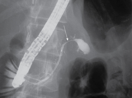
A 39-year-old woman presented with mild unspecific abdominal pain for about 1 mo. In a local hospital, she initially had ultrasound of the abdomen, which identified a dilated main pancreatic duct from body to tail of the pancreas. She was referred to our hospital for evaluation of the pancreatic tumor. Twelve years previously, the patient underwent extraction of the left adrenal gland for primary aldosteronism. Otherwise, her past medical history was noncontributory. By conventional ultrasonography, the tumor was revealed as a hypoechoic area in the body of the pancreas. Endoscopic ultrasonography (EUS) showed a homogeneous solid mass with a regular border that appeared hypoechoic compared with the normal pancreatic parenchyma (Figure 1). Computed tomography (CT) demonstrated a 2 cm × 2 cm low-density lesion located in the body of the pancreas with dilatation of the main pancreatic duct. The early phase of dynamic CT revealed a slightly less enhanced mass in the pancreatic body compared to normal pancreatic tissues (Figure 2A). However, the tumor demonstrated gradual enhancement at the delayed phase of dynamic CT (Figure 2B). On magnetic resonance imaging (MRI), the tumor of the pancreas body was hypointense on a T1-weighted image (Figure 3A). In contrast, the peripheral and central areas of the tumor were, respectively, hypointense and hyperintense on aT2-weighted image (Figure 3B). The mass did not infiltrate the portal vein or celiac artery. The patient underwent endoscopic retrograde cholangiopancreatography, which showed a normal proximal pancreatic duct and a stricture in the midpancreatic duct with a dilated distal pancreatic duct (Figure 4). Cytological examination on material from the region of the narrowing was negative for malignant cells. Routine laboratory studies were normal. Carcinoembryonic antigen and cancer antigen 19-9 remained in the normal range. The preoperative differential diagnosis was pancreatic tumor including pancreatic adenocarcinoma. Laparotomy revealed that the tumor originated from the pancreatic body. There was no extension to adjacent organs, and no metastatic lesions were found. Distal pancreatectomy and splenectomy were performed. Histological examination confirmed that the tumor was completely resected. The margin was free of tumor cells, and none of 7 regional lymph nodes examined showed metastasis. The post-operative course was uneventful. Macroscopically, approximate 22mm × 20mm × 20 mm in diameter of whitish tumor was located in pancreatic body. The tumor encircled and narrowed the main pancreatic duct, and its upstream main pancreatic duct was dilated. Microscopic study showed a well-limited nodule made up of large clusters of benign cells with small nuclei and abundant granular cytoplasm (Figure 5A), which were weakly positive with the periodic acid-Schiff staining. S-100 protein staining was also positive in the cell cytoplasm by immunohistochemistry (Figure 5B). The final diagnosis was a granular cell tumor of the pancreas narrowing the main pancreatic duct.
Figure 1.
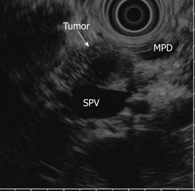
EUS image of pancreatic GCT. The tumor showed a homogeneous pattern and regular borders (arrow). EUS: Endoscopic ultrasonography; GCT: Granular cell tumor; MPD: main pancreatic duct; PV: portal vein.
Figure 2.
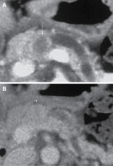
CT image of pancreatic GCT. A: CT showing poor enhancement of the tumor compared with that of the surrounding pancreatic parenchyma at early phase o dynamic CT (arrow); B: CT showing gradual enhancement of the tumor at delayed phase (arrowhead). CT: Computed tomography.
Figure 3.
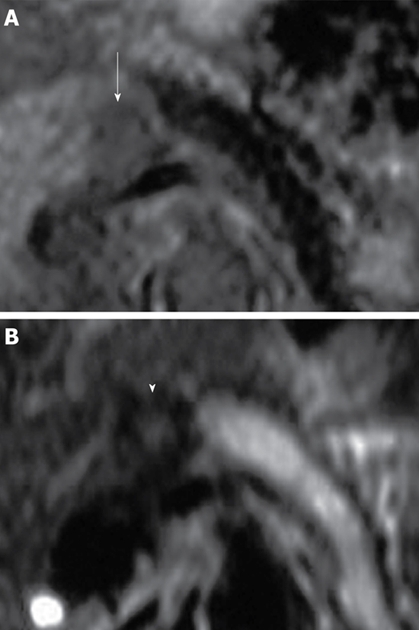
MRI findings of pancreatic GCT. A: The tumor showed a hypointense mass in the T1-weighted image (arrow); B: The surrounding and center of the tumor were hypointense and hyperintense on T2-weighted image, respectively (arrowhead). MRI: magnetic resonance imaging.
Figure 4.
ERCP showing the stricture and the dilatation in the distal pancreatic duct (arrow). ERCP: Endoscopic retrograde cholangiopancreatography.
Figure 5.
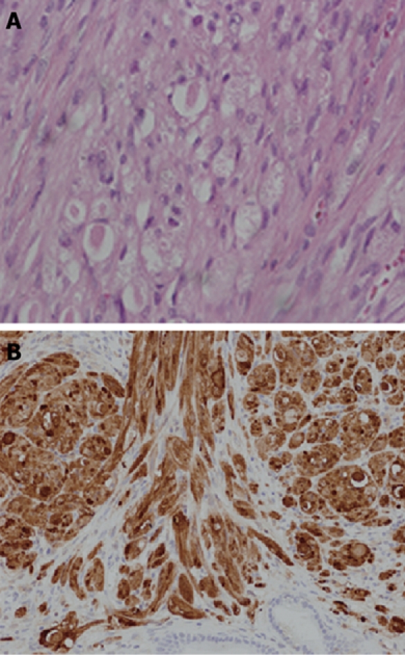
Histological findings. A: Microscopic study showed a well-limited nodule made of large clusters of benign cells with small nuclei and abundant granular cytoplasm; B: S-100 protein staining was positive in the cell cytoplasm.
DISCUSSION
The first reported case of granular cell tumor was in 1926 by Abrikossoff[9]. The tumor was found in the skeletal muscle of the tongue. Although this type of tumor is known to arise in every part of the body, GCT of the pancreas is very rare. Only six cases of GCT of the pancreas had been reported[10-15] previously, and the characteristics of these cases are sumarized in Table 1. Because of the rarity of pancreatic GCT, the characteristic epidemiology, clinical symptoms and radiological findings cannot be clarified. Although GCT is usually benign, malignant cases have been reported in subcutaneous leg tissue and esophagus (1%-2% of all GCT)[16]. There are reports of cases that have recurred or metastasized despite having a benign histological appearance. Although the morphology cannot reliably predict the biological behavior of GCT, local recurrence, rapid growth to a size greater than 4 cm, and an infiltrative pattern of growth should raise concerns about the possibility of malignancy[17-19].
Table 1.
Summary of the characteristics of the 6 cases of the granu;ar cell tumor of the pancreas found in the literature
| Author | Age | Sex | Locarization | Size (mm) | Treatment |
| Wellman et al[10] | 29 | M | Head | 6 × 4 × 3 | - |
| Sekes et al[11] | 31 | F | Head | 5 | Pancreaticojejunostomy |
| Seidler et al[12] | 62 | F | Tail | 7 × 5 | Distal pancreatectomy |
| Bin-Sagheer et al[13] | 50 | F | Body-Tail | - | Distal pancreatectomy |
| Méklati et al[14] | 26 | F | Body-Tail | 5 | Distal pancreatectomy |
| Nojiri et al[15] | 58 | M | Head | 13 | Pancreatoduodenectomy |
| Present case | 39 | F | Body | 20 | Distal pancreatectomy |
Histopathologically, the present case of GCT showed diffuse oval tumor cells with low grade atypia and eosinophilic granules were found within the tumor cells. Positive PAS staining and immunohistochemical staining for S-100 protein and neuron specific enolase in the tumor provided the evidence for the diagnosis of GCT[20,21].
The preoperative diagnosis of pancreatic GCT is very difficult since, as mentioned, the characteristics of pancreatic GCT have not so far been clarified because this tumor is very rare. An accurate preoperative diagnosis of this tumor could not be made in all patients of pancreatic GCT. In 6 cases of pancreatic GCT, 3 patients had been misdiagnosed as having pancreatic cancer and resected surgically[12,13,15]. We also misdiagnosed this tumor as pancreatic cancer since it was seen as a low density mass in the pancreas which showed marked delayed enhancement during dynamic CT. In addition, ERCP demonstrated obstruction of the main pancreatic duct by the tumor. Though obstruction of the main pancreatic duct is one of the characteristics of malignant pancreatic tumors, this could not distinguish between malignant pancreatic tumors and pancreatic GCT since pancreatic GCT also exhibits obstruction of the main pancreatic duct[11-13]. There were, however, image findings distinct from those generally observed in pancreatic carcinoma. For example, the tumor showed a mass with a regular border by EUS and with different intensity between the peripheral and central area on the T2-weighted image of MRI. To our knowledge, there are no previous reports about the characteristics of MRI images of pancreatic GCT. In another organ, Kudawara described a GCT of the subcutis of the trunk showing as hypointense mass on T2-weighted images since the tumor had abundant interstitial collagen fibers and a smaller amount of cellular components[22]. In contrast, Mukherji described GCT of the subglottic region appearing as heterogeneously increased signal intensity on T2-weighted images[23]. These findings of MRI in other organs were inconsistent with those of our case. Therefore, it is difficult to determine the characteristics of GCT since there are differences in the cellular density or surrounding area in every organ. Recently, it was reported that EUS- or CT-guided FNA is helpful for making the diagnosis of a pancreatic tumor[24,25]. However, FNA could not confirm the final diagnosis of GCT[14]. The final and exact diagnosis depends on the histopathological testing of the tissue specimen.
In conclusion, we experienced a case of pancreatic GCT with obstruction of the pancreatic duct. CT may be the best method to detect pancreatic GCT with respect to the location and size of the tumor, but accurate preoperative diagnosis remains very difficult. Although GCT is a rare disease, we should consider the possibility of GCT in the differential diagnosis of less enhanced tumors of the pancreas with pancreatic duct obstruction.
Footnotes
Peer reviewers: Hao-Dong Xu, MD, PhD, Associate Professor, Department of Pathology and Laboratory Medicine, Aab Cardiovascular Research Institute, 601 Elmwood Ave. Box 626,Rochester, NY 14642, United States; Gary Y Yang, Associate Professor, Director, GI Radiation Medicine, Department of Radiation Medicine, Roswell Park Cancer Institute, Elm and Carlton Streets, Buffalo, NY 14263, United States.
S- Editor Li LF L- Editor Hughes D E- Editor Yang C
References
- 1.El Aouni N, Laurent I, Terrier P, Mansouri D, Suciu V, Delaloge S, Vielh P. Granular cell tumor of the breast. Diagn Cytopathol. 2007;35:725–727. doi: 10.1002/dc.20736. [DOI] [PubMed] [Google Scholar]
- 2.Menon G, Easwer HV, Radhakrishnan VV, Nair S. Symptomatic granular cell tumour of the pituitary. Br J Neurosurg. 2008;22:126–130. doi: 10.1080/02688690701604566. [DOI] [PubMed] [Google Scholar]
- 3.Markesbery WR, Duffy PE, Cowen D. Granular cell tumors of the central nervous system. J Neuropathol Exp Neurol. 1973;32:92–109. doi: 10.1097/00005072-197301000-00006. [DOI] [PubMed] [Google Scholar]
- 4.Thomas de Montpréville V, Dulmet EM. Granular cell tumours of the lower respiratory tract. Histopathology. 1995;27:257–262. doi: 10.1111/j.1365-2559.1995.tb00218.x. [DOI] [PubMed] [Google Scholar]
- 5.Onoda N, Kobayashi H, Satake K, Sowa M, Chung KH, Kitada T, Seki S, Wakasa K. Granular cell tumor of the duodenum: a case report. Am J Gastroenterol. 1998;93:1993–1994. doi: 10.1111/j.1572-0241.1998.00566.x. [DOI] [PubMed] [Google Scholar]
- 6.Tohnosu N, Matsui Y, Ozaki M, Koide Y, Okuyama K, Kouzu T, Onoda S, Isono K, Horie H. Granular cell tumor of the esophagus--report of a case and review of the literature. Jpn J Surg. 1991;21:444–449. doi: 10.1007/BF02470973. [DOI] [PubMed] [Google Scholar]
- 7.Johnston J, Helwig EB. Granular cell tumors of the gastrointestinal tract and perianal region: a study of 74 cases. Dig Dis Sci. 1981;26:807–816. doi: 10.1007/BF01309613. [DOI] [PubMed] [Google Scholar]
- 8.Seo IS, Azzarelli B, Warner TF, Goheen MP, Senteney GE. Multiple visceral and cutaneous granular cell tumors. Ultrastructural and immunocytochemical evidence of Schwann cell origin. Cancer. 1984;53:2104–2110. doi: 10.1002/1097-0142(19840515)53:10<2104::aid-cncr2820531019>3.0.co;2-f. [DOI] [PubMed] [Google Scholar]
- 9.Abrikossoff A. Über Myome ausgehend von der quergestreiften willkürlichen Muskulatur. Virchows Archiv. 1926;260:215–233. [Google Scholar]
- 10.Wellmann KF, Tsai CY, Reyes FB. Granular-cell myoblastoma in pancreas. N Y State J Med. 1975;75:1270. [PubMed] [Google Scholar]
- 11.Sekas G, Talamo TS, Julian TB. Obstruction of the pancreatic duct by a granular cell tumor. Dig Dis Sci. 1988;33:1334–1337. doi: 10.1007/BF01536688. [DOI] [PubMed] [Google Scholar]
- 12.Seidler A, Burstein S, Drweiga W, Goldberg M. Granular cell tumor of the pancreas. J Clin Gastroenterol. 1986;8:207–209. doi: 10.1097/00004836-198604000-00024. [DOI] [PubMed] [Google Scholar]
- 13.Bin-Sagheer ST, Brady PG, Brantley S, Albrink M. Granular cell tumor of the pancreas: presentation with pancreatic duct obstruction. J Clin Gastroenterol. 2002;35:412–413. doi: 10.1097/00004836-200211000-00014. [DOI] [PubMed] [Google Scholar]
- 14.Méklati el-HM, Lévy P, O'Toole D, Hentic O, Sauvanet A, Ruszniewski P, Couvelard A, Vullierme MP, Caujolle B, Palazzo L. Granular cell tumor of the pancreas. Pancreas. 2005;31:296–298. doi: 10.1097/01.mpa.0000178282.58158.bf. [DOI] [PubMed] [Google Scholar]
- 15.Nojiri T, Unemura Y, Hashimoto K, Yamazaki Y, Ikegami M. Pancreatic granular cell tumor combined with carcinoma in situ. Pathol Int. 2001;51:879–882. doi: 10.1046/j.1440-1827.2001.01286.x. [DOI] [PubMed] [Google Scholar]
- 16.Vance SF 3rd, Hudson RP Jr. Granular cell myoblastoma. Clinicopathologic study of forty-two patients. Am J Clin Pathol. 1969;52:208–211. doi: 10.1093/ajcp/52.2.208. [DOI] [PubMed] [Google Scholar]
- 17.Orlowska J, Pachlewski J, Gugulski A, Butruk E. A conservative approach to granular cell tumors of the esophagus: four case reports and literature review. Am J Gastroenterol. 1993;88:311–315. [PubMed] [Google Scholar]
- 18.Klima M, Peters J. Malignant granular cell tumor. Arch Pathol Lab Med. 1987;111:1070–1073. [PubMed] [Google Scholar]
- 19.Jardines L, Cheung L, LiVolsi V, Hendrickson S, Brooks JJ. Malignant granular cell tumors: report of a case and review of the literature. Surgery. 1994;116:49–54. [PubMed] [Google Scholar]
- 20.Cavaliere A, Sidoni A, Ferri I, Falini B. Granular cell tumor: an immunohistochemical study. Tumori. 1994;80:224–228. doi: 10.1177/030089169408000312. [DOI] [PubMed] [Google Scholar]
- 21.Mittal KR, True LD. Origin of granules in granular cell tumor. Intracellular myelin formation with autodigestion. Arch Pathol Lab Med. 1988;112:302–303. [PubMed] [Google Scholar]
- 22.Kudawara I, Ueda T, Yoshikawa H. Granular cell tumor of the subcutis: CT and MRI findings. A report of three cases. Skeletal Radiol. 1999;28:96–99. doi: 10.1007/s002560050481. [DOI] [PubMed] [Google Scholar]
- 23.Mukherji SK, Castillo M, Rao V, Weissler M. Granular cell tumors of the subglottic region of the larynx: CT and MR findings. AJR Am J Roentgenol. 1995;164:1492–1494. doi: 10.2214/ajr.164.6.7754900. [DOI] [PubMed] [Google Scholar]
- 24.Rösch T, Braig C, Gain T, Feuerbach S, Siewert JR, Schusdziarra V, Classen M. Staging of pancreatic and ampullary carcinoma by endoscopic ultrasonography. Comparison with conventional sonography, computed tomography, and angiography. Gastroenterology. 1992;102:188–199. doi: 10.1016/0016-5085(92)91800-j. [DOI] [PubMed] [Google Scholar]
- 25.Yasuda K, Mukai H, Fujimoto S, Nakajima M, Kawai K. The diagnosis of pancreatic cancer by endoscopic ultrasonography. Gastrointest Endosc. 1988;34:1–8. doi: 10.1016/s0016-5107(88)71220-1. [DOI] [PubMed] [Google Scholar]


