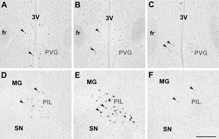
Figure 2.
Bright-field photomicrographs of in situ hybridization histochemical sections demonstrate the expression of TIP39 mRNA in the PVG (A–C) and the PIL (D–F). Arrowheads indicate some examples of the autoradiography signal above TIP39-expressing neurons. In the PVG, most TIP39 neurons have a weak autoradiography signal in control female (A) and lactating mother (B) as well as in a pup-deprived mother (C). In contrast, the low level of TIP39 mRNA in the control female (D) is increased in all parts of the PIL in lactating mother (E), whereas removing the pups reduced the level of TIP39 mRNA as indicated by the low density of TIP39-expressing cells and their weak labeling (F) similar to the image in D. fr, Fasciculus retroflexus; MG, medial geniculate body; SN, substantia nigra; 3V, third ventricle. Scale bar, 500 μm.

