Abstract
It is rare for foreign bodies to be found in the parapharyngeal space due to the protection of the mandibular ramus and zygomatic bone. The authors describe a rare case of a patient with an unusual penetrating neck injury caused by broken windshield glass in a traffic accident, which lodged in the parapharyngeal space and punctured the internal jugular veins and cranial nerves. 3 weeks later, a delayed exploration was performed on the patient after detailed evaluation of the relationship between the foreign body and the great vessels. The authors removed the glass fragment easily with no active bleeding because it had been surrounded by a fibrous envelope. This experience indicates that increasing the duration of foreign body retention in the parapharyngeal space may be helpful, allowing fibrosis to surround the foreign body, reducing the risk of active bleeding when it is removed.
Keywords: penetrating injury, neck, parapharyngeal space, foreign body, glass fragment
Introduction
Penetrating neck injuries represent a rare but complex variety of trauma. Occasionally, the penetrating body can be retained within the anatomic regions crossed. Most of these injuries are secondary to a traffic or work accident or sustained during an attack3, 4, 6–9. The clinical situation may be accompanied by various pathological conditions such as bone fractures9, vascular injury or neurological deficits3, 5. The leading cause of death in penetrating neck trauma is vascular injury.
Craniofacial bones, such as the mandibular and zygomatic bones, may provide some protection for vital structures in the parapharyngeal space such as the internal carotid artery, internal jugular vein and cranial nerves. It is rare for foreign bodies to be found in the parapharyngeal space. A few cases have been reported of foreign bodies entering from the skin surface and becoming lodged in the parapharyngeal space, related to stab wounds or work accidents, but no involvement with adjacent great vessels has been reported2–5, 9. The authors present a rare case of a patient with an unusual penetrating neck injury caused by broken windshield glass in a traffic accident. The glass lodged in the parapharyngeal space and punctured the internal jugular vein and cranial nerves (IX, X, XI, XII).
Case report
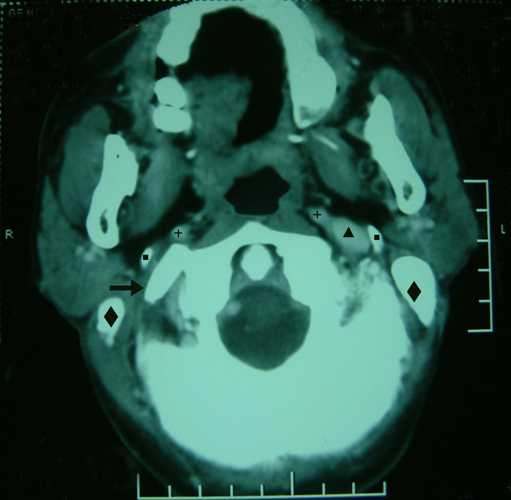
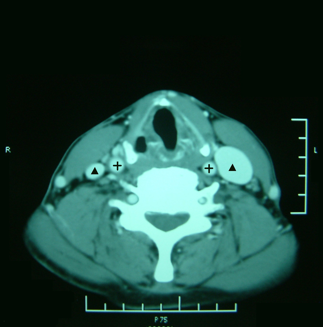
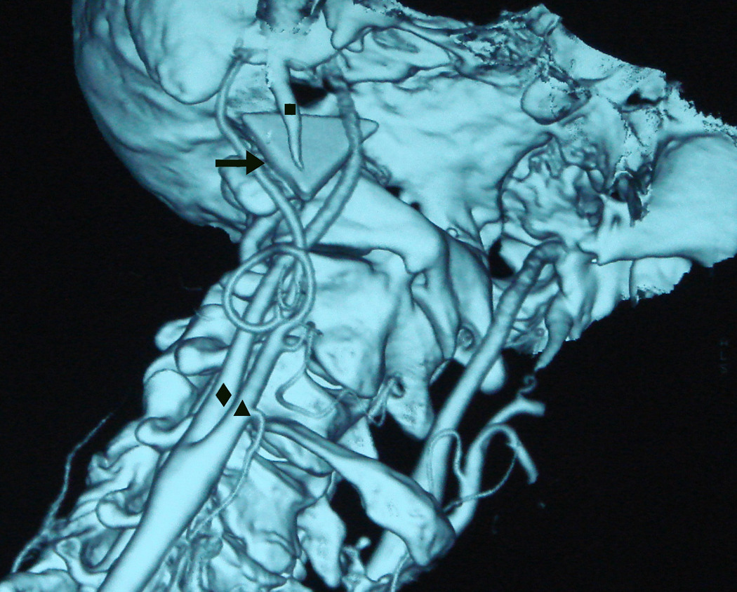
The patient was a healthy 64-year-old man. While driving a pickup truck on 12 May 2008 he collided with a lorry. He was thrown off the pickup truck and lost consciousness. He was sent to the local hospital with a wound located below his right mastoid process horizontally and had some bleeding from the wound. No other abnormalities such as brain damage, bone fracture or damage to abdominal organs were observed. The wound was treated briefly with debridement at the local hospital and it was not realized that a glass fragment had become embedded in the wound. After treatment he regained consciousness. Subsequently, he complained of difficulty swallowing and hoarseness. He was referred to the authors’ hospital on 26 May 2008. Close inspection of his upper neck showed there was no haematoma, but a 4 cm healing wound was present below his right mastoid process. Oral examination showed his tongue deviating rightwards on protrusion. Touch sensation to the posterior third of his right tongue was absent. Flexible fibreoptic endoscopy of the laryngopharynx revealed an immobile right vocal cord in the paramedian position. A plain lateral radiograph of the neck showed no radio-opaque foreign body in the soft tissues of the neck. Owing to the high suspicion of a foreign body impacted in the neck, a computed tomography (CT) scan was carried out. A contrast-enhanced CT scan showed a radio-opaque foreign body in the right parapharyngeal space at the level of the C1 vertebra traversing the internal jugular vein and its medial edge 1–2 mm from the internal carotid artery (Fig. 1). The right internal jugular vein at the level of the C1 vertebra was not shown by contrast material and was much smaller than the left internal jugular vein at the level of the thyroid cartilage (Fig. 2), suggesting the right internal jugular vein was occluded by the foreign body. Three-dimensional (3D) CT indicated that a triangular foreign body was lodged in the right parapharyngeal space and did not involve the right internal carotid artery (Fig. 3). To prevent secondary injury to the internal carotid artery due to neck movements, a cervical collar was put on the patient’s neck.
Figure 1.

Axial contrast-enhanced CT at the level of the C1 vertebra showing the relationship between the styloid process (■), the internal carotid artery (+) and the foreign body (arrow). The right internal jugular vein is not shown by contrast material contrasted with the left internal jugular vein (▲).The mastoid process is indicated by ◆.
Figure 2.

Axial contrast-enhanced CT at the level of the thyroid cartilage showing the common carotid artery (+) and the internal jugular vein (▲). The right internal jugular vein is much smaller than the left internal jugular vein.
Figure 3.

3D CT showing the relationship between the styloid process (■), the foreign body (arrow), the internal carotid artery (◆) and the external carotid artery (▲).
On 4 June 2008, an exploration was performed, under general anaesthesia, through a transverse submandibular incision on the right side. After isolating the right sternocleidomastoid muscle and retracting it laterally, the right carotid sheath was opened. The common carotid artery, vagus nerve, and hypoglossal nerve were found to be intact in the carotid triangle, but the internal jugular vein was very small in caliber (Fig 2). The digastric muscle was transected and its bellies retracted. Upward dissection along the internal carotid artery was made to the foreign body. A glass fragment with surrounding fibrosis was noted to have penetrated and occluded the internal jugular vein at the level of the C1 vertebra. The medial edge of the foreign body was lying in close proximity to the right internal carotid artery. A triangle (2.6×2.4×2.3 cm) of a glass fragment was successfully removed without any active bleeding. The right internal jugular vein was ligated. The authors did not explore the cranial nerves in the parapharyngeal space because of skull base and mandibular limitations and tissue reaction to the foreign body. The digastric muscle was sutured back, and the wound was closed in layers after inserting a drain. Postoperative recovery was uneventful and the patient was discharged on the seventh day.
The patient was last seen 3 months after the operation when his dysphagia, right hemiglossoplegia and hoarseness had not improved. His right sternocleidomastoid and trapezius muscles had become severely atrophied. At his telephone follow-up on 22 April 2010, he complained that there were no significant changes in the signs and symptoms of hoarseness, tongue deviating rightwards on protrusion, and the atrophy of the right sternocleidomastoid and trapezius muscles, but his difficulty in swallowing had been alleviated.
Discussion
Traffic accidents are one of the main causes for glass foreign bodies in the head and neck1, 7, 8. Superficial foreign bodies may be diagnosed and removed relatively easily, but sometimes locating a foreign body can be challenging. This patient had a penetrating neck injury with signs and symptoms of dysfunction of four cranial nerves, such as difficulty swallowing and sensory loss on the root of the right tongue (IX), hoarseness and right vocal cord palsy (X), atrophy of the right sternocleidomastoid and trapezius muscles (XI), and deviation of the tongue rightwards on protrusion (XII). The index of suspicion for retained objects in the neck should be high. Plain radiographs are cheap, straightforward and useful diagnostic procedures when dealing with patients who present acutely with penetrating neck trauma4, 8. The drawbacks are that these procedures are successful only if the foreign body is radio-opaque, and radiation exposure is required. Foreign bodies can be difficult to visualise because of the opacity of the adjacent bones, as observed in this patient. Angiography may be valuable in excluding major vascular injury,6 but it is invasive. Ultrasound was not suitable in this case because the object lay deep to the parotid gland, within the parapharyngeal space and beyond the normal penetrative range of superficial high-resolution ultrasound. The composition of the foreign body was initially unknown, so magnetic resonance imaging and magnetic resonance angiography were not used for fear that a metallic foreign body could be moved on examination. Contrast-enhanced and 3D CT examination gave more information about the exact location of the foreign body and the relationship between the foreign body and the surrounding vessels. This information enabled the authors to locate the foreign body with ease on exploration of the parapharyngeal space.
Generally, penetrating foreign bodies in the neck require urgent surgical exploration, identification and removal to prevent secondary complications of haemorrhage/haematoma, infection, neurovascular compromise, and the potential migration into the vascular structures or the aerodigestive tract. There are no defined criteria regarding the management of cervical foreign bodies.
Patients with active bleeding, who are in shock, or who have an expanding haematoma or airway compromise, require immediate surgical exploration. In the present patient, broken windshield glass had punctured the internal jugular vein and cranial nerves within the parapharyngeal space. There was no active bleeding or haematoma in the wound, but neurological deficits were present. The authors thought that the foreign body had acted as a tampon, arresting bleeding from the internal jugular vein. Delayed exploration avoided the potential risk of active bleeding from the internal jugular vein on removal of the foreign body. Exploration revealed that the foreign body was surrounded by a fibrous envelope and it was removed with no active bleeding.
In conclusion, the authors have reported their experience of a rare case of broken windshield glass lodged in the parapharyngeal space causing injury to the internal jugular vein and cranial nerves (IX, X, XI, XII). Preoperative assessment is important to allow planning for the safe and successful surgical removal of a foreign body from the parapharyngeal space. This experience indicates that increased duration of foreign body retention in the parapharyngeal space may be helpful to allow fibrosis to surround the foreign body, reducing the risk of active bleeding when it is removed.
Footnotes
Publisher's Disclaimer: This is a PDF file of an unedited manuscript that has been accepted for publication. As a service to our customers we are providing this early version of the manuscript. The manuscript will undergo copyediting, typesetting, and review of the resulting proof before it is published in its final citable form. Please note that during the production process errors may be discovered which could affect the content, and all legal disclaimers that apply to the journal pertain.
Declarations
Funding: None
Competing Interests: None declared
Ethical Approval: Not required
References
- 1.Agrillo A, Sassano P, Mustazza MC, Filiaci F. Complex-type penetrating injuries of craniomaxillofacial region. J Craniofac Surg. 2006;17:442–446. doi: 10.1097/00001665-200605000-00010. [DOI] [PubMed] [Google Scholar]
- 2.Dort JC, Robertson D. Nonmetallic foreign bodies of the skull base: a diagnostic challenge. J Otolaryngol. 1995;24:69–72. [PubMed] [Google Scholar]
- 3.Enomoto K, Nishimura H, Inohara H, Murata J, Horii A, Doi K, Kubo T. A rare case of a glass foreign body in the parapharyngeal space: pre-operative assessment by contrast-enhanced CT and three-dimensional CT images. Dentomaxillofac Radiol. 2009;38:112–115. doi: 10.1259/dmfr/69946733. [DOI] [PubMed] [Google Scholar]
- 4.Islam S, Esmil T, Umapathy N, Hoffman GR. Foreign body (metal key) impacted in the upper neck. Injury Extra. 2006;37:109–112. [Google Scholar]
- 5.Khadivi E, Bakhshaee M, Khazaeni K. A rare penetrating neck trauma to zone III. Emerg Med J. 2007;24:840. doi: 10.1136/emj.2006.044586. [DOI] [PMC free article] [PubMed] [Google Scholar]
- 6.Lu Q, Bao J, Pei Y, Jing Z. Temporary balloon occlusion of both the superior vena cava and the internal jugular vein to achieve removal of a migrated foreign body: Case report. Ann Vasc Surg. 2009;23:687. doi: 10.1016/j.avsg.2009.03.010. e9–13. [DOI] [PubMed] [Google Scholar]
- 7.Narita N, Yamada T, Imoto Y, Ogi K, Sakashita M, Ito Y, Kouraba S, Yasuta M, Tsuzuki H, Fujieda S. Treatment of scattered glass foreign bodies in both the superficial and deep neck: a case report. Auris Nasus Larynx. 2005;32:295–299. doi: 10.1016/j.anl.2005.03.003. [DOI] [PubMed] [Google Scholar]
- 8.Ozturk K, Keles B, Cenik Z, Yaman H. Penetrating zone II neck injury by broken windshield. Int Wound J. 2006;3:63–66. doi: 10.1111/j.1742-4801.2006.00177.x. [DOI] [PMC free article] [PubMed] [Google Scholar]
- 9.Zhao X, Liu J, Guo Y. Removal of a large metallic foreign body from the base of the left skull. J Otolaryngol. 2004;33:120–122. doi: 10.2310/7070.2004.02097. [DOI] [PubMed] [Google Scholar]


