Abstract
Background and objectives: Mutations in podocyte genes are associated with steroid-resistant nephrotic syndrome (SRNS), mostly affecting younger age groups. To date, it is unclear whether these patients benefit from intensified immunosuppression with cyclosporine A (CsA). The aim of this study was to evaluate the influence of podocyte gene defects in congenital nephrotic syndrome (CNS) and pediatric SRNS on the efficacy of CsA therapy and preservation of renal function.
Design, settings, participants, & measurements: Genotyping was performed in 91 CNS/SRNS patients, irrespective of age at manifestation or response to CsA.
Results: Mutations were identified in 52% of families (11 NPHS1, 17 NPHS2, 11 WT1, 1 LAMB2, 3 TRPC6). Sixty-eight percent of patients with nongenetic SRNS responded to CsA, most of them achieved complete remission. In contrast, none of the patients with genetic CNS/SRNS experienced a complete remission and only two (17%) achieved a partial response, both affected by a WT1 mutation. Preservation of renal function was significantly better in children with nongenetic disease after a mean follow-up time of 8.6 years (ESRD in 29% versus 71%).
Conclusions: The mutation detection rate in our population was high (52%). Most patients with genetic CNS/SRNS did not benefit from CsA with significantly lower response rates compared with nongenetic patients and showed rapid progression to end-stage renal failure. These data strongly support the idea not to expose CNS/SRNS patients with inherited defects related to podocyte function to intensified immunosuppression with CsA.
Defects in various genes have been associated with pediatric congenital (CNS) and steroid-resistant nephrotic syndrome (SRNS). This novel insight moved the podocyte into the center of interest regarding the pathophysiology of proteinuria. Among these genes are NPHS1, NPHS2, WT1, LAMB2, TRPC6, and PLCE1. NPHS1 and NPHS2 encode nephrin and podocin, respectively, which have important roles for the organization of the slit diaphragm. Recessive mutations in NPHS1 and NPHS2 cause congenital and childhood SRNS (1,2). WT1 is a major mediator of podocyte differentiation and involved in the pathogenesis of isolated diffuse mesangial sclerosis (DMS) and Denys–Drash and Frasier syndromes (3). LAMB2 encodes laminin β2, one component of the heterotrimeric laminins that link the podocyte to the glomerular basement membrane. Recessive mutations in LAMB2 cause early-onset nephrotic syndrome and DMS in conjunction with distinct ocular symptoms (Pierson syndrome) (4). Dominant mutations in TRPC6, encoding the cation channel TRPC6, are associated with late-onset focal segmental glomerulosclerosis (FSGS) (5,6). PLCE1 is involved in early-onset recessive nephrotic syndrome (NPHS3) and encodes phospholipase C ε-1, implicated in podocyte signaling processes (7).
Therapy of SRNS is demanding. Numerous immunosuppressive agents have shown to be effective in a subset of SRNS patients (8,9). Cyclosporine A (CsA) is used preferentially and several studies demonstrated CsA-induced remission rates (including partial remission) of up to 70% in pediatric SRNS patients (10–12). Similar response rates were reported in a recent multicenter trial (8). CsA is a calcineurin inhibitor and its antiproteinuric properties were attributed to this immunosuppressive effect (13,14). As a consequence, it was discussed to spare children with hereditary SRNS from intensified immunosuppression with CsA. However, the systematic analysis of genes implicated in CNS/SRNS with respect to the effectiveness of intensified immunosuppressive therapy is still lacking, especially mutational analysis in patients that responded to CsA. Few patients with proven hereditary SRNS and partial remission induced by CsA have been reported (7,15–18). Hinkes et al. report one steroid- and one CsA-sensitive (DMS) patient with homozygous PLCE1 mutation in the initial publication of PLCE1 involved in nephrotic syndrome (7). However, these findings have not been confirmed in subsequent studies.
The mechanisms of remission induction in this subset of patients are still unclear, and in addition to effects on vascular perfusion and the immune system, direct effects on the podocyte architecture are discussed. Following this idea, Faul et al. demonstrated a direct effect of CsA on the stabilization of the podocyte actin cytoskeleton, thus suggesting a benefit of CSA treatment also for patients with hereditary SRNS (19). However, clinical studies supporting this hypothesis are lacking so far.
We therefore sought to examine these potential genotype/phenotype relationships in a retrospective study of all patients with CNS and SRNS treated at the University Children's Hospitals of Essen and Münster. Specifically, we assessed clinical data and performed mutational analysis for NPHS1, NPHS2, WT1, LAMB2, TRPC6, and PLCE1, stratified according to the clinical course of the patients.
Patients and Methods
Patients and Data Recruitment
Between 1999 and 2009, 91 consecutive patients (44 males) of 82 families with CNS (age at onset <3 months; [20]) or primary SRNS (age at onset >3 months; [20]) were treated at the University Children's Hospitals of Essen and Münster. Of these were four pairs of siblings (MS3/MS4, MS6/MS7, MS11/MS12, MS13/MS14) and one sibship of Sinti origin with six affected children (E5/E6/E8/E12/E14/E15). CNS and SRNS patients were included in the study presented here because of the observation that from a genetic point of view, it seems arbitrary to distinguish between congenital and infantile nephrotic syndrome. The most important genes (NPHS1, NPHS2, WT1) are associated with a broad phenotypic spectrum of nephrotic syndrome similar to a phenotypic continuum (15,21–23). Patients with secondary nephrotic syndrome or secondary steroid resistance were excluded from the study.
Standard steroid treatment and steroid response were defined according the criteria of the German Society of Pediatric Nephrology (24) and the International Study of Kidney Disease in Children (20). Six patients received additional steroid pulses. Complete remission was defined as proteinuria <4 mg/m2 per hour or trace of protein on dipstick analysis and normalization of serum albumin (>3.5 g/dl). Partial remission was defined as proteinuria between 4 and 40mg/m2 per hour and normalization of serum albumin. Steroid resistance was defined as failure of induction of complete remission after 4 weeks of standard therapy with prednisone and optionally steroid pulses.
Mutational Analysis
Genomic DNA was isolated from peripheral blood. Mutational analysis was performed for NPHS1 (NCBI AccN° NG_013356.1), NPHS2 (AccN° NM_014625.2), exon 8 and 9 of WT1 (AccN° NG_009272.1), LAMB2 (AccN° NM_002292.3), TRPC6 (AccN° NM_004621.5), and PLCE1 (AccN° NM_016341.3). Patients without pathogenic mutations in WT1 or NPHS2 received subsequent analysis of NPHS1 (age at onset <6 years) or TRPC6 (age at onset >6 years). In patients with DMS, we additionally performed analysis of PLCE1, and in patients with ocular symptoms we performed analysis of LAMB2. Novel mutations were excluded in at least 100 healthy individuals.
Statistical Analyses
Auxologic parameters were described as mean ± SD. Comparisons of CsA response rates and renal survival between the different patient groups were performed using Fisher's exact test. The level of statistical significance was predefined as α = 0.05. Survival data were analyzed by Kaplan–Meier survival curves. The statistical analysis was performed using GraphPad Prism (version 5.01 for Windows, GraphPad Software, San Diego, CA).
Results
In total, we enrolled 91 patients with CNS/SRNS. Twenty-six patients presented with CNS, and 65 patients presented with primary SRNS. The overall mutation detection rate was 52% (100% in CNS and 38% in SRNS). In detail, we identified 11 NPHS1, 17 NPHS2, 11 WT1, 1 LAMB2, and 3 TRPC6 mutations. No mutations were identified in PLCE1. Results of mutational analysis and clinical data are presented in Tables 1 and 2. Clinical data of patients with nongenetic SRNS are summarized in Supplement 1.
Table 1.
Overview of mutational analysis and genotypic and phenotypic data
| Patient | Gender | Nucleotide Changea | Effect on Protein | Age at Onset (CNS/years) | Response to CsA (Y/N/0) | Kidney Biopsy | Time to ESRD (months) | Renal Outcome | Reference |
|---|---|---|---|---|---|---|---|---|---|
| NPHS1 | |||||||||
| E1 | M | 515_517del (H) | T172del | CNS | 0 | Finnish type | 70 | RTx | 25 |
| E3 | M | 614_621delCACCCCGG 613_622insTT (H) | T205I,del206,207 | CNS | 0 | ND | — | CRI | 25 |
| E4 | F | 2339G>A (h);2928-3C>G (h) | G796R;Splicing of exon 22 | CNS | 0 | DMS | 22 | RTx | Novel;novel |
| E5 | M | DelTCAinsCC2617 (H);2552C>T (H) | L904X and A851V | CNS | N | Finnish type | 40 | RTx | 40 |
| E6 | F | DelTCAinsCC2617 (H);2552C>T (H) | L904X and A851V | CNS | 0 | ND | — | CRI | 40 |
| E8 | F | DelTCAinsCC2617 (H);2552C>T (H) | L904X and A851V | CNS | 0 | ND | — | CRI | 40 |
| E12 | F | DelTCAinsCC2617 (H);2552C>T (H) | L904X and A851V | CNS | N | ND | 49 | RTx | 40 |
| E14 | F | DelTCAinsCC2617 (H);2552C>T (H) | L904X and A851V | CNS | 0 | MC | 23 | RTx | 40 |
| E15 | M | DelTCAinsCC2617 (H);2552C>T (H) | L904X and A851V | CNS | 0 | ND | 23 | RTx | 40 |
| E7 | F | 1699T>A (H) | C567S | CNS | 0 | DMS | 38 | RTx | Novel |
| E16 | M | 3286G>T (H);563A>T (H); | G1096C;N188I | CNS | 0 | ND | — | Normal | Novel;21 |
| MS1 | F | 2816-4_2822del (H) | Splicing of exon 21 | CNS | 0 | Finnish type | 72 | ESRD | Novel |
| MS2 | F | 614_621delCACCCCGG 613_622insTT (H) | T205I,del206,207 | CNS | N | ND | 24 | RTx | 25 |
| MS9 | F | 515_517del (H) | T172del | CNS | 0 | DMS | 25 | RTx | 25 |
| MS10 | F | 1713del (H) | S571RfsX51 | CNS | 0 | Finnish type | 7 | RTx | Novel |
| MS41 | M | 320C>A (h);1868G>T (h) | A107E;C623F | 0 9/12 | 0 | Finnish type | 107 | RTx | Novel;25 |
| NPHS2 | |||||||||
| E9 | M | 413G>A (H) | R138Q | CNS | N | MC | 82 | RTx | 2 |
| E10 | F | 871C>T (h);686G>A (h) | R291W;R229Q | CNS | 0 | MC | — | Normal | 2;41 |
| E13 | M | 413G>A (h);412C>T (h) | R138Q;R138X | CNS | 0 | ND | 199 | ESRD | 2 |
| E17 | F | 503G>A (H) | R168H | 2 9/12 | 0 | Diffuse mesangioproliferation | — | CRI | 26 |
| E22 | M | 413G>A (h);419delG (h) | R138Q;G140DfsX41 | 3 6/12 | 0 | MC | — | Normal | 2 |
| E27 | M | 890C>T (h);686G>A (h) | A297V;R229Q | 11 9/12 | 0 | FSGS | — | Normal | 22;41 |
| E29 | M | 460_467insT (H) | V165X | 0 11/12 | N | FSGS | 104 | RTx | 41 |
| E41 | M | 413G>A (H) | R138Q | 0 8/12 | 0 | MC | 103 | RTx | 2 |
| E43 | F | 413G>A (H) | R138Q | 0 9/12 | 0 | ND | 65 | RTx | 2 |
| MS3 | F | 413G>A (H) | R138Q | CNS | 0 | FSGS | 86 | RTx | 2 |
| MS4 | F | 413G>A (H) | R138Q | CNS | N | FSGS | 40 | RTx | 2 |
| MS8 | M | 413G>A (H) | R138Q | CNS | 0 | DMS | 83 | RTx | 2 |
| MS16 | F | 413G>A (h);868G>A (h) | R138Q;V290M | 11 0/12 | 0 | FSGS | — | Normal | 2;41 |
| MS19 | M | 413G>A (H) | R138Q | 0 5/12 | 0 | ND | — | Normal | 2 |
| MS21 | M | 460_467insT (h);413G>A (h) | V165X;R138Q | 2 0/12 | N | FSGS | 46 | RTx | 41;2 |
| MS23 | F | 868G>A (H) | V290M | 0 11/12 | 0 | MC | — | Normal | 41 |
| MS40 | F | 983A>G (h);686G>A (h) | Q328R;R229Q | 8 10/12 | 0 | FSGS | — | Normal | 15;41 |
| MS44 | F | 929A>T (h);686G>A (h) | E310V;R229Q | 4 0/12 | N | FSGS | 102 | RTx | 15;41 |
| WT1 | |||||||||
| E2 | M (XY) | 1180C>T (h) | R394W | CNS | 0 | DMS | 0 | ESRD | 27 |
| E11 | M (XY) | 1181G>A (h) | R394Q | CNS | 0 | FSGS | 4 | RTx | 3 |
| E19 | M (XY) | 1180C>T (h) | R394W | 0 7/12 | 0 | DMS | 0 | RTx | 27 |
| E26 | F (XX) | 1180C>T (h) | R394W | 4 2/12 | 0 | FSGS | 2 | RTx | 27 |
| E37 | M(F)(XY) | 1180C>T (h) | R394W | 4 11/12 | 0 | DMS | 0 | RTx | 27 |
| E38 | F (XY) | 1180C>T (h) | R394W | 0 7/12 | 0 | FSGS | 0 | RTx | 27 |
| MS5 | F (XX) | 1162C>T (h) | C388R | CNS | 0 | DMS | 21 | RTx | 42 |
| MS18 | F (XX) | IVS9 + 4C>T (h) | Splicing of exon 9 | 4 0/12 | 0 | FSGS | — | Normal | 43 |
| MS24 | F (XX) | 1190A>C (h) | H397P | 1 3/12 | Y (partial) | FSGS | 97 | RTx | 42 |
| MS30 | F (XY) | IVS9 + 4C>T (h) | Splicing of exon 9 | 5 1/12 | Y (partial) | FSGS | — | Normal | 43 |
| MS32 | F (XX) | 1180C>T (h) | R394W | 0 6/12 | 0 | ND | 1 | RTx | 27 |
| LAMB2 | |||||||||
| MS6 | M | 961T>C (h);4140C>A (h) and 4177C>T (h) | C321R;N1380K and L1393F | CNS | 0 | DMS | 7 | RTx | 28 |
| MS7 | F | 961T>C (h);4140C>A (h) and 4177C>T (h) | C321R;N1380K and L1393F | CNS | 0 | DMS | 32 | RTx | 28 |
| TRPC6 | |||||||||
| E31 | M | 495T>C (h) | M132T | 8 8/12 | N | FSGS | 12 | RTx | 29 |
| E35 | M | 265delA (h) | 89fsX8 | 7 11/12 | N | FSGS | 64 | ESRD | Novel |
| E40 | M | 2270G>A (h) | G757D | 1 | 0 | FSGS | — | Normal | Novel |
H, homozygous; h, heterozygous; N, no response to CsA treatment; Y, response to CsA treatment; 0, patient did not receive CsA; ND, no data; MC, minimal change glomerulopathy; CRI, chronic renal insufficiency; RTx, renal transplant; Normal, normal renal function. Siblings are in bold; sequence variations of unknown significance are presented in italic letters.
Karyotype (in parentheses).
Table 2.
Overview of NPHS2 mutational analysis and genotypic and phenotypic data in patients with heterozygous NPHS2 mutations
| Patient | Gender | Nucleotide Change | Effect on Protein | Age at Onset (years) | Response to CsA (Y/N/0) | Kidney Biopsy | Time to ESRD (months) | Renal Outcome | Reference |
|---|---|---|---|---|---|---|---|---|---|
| MS11 | M | 413G>A (h) | R138Q;wt | 7 0/12 | Y | MC | ND | RTx | 2 |
| MS12 | M | 413G>A (h) | R138Q;wt | 6 0/12 | Y (partial) | FSGS | 96 | RTx | 2 |
| MS13 | F | 593A>C (h) | E198A;wt | 8 5/12 | 0 | ND | — | Normal | Novel |
| MS14 | M | 593A>C (h) | E198A;wt | 2 7/12 | 0 | FSGS | — | Normal | Novel |
| MS31 | M | 686G>A (h) | R229Q;wt | 2 5/12 | Y | FSGS | — | Normal | 41 |
| MS33 | F | 686G>A (h) | R229Q;wt | 6 3/12 | 0 | FSGS | — | Normal | 41 |
| MS38 | F | 686G>A (h) | R229Q;wt | 11 7/12 | N | MC | — | CRI | 41 |
| MS43 | M | 686G>A (h) | R229Q;wt | 6 3/12 | Y | FSGS | — | Normal | 41 |
Siblings are in bold.
Mutational Analysis
NPHS1
Homozygous NPHS1 mutations were detected in 14 patients from nine families and compound heterozygous mutations in 2 unrelated patients. Among these, seven NPSH1 mutations were novel (Table 2). One large Sinti family with six affected children presented with double homozygous sequence variations (L904X and A851V, a sequence variation of unknown significance). Double homozygous sequence variations in NPHS1 were also identified in another CNS patient of Turkish origin, (G1096C, a novel mutation affecting the donor splice site of exon 24 and N188I, a sequence variation of unknown significance [21]). One SRNS patient (MS35) was affected by two heterozygous NPHS1 variants of unknown significance in compound heterozygous state: R408Q (25) and 3286 + 89C>T (novel). Because of the lack of evidence for pathogenicity of both variants, this patient was assigned to the nongenetic group.
NPHS2
We identified homozygous NPHS2 mutations in 10 patients from nine families and compound heterozygous mutations in 8 unrelated patients. The non-neutral polymorphism R229Q was identified in four patients with an additional pathogenic heterozygous mutations in NPHS2.
The sequence variation A242V, a polymorphism in individuals of African origin (26), was present in the homozygous state in one Congonese patient. Heterozygous NPHS2 mutations or sequence variations were identified in eight patients of the study group (Table 2): R138Q in one pair of siblings, E198A in a second pair of siblings, and the non-neutral polymorphism R229Q in four additional patients. These patients and the Congonese patient were assigned to the nongenetic group.
WT1
We identified 11 dominant WT1 mutations. Nine patients were affected by a missense mutation in WT1, among these, six patients with the frequent hot-spot mutation, R394W, which is typically associated with the development of DDS (27). Five of the nine patients with a WT1-missense mutation had a XY karyotype (isolated DMS = 1 [E2]; hypospadia n = 2 [E11, E19]; intersex genitalia n = 1 [E37]; complete sex reversal n = 1 [E38]). A typical Frasier splice-site mutation (IVS9 + 4C>T) was identified in two patients: one female patient with XX karyotype and FSGS (MS18) and one phenotypically female patient with XY karyotype, FSGS, and development of dysgerminoma (MS30).
LAMB2
One pair of siblings presented with Pierson syndrome, characterized by CNS and microcoria. The clinical case has been described by Hasselbacher et al. (28). Three heterozygous LAMB2 sequence variations were identified—a paternally inherited missense mutation (C321R) and two maternally inherited sequence alterations (N1380K and L1393F).
TRPC6
Dominant TRPC6 mutations were identified in three patients—among these, two novel mutations (G757D and 89fsX8). The third mutation (M132T) has been identified in a patient recently published by Heeringa et al. (29). The patients presented with an onset of disease at 1, 7, and 8 years, respectively.
Clinical Characteristics and Genotype-Phenotype Correlations
The analysis of clinical outcome was performed after stratification of the patients into three groups according to the international classification (20) and the results of genetic testing: patients in whom we could not identify a mutation were assigned to the nongenetic SRNS group and patients with mutations were divided into CNS with early-onset (< 3 months of age) and genetic SRNS with onset >3 months. Genotype-phenotype correlations are depicted in Table 3. The clinical data of all study patients was assessed over a mean observation time of 103.0 ± 68.2 months. Forty-three patients received CsA subsequent to the diagnosis of CNS/SRNS. The mean dose at 6 months of therapy was 6.5 ± 2.9 mg/kg per day. The doses were adjusted to achieve trough levels between 80 and 120 ng/ml. Renal outcome was classified at the end of observation and categorized into normal renal function (GFR >90 ml/min per 1.73 m2), chronic renal insufficiency stage II to IV (GFR 15 to 90 ml/min per 1.73 m2), ESRD (GFR <15 ml/min per 1.73 m2), or patients who received a renal transplant.
Table 3.
Genotype-phenotype correlations in CNS/SRNS
| Nongenetic |
Genetic |
||
|---|---|---|---|
| SRNS (age at onset >3 months) (n = 41) | CNS (n = 26) | SRNS (age at onset >3 months) (n = 24) | |
| Gender (male/female) | 21/20 | 11/15 | 12/12 |
| Age at onset (mean ± SD; months) | 77.6 ± 53.2 | PP | 43.5 ± 42.8 |
| Immunosuppression with CsA | 76% | 19% (P < 0.0001)b | 29% (P = 0.0005)c |
| Response to CsA | 68% | 0% (P = 0.008)b | 29% (P = 0.09)c |
| Observation period (mean ± SD; months) | 78.8 ± 59.7 | 118.3 ± 65.9 | 125.0 ± 74.5 |
| Time to develop ESRD (mean ± SD; months)a | 50.1 ± 47.0 | 37.4 ± 27.6 | 44.7 ± 44.0 |
| Development of ESRDa | 29% | 84% (P < 0.0001)b | 58% (P = 0.04)c |
| Normal renal function at end of observationa | 63% | 8% (P < 0.0001)b | 38% (P = 0.07)c |
| Recurrence after RTx (Y/N) | 9% | 6% | 0% |
PP, postnatal onset.
One patient of the CNS group with CRI died early and was excluded from renal outcome analysis.
Statistical significance of differences between CNS versus nongenetic SRNS.
Statistical significance of differences between genetic SRNS versus nongenetic SRNS.
Patients without Mutations in the Analyzed Genes
Forty-one patients presented with SRNS without mutations in the analyzed genes. Mean age at onset was 77.6 ± 53.2 months. Kidney biopsies were performed in 40 of 41 patients and showed DMS (n = 1), minimal change glomerulopathy (n = 10), FSGS (n = 28), and mesangial proliferation (n = 1).
Thirty-one of 41 patients received CsA. The mean duration of therapy was 39.4 ± 40.5 months. Complete remission was observed in 17 of 31 patients (55%) and partial response in 4 of 31 patients (13%). Ten patients (32%) did not respond to CsA treatment. Thirty-four patients received ACE inhibitors (ACEIs) or angiotensin receptor blockers (ARBs) as antiproteinuric therapy. Three patients developed chronic renal insufficiency (7%), 12 (29%) progressed to ESRD (11 with renal transplant), and 26 (63%) preserved a normal renal function. Mean time to ESRD was 50.1 ± 47.0 months.
Patients with CNS
Twenty-six patients were diagnosed with CNS. Five patients received CsA, but none of them responded to this therapy. Twenty-one patients received a therapy with an ACEI or ARB. Mean time to ESRD was 37.4 ± 27.6 months. One patient died at the age of 1 year with an impaired renal function.
Patients with a Hereditary Basis of SRNS
Twenty-four patients presented with genetic SRNS (age at onset >3 months). Seven of 24 patients received CsA. The mean duration of therapy was 32.0 ± 26.1 months (at least 10 months). Immunosuppressive therapy with CsA led to a partial response in two patients with WT1 mutation: one phenotypically female Frasier patient (karyotype XY) affected by a WT1 splice-site mutation (MS30) and one patient (karyotype XX) with isolated FSGS affected by a WT1 missense mutation (MS24). None of the patients with hereditary SRNS (age at onset >3 months) showed a complete response to CsA. Seventeen patients received an ACEI and four patients an ARB as antiproteinuric therapy. Mean time to ESRD was 45.9 ± 45.2 months.
Response to CsA Treatment
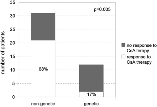
Response to CsA treatment was significantly better in patients without mutations in podocyte genes than in patients with genetic CNS/SRNS (68% versus 17%, P = 0.005; Figure 1).
Figure 1.
Response to CsA treatment in patients with and without pathogenic mutations in podocyte genes (17% versus 68%; P = 0.005; Fisher's exact test). Response was defined as induction of a complete or partial remission.
Only two patients with genetic SRNS (affected by mutations in WT1) showed a partial response to CsA with a reduction of proteinuria and normalization of serum albumin.
Renal Function
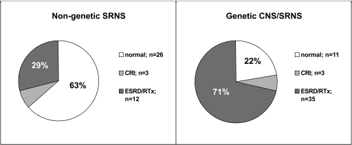
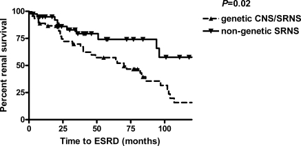
In patients without mutations, development of ESRD was 29% compared with 71% in patients with genetic disease (84% CNS, 58% SRNS). Of note, at the end of observation, a normal renal function was preserved in 63% of patients without mutations compared with 22% of patients with genetic disease (P = 0.0001) (Figure 2). The time course is reflected by Kaplan–Meier survival curves, which show a significantly slower progression to ESRD in patients without mutations compared with patients with genetic disease (P = 0.02) (Figure 3).
Figure 2.
Renal survival in patients with and without pathogenic mutations in podocyte genes. Development of ESRD was 29% in patients without pathogenic mutation compared with 71% in patients with proven mutation (P = 0.0001; Fisher's exact test). Sixty-three percent (nongenetic) compared with 22% (genetic) of the patients preserved a normal renal function over the mean follow-up time of the study (8.6 years).
Figure 3.
The Kaplan–Meier survival curve shows the progression to ESRD over time (months). Patients without a mutation in podocyte genes show a slower progression to ESRD over time (P = 0.02).
Discussion
The mutation detection rate in the study presented here was high (52%; 100% in patients with congenital disease; 38% in patients with SRNS [age at onset >3 months]). These results confirm that a substantial proportion of pediatric CNS/SRNS patients have a genetic basis of the disease. Therapy for patients with hereditary SRNS varies considerably, especially with respect to the use of immunosuppressants. However, the benefit of intensified immunosuppressive therapy in these patients has not yet been demonstrated by a systematic analysis. The clinical experience is limited to single-center observations demonstrating a partial response to CsA in selected patients (7,15–18).
In contrast to former studies, we here explicitly included patients that responded to CsA but had not yet been offered genetic testing. The distribution of identified mutations revealed a predominance of autosomal recessive mutations in NPHS1 and NPHS2, especially in patients with congenital disease.
Heterozygous NPHS2 mutations or polymorphisms were detected in eight patients. Because of the recessive nature of NPHS2-associated disease, these individuals were assessed as patients with nongenetic SRNS. Four of these patients presented with a heterozygous R229Q variant, a polymorphism with an allele frequency of 0.02 to 0.06 in the European population (30,31). Its relevance is controversially discussed. In vitro studies demonstrated a reduced binding capacity to nephrin for R229Q-podocin (31), and some studies point to an association of R229Q and development of microalbuminuria in the general population (32) or an enhanced allele frequency of R229Q in nongenetic FSGS patients (31,33).
Machuca et al. reported a large number of FSGS patients heterozygous for R229Q in association with a second pathogenic mutation in NPHS2; these patients typically present with later onset of disease and slower progression to ESRD (34). We identified four patients with R229Q in a compound heterozygous state.
Two pairs of siblings exhibited only one pathogenic NPHS2 sequence variation in the heterozygous state. One pair of siblings was affected by a heterozygous R138Q mutation, a mutation shown to be associated with mistrafficking of the mutated podocin protein and to be associated with a severe phenotype (26,35,36). The other pair of siblings showed a novel heterozygous mutation (E198A). Because both pairs of siblings present a familial disorder, we have to consider to have missed the second mutation in NPHS2. Alternatively, the presence of additional mutations in other genes involved in SRNS has to be taken into account to be disease-causing in these two families. Eventually, these heterozygous mutations act as a modifier of disease severity.
CsA response rate in patients with nongenetic SRNS in the study presented here largely exceeds those of other studies that reported complete remission rates of 12% and 13%, respectively, and partial remission in 57% and 47% of patients, respectively (8,10). However, in the study of Cattran et al. (10), patients have not been stratified according to the origin of disease (immunological/hereditary). The study of Plank et al. (8) excluded patients with mutations in NPHS2 and WT1, but the number of patients included was rather small. The study presented here reveals that response rates are significantly higher in nongenetic disease (complete remission in 55%, partial remission in 13%) compared with hereditary CNS/SRNS (complete remission in 0% and partial remission in 17%; in total, 68% versus 17%; P < 0.0001).
In addition to the beneficial effects on the permselectivity of the glomerular barrier and renal plasma flow (37), antiproteinuric properties of CsA were mainly related to its immunmodulatory action on T cells. Recently, it was demonstrated that the antiproteinuric properties of CsA may also result from a direct stabilization of the actin cytoskeleton (19). The hypothesis of these authors suggests that CsA might be a promising therapeutic agent for SRNS patients of all entities, immunological as well as hereditary, and it is supported by a few case reports describing a (partial) response to CsA treatment in patients with WT1, NPHS2, and PLCE1 mutations (7,15–18). In the study by Ruf et al. reporting NPHS2 mutational analysis in 190 SRNS patients, 29 patients with mutations in NPHS2 were included who have received CsA or cyclophosphamide. Of these, five individuals exhibited partial response upon this treatment, one with congenital onset of the disease (15). In the study presented here, no significant benefit of CSA treatment in patients with hereditary CNS/SRNS was evident. Only 2 of 12 treated patients responded partially to CsA, both affected by a mutation in WT1. Together with the observations of Gellermann et al. reporting partial remission in three patients affected by WT1 mutations, the impression arises that CsA therapy in a subset of WT1 mutation carriers might reduce proteinuria to a certain extent.
With respect to the evaluation of the renal outcome, our data demonstrate that patients without proven mutations in podocyte genes present with more than a halfened risk to develop ESRD compared with the patients with genetic disease (29% versus 71%; P = 0.0001). Vice versa, our data revealed that the chance to preserve a normal renal function over the mean observation period of 8.6 years was 63% for nongenetic patients compared with 22% for patients with genetic disease. Previous studies reported similar rates of ESRD in pediatric SRNS cohorts with 30% to 75% but did not stratify the patients according to hereditary or immunological background of the disease (38,39). Of 14 patients who experienced a complete remission of proteinuria under CsA therapy, 13 patients maintained a normal renal function. These data confirm in children the observation in adult patients that remission of proteinuria is a significant predictor of renal survival (10). However, the benefit of CsA-induced partial remission on renal survival remains unclear, especially in patients with hereditary disease. CsA, despite its effects on proteinuria, may also have nephrotoxic side effects, which might even be more pronounced in a kidney predamaged by a (genetic) structural defect. Half of the patients of the study presented here with partial remission induction under CsA with hereditary or nonhereditary disease reached ESRD. Three of the five patients with NPHS2 mutations and partial remission reported by Ruf et al. (15) developed ESRD. On the contrary, the patients with hereditary disease and partial response reported by Malina et al., Gellermann et al., Hinkes et al., and Caridi et al. (7,16–18) presented with a more favorable renal outcome. However, because of the small number of reported patients, it remains to be demonstrated that a partial response is accompanied by a better overall renal outcome.
In conclusion, our retrospective evaluation presents a detailed analysis of CsA treatment efficacy and renal outcome in a considerable number of CNS/SRNS patients with respect to the patients' disease entity (hereditary or nonhereditary). The different background was shown to be causative for distinct differences in response rates to immunosuppressive treatment and renal survival. Therefore, our data may help to facilitate future therapy decisions with respect to the initiation of long-term immunosuppressive therapy in SRNS patients and to avoid it whenever unnecessary.
Disclosures
None.
Acknowledgments
This work was supported by the German Society of Pediatric Nephrology and the Alfried Krupp von Bohlen und Halbach Foundation. Further support was received from the National Institutes of Health (R01-DK076683 and RC1-DK086542) to F.H., who is an investigator of the Howard Hughes Medical Institute, a Doris Duke Distinguished Clinical Scientist, and a Frederick G. L. Huetwell Professor.
Footnotes
Published online ahead of print. Publication date available at www.cjasn.org.
Supplemental information for this article is available online at http://www.cjasn.org/.
References
- 1.Kestilä M, Lenkkeri U, Männikkö M, Lamerdin J, McCready P, Putaala H, Ruotsalainen V, Morita T, Nissinen M, Herva R, Kashtan CE, Peltonen L, Holmberg C, Olsen A, Tryggvason K: Positionally cloned gene for a novel glomerular protein—nephrin—is mutated in congenital nephrotic syndrome. Mol Cell 1: 575–582, 1998 [DOI] [PubMed] [Google Scholar]
- 2.Boute N, Gribouval O, Roselli S, Benessy F, Lee H, Fuchshuber A, Dahan K, Gubler M-C, Niaudet P, Antignac C: NPHS2, encoding the glomerular protein podocin, is mutated in autosomal recessive steroid-resistant nephrotic syndrome. Nat Genet 24: 349–354, 2000 [DOI] [PubMed] [Google Scholar]
- 3.Jeanpierre C, Denamur E, Henry I, Cabanis MO, Luce S, Cécille A, Elion J, Peuchmaur M, Loirat C, Niaudet P, Gubler MC, Junien C: Identification of constitutional WT1 mutations, in patients with isolated diffuse mesangial sclerosis, and analysis of genotype/phenotype correlations by use of a computerized mutation database. Am J Hum Genet 62: 824–833, 1998 [DOI] [PMC free article] [PubMed] [Google Scholar]
- 4.Zenker M, Aigner T, Wendler O, Tralau T, Müntefering H, Fenski R, Pitz S, Schumacher V, Royer-Pokora B, Wühl E, Cochat P, Bouvier R, Kraus C, Mark K, Madlon H, Dötsch J, Rascher W, Maruniak-Chudek I, Lennert T, Neumann LM, Reis A: Human laminin beta2 deficiency causes congenital nephrosis with mesangial sclerosis and distinct eye abnormalities. Hum Mol Genet 13: 2625–2632, 2004 [DOI] [PubMed] [Google Scholar]
- 5.Reiser J, Polu KR, Möller CC, Kenlan P, Altintas MM, Wei C, Faul C, Herbert S, Villegas I, Avila-Casado C, McGee M, Sugimoto H, Brown D, Kalluri R, Mundel P, Smith PL, Clapham DE, Pollak MR: TRPC6 is a glomerular slit diaphragm-associated channel required for normal renal function. Nat Genet 37: 739–744, 2005 [DOI] [PMC free article] [PubMed] [Google Scholar]
- 6.Winn MP, Conlon PJ, Lynn KL, Farrington MK, Creazzo T, Hawkins AF, Daskalakis N, Kwan SY, Ebersviller S, Burchette JL, Pericak-Vance MA, Howell DN, Vance JM, Rosenberg PB: A mutation in the TRPC6 cation channel causes familial focal segmental glomerulosclerosis. Science 308: 1801–1804, 2005 [DOI] [PubMed] [Google Scholar]
- 7.Hinkes B, Wiggins RC, Gbadegesin R, Vlangos CN, Seelow D, Nürnberg G, Garg P, Verma R, Chaib H, Hoskins BE, Ashraf S, Becker C, Hennies HC, Goyal M, Wharram BL, Schachter AD, Mudumana S, Drummond I, Kerjaschki D, Waldherr R, Dietrich A, Ozaltin F, Bakkaloglu A, Cleper R, Basel-Vanagaite L, Pohl M, Griebel M, Tsygin AN, Soylu A, Müller D, Sorli CS, Bunney TD, Katan M, Liu J, Attanasio M, O'Toole JF, Hasselbacher K, Mucha B, Otto EA, Airik R, Kispert A, Kelley GG, Smrcka AV, Gudermann T, Holzman LB, Nürnberg P, Hildebrandt F: Positional cloning uncovers mutations in PLCE1 responsible for a nephrotic syndrome variant that may be reversible. Nat Genet 38: 1397–1405, 2006 [DOI] [PubMed] [Google Scholar]
- 8.Plank C, Kalb V, Hinkes B, Hildebrandt F, Gefeller O, Rascher W; Arbeitsgemeinschaft für Pädiatrische Nephrologie: Cyclosporin A is superior to cyclophosphamide in children with steroid-resistant nephrotic syndrome—A randomized controlled multicentre trial by the Arbeitsgemeinschaft für Pädiatrische Nephrologie. Pediatr Nephrol 23: 1483–1493, 2008 [DOI] [PMC free article] [PubMed] [Google Scholar]
- 9.Moudgil A, Bagga A, Jordan SC: Mycophenolate mofetil therapy in frequently relapsing steroid-dependent and steroid-resistant nephrotic syndrome of childhood: Current status and future directions. Pediatr Nephrol 20: 1376–1381, 2005 [DOI] [PubMed] [Google Scholar]
- 10.Cattran DC, Appel GB, Hebert LA, Hunsicker LG, Pohl MA, Hoy WE, Maxwell DR, Kunis CL: A randomized trial of cyclosporine in patients with steroid-resistant focal segmental glomerulosclerosis. North America Nephrotic Syndrome Study Group. Kidney Int 56: 2220–2226, 1999 [DOI] [PubMed] [Google Scholar]
- 11.Ponticelli C, Rizzoni G, Edefonti A, Altieri P, Rivolta E, Rinaldi S, Ghio L, Lusvarghi E, Gusmano R, Locatelli F, et al. : A randomized trial of cyclosporine in steroid-resistant idiopathic nephrotic syndrome. Kidney Int 43: 1377–1384, 1993 [DOI] [PubMed] [Google Scholar]
- 12.Ehrich JH, Geerlings C, Zivicnjak M, Franke D, Geerlings H, Gellermann J: Steroid-resistant idiopathic childhood nephrosis: Overdiagnosed and undertreated. Nephrol Dial Transplant 22: 2183–2193, 2007 [DOI] [PubMed] [Google Scholar]
- 13.Tejani A, Ingulli E: Current concepts of pathogenesis of nephrotic syndrome. Contrib Nephrol 114: 1–5, 1995 [DOI] [PubMed] [Google Scholar]
- 14.Tejani A, Ingulli E: Cyclosporin in steroid-resistant idiopathic nephrotic syndrome. Contrib Nephrol 114: 73–77, 1995 [DOI] [PubMed] [Google Scholar]
- 15.Ruf RG, Lichtenberger A, Karle SM, Haas JP, Anacleto FE, Schultheiss M, Zalewski I, Imm A, Ruf EM, Mucha B, Bagga A, Neuhaus T, Fuchshuber A, Bakkaloglu A, Hildebrandt FArbeitsgemeinschaft Für Pädiatrische Nephrologie Study Group: Patients with mutations in NPHS2 (podocin) do not respond to standard steroid treatment of nephrotic syndrome. J Am Soc Nephrol 15: 722–732, 2004 [DOI] [PubMed] [Google Scholar]
- 16.Malina M, Cinek O, Janda J, Seeman T: Partial remission with cyclosporine A in a patient with nephrotic syndrome due to NPHS2 mutation. Pediatr Nephrol 24: 2051–2053, 2009 [DOI] [PubMed] [Google Scholar]
- 17.Gellermann J, Stefanidis CJ, Mitsioni A, Querfeld U. Successful treatment of steroid-resistant nephrotic syndrome associated with WT1 mutations. Pediatr Nephrol 25: 1285–1289, 2010 [DOI] [PubMed] [Google Scholar]
- 18.Caridi G, Bertelli R, Carrea A, Di Duca M, Catarsi P, Artero M, Carraro M, Zennaro C, Candiano G, Musante L, Seri M, Ginevri F, Perfumo F, Ghiggeri GM: Prevalence, genetics, and clinical features of patients carrying podocin mutations in steroid-resistant nonfamilial focal segmental glomerulosclerosis. J Am Soc Nephrol 12: 2742–2746, 2001 [DOI] [PubMed] [Google Scholar]
- 19.Faul C, Donnelly M, Merscher-Gomez S, Chang YH, Franz S, Delfgaauw J, Chang JM, Choi HY, Campbell KN, Kim K, Reiser J, Mundel P: The actin cytoskeleton of kidney podocytes is a direct target of the antiproteinuric effect of cyclosporine A. Nat Med 14: 931–938, 2008 [DOI] [PMC free article] [PubMed] [Google Scholar]
- 20.International Study of Kidney Disease in Children The primary nephrotic syndrome in children. Identification of patients with minimal change nephrotic syndrome from initial response to prednisone. J Pediatr 98: 561–564, 1981 [DOI] [PubMed] [Google Scholar]
- 21.Koziell A, Grech V, Hussain S, Lee G, Lenkkeri U, Tryggvason K, Scambler P: Genotype/phenotype correlations of NPHS1 and NPHS2 mutations in nephrotic syndrome advocate a functional inter-relationship in glomerular filtration. Hum Mol Genet 11: 379–388, 2002 [DOI] [PubMed] [Google Scholar]
- 22.Caridi G, Bertelli R, Di Duca M, Dagnino M, Emma F, Onetti Muda A, Scolari F, Miglietti N, Mazzucco G, Murer L, Carrea A, Massella L, Rizzoni G, Perfumo F, Ghiggeri GM: Broadening the spectrum of diseases related to podocin mutations. J Am Soc Nephrol 14: 1278–1286, 2003 [DOI] [PubMed] [Google Scholar]
- 23.Philippe A, Nevo F, Esquivel EL, Reklaityte D, Gribouval O, Tête MJ, Loirat C, Dantal J, Fischbach M, Pouteil-Noble C, Decramer S, Hoehne M, Benzing T, Charbit M, Niaudet P, Antignac C: Nephrin mutations can cause childhood-onset steroid-resistant nephrotic syndrome. J Am Soc Nephrol 19: 1871–1878, 2008 [DOI] [PMC free article] [PubMed] [Google Scholar]
- 24.Ehrich JMM, Brodehl JArbeitsgemeinschaft für Pädiatrische Nephrologie/APN Long versus standard prednisone therapy for initial therapy of idiopathic nephrotic syndrome in children. Eur J Pediatr 152: 357–361, 1993 [DOI] [PubMed] [Google Scholar]
- 25.Lenkkeri U, Männikkö M, McCready P, Lamerdin J, Gribouval O, Niaudet PM, Antignac CK, Kashtan CE, Homberg C, Olsen A, Kestilä M, Tryggvason K: Structure of the gene for congenital nephrotic syndrome of the Finnish type (NPHS1) and characterization of mutations. Am J Hum Genet 64: 51–61, 1999 [DOI] [PMC free article] [PubMed] [Google Scholar]
- 26.Weber S, Gribouval O, Esquivel EL, Morinière V, Tête MJ, Legendre C, Niaudet P, Antignac C: NPHS2 mutation analysis shows genetic heterogeneity of steroid-resistant nephrotic syndrome and low post-transplant recurrence. Kidney Int 66: 571–579, 2004 [DOI] [PubMed] [Google Scholar]
- 27.Baird PN, Santos A, Groves N, Jadresic L, Cowell JK: Constitutional mutations in the WT1 gene in patients with Denys–Drash syndrome. Hum Mol Gen 1: 301–305, 1992 [DOI] [PubMed] [Google Scholar]
- 28.Hasselbacher K, Wiggins RC, Matejas V, Hinkes BG, Mucha B, Hoskins BE, Ozaltin F, Nürnberg G, Becker C, Hangan D, Pohl M, Kuwertz-Bröking E, Griebel M, Schumacher V, Royer-Pokora B, Bakkaloglu A, Nürnberg P, Zenker M, Hildebrandt F: Recessive missense mutations in LAMB2 expand the clinical spectrum of LAMB2-associated disorders. Kidney Int 70: 1008–1012, 2006 [DOI] [PubMed] [Google Scholar]
- 29.Heeringa SF, Möller CC, Du J, Yue L, Hinkes B, Chernin G, Vlangos CN, Hoyer PF, Reiser J, Hildebrandt F: A novel TRPC6 mutation that causes childhood FSGS. PLoS One 10: e7771, 2009 [DOI] [PMC free article] [PubMed] [Google Scholar]
- 30.Franceschini N, North KE, Kopp JB, McKenzie L, Winkler C: NPHS2 gene, nephrotic syndrome and focal segmental glomerulosclerosis: a HuGE review. Genet Med 8: 63–75, 2006 [DOI] [PubMed] [Google Scholar]
- 31.Tsukaguchi H, Sudhakar A, Le TC, Nguyen T, Yao J, Schwimmer JA, Schachter AD, Poch E, Abreu PF, Appel GB, Pereira AB, Kalluri R, Pollak MR: NPHS2 mutations in late-onset focal segmental glomerulosclerosis: R229Q is a common disease-associated allele. J Clin Invest 110: 1659–1666, 2002 [DOI] [PMC free article] [PubMed] [Google Scholar]
- 32.Pereira AC, Pereira AB, Mota GF, Cunha RS, Herkenhoff FL, Pollak MR, Mill JG, Krieger JE: NPHS2 R229Q functional variant is associated with microalbuminuria in the general population. Kidney Int 65: 1026–1030, 2004 [DOI] [PubMed] [Google Scholar]
- 33.McKenzie LM, Hendrickson SL, Briggs WA, Dart RA, Korbet SM, Mokrzycki MH, Kimmel PL, Ahuja TS, Berns JS, Simon EE, Smith MC, Trachtman H, Michel DM, Schelling JR, Cho M, Zhou YC, Binns-Roemer E, Kirk GD, Kopp JB, Winkler CA: NPHS2 variation in sporadic focal segmental glomerulosclerosis. J Am Soc Nephrol 18: 2987–2995, 2007 [DOI] [PMC free article] [PubMed] [Google Scholar]
- 34.Machuca E, Hummel A, Nevo F, Dantal J, Martinez F, Al-Sabban E, Baudouin V, Abel L, Grünfeld J-P, Antignac C: Clinical and epidemiological assessment of steroid-resistant nephrotic syndrome associated with the NPHS2 R229Q variant. Kidney Int 75: 727–735, 2009 [DOI] [PubMed] [Google Scholar]
- 35.Roselli S, Moutkine I, Gribouval O, Benmerah A, Antignac C: Plasma membrane targeting of podocin through the classical exocytic pathway: Effect of NPHS2 mutations. Traffic 5: 37–44, 2004 [DOI] [PubMed] [Google Scholar]
- 36.Philippe A, Weber S, Esquivel EL, Houbron C, Hamard G, Ratelade J, Kriz W, Schaefer F, Gubler MC, Antignac C: A missense mutation in podocin leads to early and severe renal disease in mice. Kidney Int 73: 1038–1047, 2008 [DOI] [PubMed] [Google Scholar]
- 37.Zietse R, Wenting GJ, Kramer P, Schalekamp MA, Weimar W: Effects of cyclosporine A on glomerular barrier function in the nephrotic syndrome. Clin Sci 82: 641–650, 1992 [DOI] [PubMed] [Google Scholar]
- 38.Mekahli D, Liutkus A, Ranchin B, Yu A, Bessenay L, Girardin E, Van Damme-Lombaerts R, Palcoux JB, Cachat F, Lavocat MP, Bourdat-Michel G, Nobili F, Cochat P: Long-term outcome of idiopathic steroid-resistant nephrotic syndrome: A multicenter study. Pediatr Nephrol 24: 1525–1532, 2009 [DOI] [PubMed] [Google Scholar]
- 39.Sorof JM, Hawkins EP, Brewer ED, Boydstun II, Kale AS, Powell DR: Age and ethnicity affect the risk and outcome of focal segmental glomerulosclerosis. Pediatr Nephrol 12: 764–768, 1998 [DOI] [PubMed] [Google Scholar]
- 40.Beltcheva O, Martin P, Lenkkeri U, Tryggvason K: Mutation spectrum in the nephrin gene (NPHS1) in congenital nephrotic syndrome. Hum Mutat 17: 368–373, 2001 [DOI] [PubMed] [Google Scholar]
- 41.Karle SM, Uetz B, Ronner V, Glaeser L, Hildebrandt F, Fuchshuber A: Novel mutations in NPHS2 detected in both familial and sporadic steroid-resistant nephrotic syndrome. J Am Soc Nephrol 13: 388–393, 2002 [DOI] [PubMed] [Google Scholar]
- 42.Ruf RG, Schultheiss M, Lichtenberger A, Karle SM, Zalewski I, Mucha B, Everding AS, Neuhaus T, Patzer L, Plank C, Haas JP, Ozaltin F, Imm A, Fuchshuber A, Bakkaloglu A, Hildebrandt F; APN Study Group: Prevalence of WT1 mutations in a large cohort of patients with steroid-resistant and steroid-sensitive nephrotic syndrome. Kidney Int 66: 564–570, 2004 [DOI] [PubMed] [Google Scholar]
- 43.Barbaux S, Niaudet P, Gubler MC, Grünfeld JP, Jaubert F, Kuttenn F, Fékété CN, Souleyreau-Therville N, Thibaud E, Fellous M, McElreavey K: Donor splice-site mutations in WT1 are responsible for Frasier syndrome. Nat Genet 17: 467–470, 1997 [DOI] [PubMed] [Google Scholar]


