Abstract
Wilson’s disease is a rare inherited disorder of copper metabolism causing severe damage to vital organs. Liver and brain disorders are the main manifestations. Severe hemolytic anemia is an unusual complication of Wilson’s disease. We present a case who developed spherocytic acute hemolytic anemia (Coomb’s negative) as the initial manifestation of Wilson’s disease. On examination Kayser- Fleischer ring was found. Laboratory data supported a diagnosis of Wilson’s disease.
Keywords: Wilson’s disease (WD), Spherocytic anemia
Wilson’s disease (hepatolenticular degeneration) is an autosomal recessive disorder of copper metabolism characterized by excessive amount of copper in liver, brain, eye and other body tissues. The main clinical symptoms are usually due to hepatic (42%) or/and neurologic (34%) involvement [1]. Rarely, Wilson’s disease is first detected during a coincident episode of acute hemolysis [1–3]. An occasional patient may present with bony deformities [4]. The authors report here a case of Wilson’s disease who presented with acute hemolytic crisis as a first manifestation, which on further evaluation revealed a diagnosis of Wilson’s disease.
Case Report
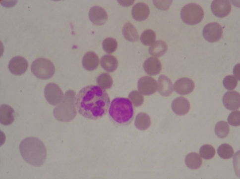
A four and half year old male child presented with abdominal distention for 7 months and pallor for 7 days. He had no fever, convulsion or autonomic disturbances. His other sibling is in good health. Physical examination revealed a body weight of 15 kg, moderate anemia and mild icterus. Abdominal examination showed enlarged liver, which was firm, non-tender and 5 cm below the right costal margin in the midclavicular line. Spleen was firm and 7 cm below the left costal margin. Haematological investigations showed hemoglobin of 3.2 gm/dl, total leukocyte count of 14,760/cu mm, differential leukocyte count was N86%, L10%, M01%, Stab 03%, hematocrit-13.2%, RBC count-1.1 × 10 12/l, MCV-113.8 fl, MCH-33.6 pg, MCHC-24.2 g/dl. Peripheral smear showed predominance of spherocytes, polychromatophils and few nucleated RBCs (Fig. 1). Corrected reticulocyte count was 14.98%. Based on these investigations a diagnosis of hemolytic anemia was made and a possibility of autoimmune hemolytic anemia or hereditary sherocytosis was suggested. The Coomb’s test was performed twice and was negative thus ruling out autoimmune hemolytic anemia. Since patient presented with jaundice, liver function tests were performed which revealed total serum bilirubin 4.0 mg/dl, conjugated and unconjugated bilirubin were 1.1 and 2.9 mg/dl respectively, A.L.T. 57 IU/L, A.S.T. 288 IU/L, alkaline phosphatase 194 IU/L, serum total protein 7.1 gm/dl, serum albumin 4.1 gm/dl with an A:G ratio 1.37. Prothrombin time was 18 s. Further evaluation showed Kayser–Fleischer (KF) rings on both corneas by slit lamp examination. Serum ceruloplasmin level was 10 mg/dl. The level of 24 h urinary copper excretion was 300 μg. He received 1 unit of packed cells and was put on penicillamine treatment (0.75 gm BD–0.25 gm OD). After 8 days of tretment, he showed correction of anemia (Hb = 7.7 gm/dl, HCT = 23.4%, Corrected Reticulocyte count = 10.11%) and discharged on penicillamine. During the followup of 5 months, his liver enzymes were decreased; however he had three episodes of hemolysis and received five blood transfusions.
Fig. 1.
P/S: Showing Spherocytes (Wright Stain × 1000)
Discussion
Wilson’s disease is a rare inherited disorder with an incidence of about 1 in 35000–100,000 live births, usually presenting between 5 and 35 years of age [5]. The gene for WD (ATP7B) has been mapped to chromosome 13 (13q14.3) [6]. The disease is not manifested clinically before 4–5 years of age because it takes time for copper to accumulate to toxic levels in the liver till such age. Hepatic manifestations of WD are more likely to occur in early childhood, while neurological symptoms are more commonly observed in adolescents [7]. Hemolytic anemia is a recognized but uncommon (10–15%) complication of this disease [1]. The prodrome to WD is occasionally a severe spherocytic hemolytic anemia. The hemolysis in Wilson’s disease is due to deficiency of ceruloplasmin, the copper transport protein which results in exessive inorganic copper in the the blood circulation, much of it accumulates in red blood cells. Although exact mechanism is not known, the increased copper accumulation in the RBC’S may damage the cell membrane, accelerate oxidation of hemoglobin and inactivate enzymes of pentose phosphate and glycolytic pathways. In present case there were increased number of spherocytes in peripheral blood possibly suggesting cell membrane damage. There were no signs of intravascular hemolysis.
Acute intravascular hemolysis and acute liver failure associated as first manifestations of WD have been reported earlier by Roche-Sicot et al. [8]. During the hepatic stage KF ring may be absent. KF rings are almost always present when the patient has neurologic symptoms. However, in our case, there were no any neurypsychiatric symptoms. The diagnosis of WD was made on the basis of KF rings; low serum ceruloplasmin and elevated basal urinary copper excretion. Among them 24 h urinary copper is the most sensitive test for the diagnosis of WD, particularly when liver biopsy can’t be performed due to coagulation abnormality [9]. But hepatic copper estimation is the most reliable test which is not easily available in India. Liver biopsy may not be possible because of bleeding problems and histological features are often not diagnostic of WD [10]. Hemolytic anemia often remits and may occasionally recur but the organ toxicity of copper (e.g. cirrhosis), generally, is the subsequent problem, unless treated.
Conclusion
Thus an acute hemolytic anemia may be the presenting episode in some patients of Wilson’s disease. So in case of spherocytic acute hemolytic anemia (Coomb’s negative) associated with liver failure; one should always suspect WD and investigate accordingly.
References
- 1.Grudeva–Popova JG, Spasova MI, Chepileva KG, Zaprianov ZH. Acute hemolytic anemia as an initial clinical manifestation of Wilson’s disease. Folia Med (Plovdiv) 2000;42(2):42–46. [PubMed] [Google Scholar]
- 2.McIntyre H, Clink HM, Levi AJ, et al. Hemolytic anemia in Wilson’s disease. N Engl J Med. 1967;276:439–444. doi: 10.1056/NEJM196702232760804. [DOI] [PubMed] [Google Scholar]
- 3.Deiss A, Lee CR, Cartwright GE. Hemolytic anemia in Wilson’s disease. Ann Intern Med. 1970;73:413–418. doi: 10.7326/0003-4819-73-3-413. [DOI] [PubMed] [Google Scholar]
- 4.Walshe JM. Wilson’s disease. The presenting symptoms. Arch Dis Child. 1962;37:253–256. doi: 10.1136/adc.37.193.253. [DOI] [PMC free article] [PubMed] [Google Scholar]
- 5.Roberts EA, Schilsky ML. A practical guidelines on Wilson’s disease. Hepatology. 2003;37(6):1475–1492. doi: 10.1053/jhep.2003.50252. [DOI] [PubMed] [Google Scholar]
- 6.Kasper DL, Braunwald E, Hauser S, Longo D, Jameson JL, Fauci AS (2005) Harrison’s principles of internal medicine, 16th edn. McGraw Hill, New York
- 7.Kalra V, Khurana D, Mittal R. Wilson’s disease-Early onset &lessons from a pediatric cohort in India. Indian Pediatr. 2000;37:595–601. [PubMed] [Google Scholar]
- 8.Roche-Sicot J, Benhemou JP. Acute intravascular hemolysis and acute liver failure associated as a first manifestation of Wilson’s disease. Ann Intern Med. 1977;86:301–303. doi: 10.7326/0003-4819-86-3-301. [DOI] [PubMed] [Google Scholar]
- 9.Yuce A, Kocak N, Demir H, Guracan F, et al. Evaluation of diagnostic parameters of Wilson’s disease in childhood. Indian J Gastroenterol. 2003;22(1):4–6. [PubMed] [Google Scholar]
- 10.Pandit A, Bavdekar A, Bhave S. Wilson’s disease. Indian J Pediatr. 2002;69(9):785–791. doi: 10.1007/BF02723693. [DOI] [PubMed] [Google Scholar]


